general characteristics
Manifestations depend on the characteristics of the olfactory disorder.
Patients may notice decreased sensitivity to smells or a complete loss of smell. Dysosmia often develops gradually: first, sensitivity to weak aromas disappears, and then even strong odorous substances (for example, ammonia) cease to be distinguished. Often, the sense of taste decreases at the same time. If the condition develops against the background of inflammatory diseases, disturbances in the sense of smell are combined with mucous discharge, nasal congestion, and a feeling of itching and burning. In other cases, increased sensitivity to aromas occurs, when even familiar odors cause discomfort to a person, causing headaches and other unpleasant sensations. When the sense of smell is distorted, patients may notice a sudden sensation of stench, which is described as the smell of feces, gasoline, or chemicals (cacosmia). Violations of the olfactory function do not pose a threat to human life, but sometimes they become the first symptoms of serious neurological diseases, so if they appear, you should seek qualified medical help.
Development mechanism
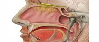
The olfactory zone of the nasal mucosa, containing specific nerve receptors, is located in the superior nasal concha. In order for a person to perceive odors, air along with odorous substances must pass through this section of the respiratory tract. In the presence of mechanical obstacles to the passage of air to anatomical structures, disturbances in the sense of smell are observed, such as a decrease in sensitivity to some or all odors. This condition is often caused by deviated nasal septum, hypertrophic rhinitis, sinusitis and adenoids.
Pathological insensitivity to odorous substances can be provoked by disturbances in different parts of the olfactory analyzer. Normally, information from the receptors of the nasal mucosa travels through special fibers to the subcortical structures and the olfactory center in the cerebral cortex. The symptom is observed with traumatic damage and rupture of the olfactory nerve due to traumatic brain injury, errors during neurosurgical operations. With unilateral damage to the nervous structures, loss of smell is determined only on the side of the pathological process.
Inhibition of sensitivity to various aromas develops during pathological processes in the mucous membrane of the nasal cavity (for example, atrophic rhinitis), when peripheral nerve receptors are destroyed. Impaired odor perception also occurs in degenerative brain diseases (Parkinson's disease, Alzheimer's disease) and brain tumors. In this case, dysosmia is caused by gradual atrophy and death of nerve cells in the olfactory zone.
A special mechanism for impaired sense of smell is characteristic of epilepsy. With this disease, patients note the appearance of unpleasant odors, which is associated with the formation of excitation zones in the brain, impulses from which spread to various parts of the cortex. Dysosmia can also occur in the absence of organic pathology - the symptom is observed in acute psychoses and hysterical states. The development of olfactory dysfunction is associated with a functional disruption of connections between parts of the peripheral and central nervous systems.
Olfactory organ
In aquatic animals at the lower stages of phylogenesis, the sense of smell and taste are poorly differentiated. The simplest form of the olfactory organ, the olfactory pits, is observed in fish. As the nasal cavity forms, olfactory cells move into its mucous membrane.
The human olfactory organ is formed in the 4th week of intrauterine development in the form of paired thickenings of the ectoderm lining the olfactory fossa. As the head develops, the olfactory pits deepen, move closer together, and take part in the formation of the nasal cavity. The rudiment of the olfactory organ is displaced into the mucous membrane of the nasal cavity. The sensory cells of the olfactory fossa are connected through processes to the olfactory bulbs.
The olfactory organ
(organum olfactus
seu
organum olfactorium)
(see
Fig. 55)
of an adult is represented by olfactory neurosensory cells embedded in the mucous membrane of the superior turbinate, superior nasal passage and the corresponding part of the septum of the nasal cavity - the so-called olfactory region.
These cells are the first neuron
of the olfactory analyzer.
Their specific irritant is volatile substances inhaled with air, which dissolve in mucus, lead to a number of cytochemical transformations and are perceived by receptors. The neurites of the olfactory cells unite into 20-40 olfactory filaments (fila olfactoria),
which pass through the openings of the cribriform plate of the ethmoid bone and end in
the olfactory bulb (bulbus olfactorius).
The body of the second neuron lies in the olfactory bulb .
The neurites of these cells form
the olfactory tract (tractus olfactorius),
which ends in the primary olfactory centers - the nerve ganglia of the olfactory triangle, the anterior perforated substance and the septum pellucidum.
The bodies of the third neurons are located in them .
reach
the cortical end of the olfactory analyzer
in various ways (see
Olfactory brain) which is located in the crus and parahippocampal gyrus.
From the neurons of the latter, impulses enter the subcortical center of smell, which is located in the nuclei of the mammillary bodies (corpora mammillaria)
of the diencephalon.
In addition to the mammillary bodies, the subcortical center of smell is also the anterior nuclei of the thalamus (nuclei anteriores thalami).
The subcortical centers of smell are connected to each other
by the mastoid-thalamic fascicle (fasciculus mammillothalamicus).
Classification
Smell disorders are divided into acquired and congenital (with Kallmann syndrome). All pathological conditions are classified into two groups: symptoms of irritation (increased sensitivity to different odors) and symptoms of loss (decreased sense of smell). To choose the right treatment method, classification according to the type of disorder is also important, according to which the following types of olfactory disorders are distinguished:
- Hyperosmia.
Sensitivity to odors of normal intensity increases with autoimmune diseases, Lyme disease, and brain pathologies (Parkinson's disease, multiple sclerosis). The symptom also occurs during pregnancy. - Hyposmia
. A decrease in the function of smell is possible both with damage to the cortical centers and with disruption of the receptors of the nasal mucosa due to acute or chronic inflammatory processes. The sign is typical for smokers. - Anosmia.
Complete loss of smell occurs after brain injuries, nasal polyps, and destructive processes in the mucous layer. The symptom is pathognomonic for intoxication of the body with salts of heavy metals and acrylates. - Parosmia
. Distorted perception of odors most often occurs during the period of convalescence after inflammatory diseases (rhinitis, sinusitis). A type of parosmia is cacosmia, which is observed with sinusitis and dental pathology. - Olfactory hallucinations (phantosmia)
. The sensation of a non-existent smell becomes a harbinger of an incipient epileptic attack. The disorder also develops with cerebral injuries and neoplasms. - Olfactory agnosia
. Familiar smells are no longer recognized when the area of the cerebral cortex responsible for the olfactory function is damaged. The symptom is determined by strokes, brain tumors, abscesses.
Depending on the location of the lesion, rhinogenic olfactory disorders are distinguished, which are associated with chronic rhinitis, deviated nasal septum, nasal polyps, and neurosensory ones. The latter, in turn, are divided into peripheral (pathology of the olfactory nerve endings in the nasal cavity) and central (damage to the brain centers of smell). Separately, combined dysosmia is distinguished, when nasal breathing disorders are combined with changes in the mucous membrane of the olfactory area.
Smell
Smell is the perception by animals and humans through the corresponding organs of a certain property (smell) of chemical compounds in the environment. O. is one of the types of chemoreception, characterized in that odorous substances (OS) are usually present in low concentrations and, as a rule, are not beneficial or harmful to the body in themselves. They serve only as signals indicating certain objects or events in the external environment. In terrestrial animals, PVs are delivered to the olfactory receptors
in the form of vapors in a stream of air or by diffusion. For aquatic animals, non-volatile substances that are devoid of smell in the usual sense can be “odorous” (for example, solutions of some amino acids excite the olfactory receptors of fish).
The role of O., as well as the level of development of the sense of smell of organs
, varies greatly among different animal species.
Thus, mammals are divided into macrosmatics, in which the organs are well developed (most species include them), microsmatics with a relatively weak development of the organs (seals, baleen whales, primates), and anosmatics, in which the typical organs of the organism are absent (toothed whales). (see Fig. 1
).
Animals use oxygen to search for and select food, track prey, escape from an enemy, and for bioorientation
and biocommunication (marking territory, finding and recognizing a sexual partner, etc.; see
Animal Communication
).
A special group of PVs consists of pheromones, which are secreted by animals, usually with the help of special glands, into the environment and regulate the behavior of representatives of the same species (all kinds of odorous marks, attracting substances - attractants, “alarm substances”, etc.).
If in vertebrates olfactory signals act, as a rule, in combination with others - visual, auditory, tactile signals, then in insects a pheromone can play the role of the only “key stimulus” that completely determines their behavior. In human life, oxygen plays a relatively modest, but important role. People with impaired oxygen more often suffer from food poisoning. The taste of food is determined to a large extent by the olfactory sensations. To study O., they use olfactometers—devices that allow one to dose the intensity and duration of exposure to PV. Thus, for many compounds, the olfactory threshold was determined, i.e., the minimum concentration of a substance at which its odor is perceived. Olfactory sensitivity to some PVs is unusually high. Thus, a person feels the presence of trinitrobutyltoluene when its content is 1 cm3
air about 5·10-15
g
(10 million molecules).
Even higher is the sensitivity of a dog to butyric acid (10 thousand molecules per 1 cm3
) or a male silkworm butterfly to bombycol, the female sex pheromone (100 molecules per 1
cm3
).
In these cases, 1 PV molecule is apparently sufficient to excite a single olfactory receptor. With prolonged or repeated exposure to PV, the olfactory threshold to this substance and, to a lesser extent, to other PV increases. This olfactory adaptation is partly associated with fatigue of the olfactory receptors and is partly formed in the brain centers of the olfactory analyzer
. Mn. In addition to the olfactory receptors, PVs also excite the sensitive endings of the trigeminal nerve fibers, which are located in the nasal mucosa and serve as nonspecific receptors of “general chemical sensitivity.” The participation of the trigeminal nerve reception in the formation of a complex sense of smell is especially pronounced for PV with a sharp irritating odor such as acetic acid and ammonia.
The number of chemical substances is huge, and the smell of each of them is unique: there are no two different chemical compounds with exactly the same smell. Various odor classification schemes have been proposed, but none of them have become generally accepted. Substances that are similar or have different chemical structures can have similar odors. The connection between odor and the chemical structure of a substance remains largely unexplored; however, in practical work with aromatic substances
a number of rules of thumb are used to sometimes predict the odor character of a new compound based on its structure.
The mechanism of the primary interaction of PV molecules with the olfactory receptor cell has not been sufficiently studied. Subsequent stages of signal transmission in the olfactory system have been better studied, mainly by recording bioelectric potentials
.
When PV acts on the olfactory epithelium, its surface becomes electronegative relative to the rest of the tissue. This electrical reaction, the so-called. electroolfactogram consists of the responses of a large number of individual receptor cells ( Fig. 2
).
In each of them, a shift in the potential of the cell membrane occurs, leading to the appearance of nerve impulses or a change in their frequency. Typically, receptors respond by increasing the frequency of impulses, while exhibiting different sensitivity to different PIs. Some substances can cause a decrease in the frequency of impulses; This receptor may be insensitive to some substances. Receptors have different selectivity for PV. The combination of olfactory receptors of many types serves as the basis for distinguishing a large number of odors. The impulse responses of nerve cells recorded in the olfactory centers of the brain have similar properties. From the antenna of insects, it is possible to register electrical potentials of two types in response to stimulation of the PV: a slow electroantennogram (analogue of an electroolfactogram) and nerve impulses of individual receptors ( Fig. 3
). In many insects, in addition to a set of receptors with low selectivity and a wide range of perceived substances, reminiscent in properties of the olfactory receptors of vertebrates, another type of receptors has been found - “narrow specialists” that react only to one substance (for example, a sex pheromone).
In older people, olfactory sensitivity decreases. During pregnancy, O. may worsen and become perverted. O. worsens or disappears with swelling and atrophic changes in the nasal mucosa, especially during ozena
, tumors or injuries to certain parts of the brain.
O. disturbances can manifest themselves as signs of irritation (hyperosmia—increased sensitivity to odors, olfactory hallucinations in some mental illnesses—the sensation of non-existent, often unpleasant odors) and loss (decreased O.— hyposmia
, loss—
anosmia
, impaired recognition of odors). Treatment: elimination of the main cause that caused the disorder O.
Lit.:
Bronshtein A.I., Taste and smell, M. - L., 1950; Minor A.V., Physiology of smell, in the book: Physiology of sensory systems, part 2, M. - L., 1972; Moncrieff RW, The chemical senses, 3 ed., L., 1967; Ottoson D., Shepherd GM, Experiments and concepts in olfactory physiology, in: Progress in brain research, v. 23, 1967; Taste and smell in vertebrates. (Ciba Foundation Symposium), L., 1970; Olfaction and taste (Proceedings of International symposium), v. 1-4, Oxf., 1963-1972.
A. V. Minor.
Rice. 2. Electrical potentials recorded from the olfactory receptors of a frog upon irritation with an odorous substance; A - total reaction of the olfactory epithelium - electroolfactogram; B - response of a single receptor obtained using an intracellular electrode. The arrows indicate the moments of short-term exposure to vapors of an odorous substance.
Rice. 1. Comparative size of the primary olfactory center - the olfactory bulb in macrosmatics (a, b) and microsmatics (c, d). In the schematic representation of the brain, the olfactory bulb is blackened (a - cat, b - fox, c - chimpanzee, d - human).
Rice. 3. Electric potentials of olfactory receptors of insects: A - electroantennogram. The antenna of a male silkworm was blown by air passing over filter paper coated with bombicol (a female attractant); curves 1-5 correspond to 10-8, 10-7, 10-6, l0-5, 10-4 g
bombicola. B - impulse activity of two olfactory receptor cells in the sensilla of Chinese oak Saturnia when stimulated by different odorous substances: 1 - terpeniol excites one cell (large impulses) and inhibits the other (small impulses); 2 - geraniol strongly inhibits the activity of both cells; 3 - isosafrole weakly inhibits both cells. At the top - ways to register biopotentials are schematically shown; black line—stimulation period.