Have you ever wondered why we need a spinal cord? Everything is clear with the head: it is responsible for cognitive functions, receives and distributes impulses throughout the body, thanks to which all our systems work. What does the spinal cord do? This is a kind of connecting link between the brain and the peripheral nervous system. And if something happens to it, the body stops working fully.
What is the spinal cord
The spinal cord, together with the brain, is part of the central nervous system of the human body. Both of them begin to form in the early stages of embryo development. Around the 20th day of pregnancy, certain folds form on the body of the embryo, which will eventually turn into what experts call the neural tube of the fetus. It represents the rudiment of the future central nervous system. During the development of the tube, its front part will turn into the brain, and the back will serve as the basis for creating the spinal cord.
Content:
- What is the spinal cord
- How does the spinal cord control the body?
- Functions of the spinal cord
- Consequences of central nervous system injury
Humanity has been studying the structure and role of the spinal cord since antiquity. For example, the ancient Greek physician Galen studied the central nervous system of animals and gladiators, analyzing how different types of injuries affect the functionality of the body. Modern scientists already know for sure that the spinal cord is a cylindrical bundle of nerve fibers that is connected to the brain and runs through the spine from the neck to the lower back. Scientists also know that in the body of an adult, this organ can reach 40-45 cm in length, 1-1.5 cm in width and weigh about 35 g. It has the shape of a thick-walled tube with a narrow channel inside, somewhat flattened in the anteroposterior direction, located inside the spinal canal, which is divided into segments (they correspond to the sections of the spine):
- 7 cervical;
- 12 breast;
- 5 lumbar;
- 5 sacral;
- 3-4 coccygeal.
Interestingly, in the adult body, the spinal cord does not occupy the entire cavity of the spinal canal, but only about 2/3 of its length. Experts explain this feature by the fact that the spinal column grows faster than the spinal cord, therefore, as a person grows, this part of the central nervous system takes up less and less space in the spinal canal. That is, in the body of adults, the spinal cord in its lower part reaches only the first lumbar vertebrae.
The spinal cord is covered by three meninges (protective membranes). The first, located closest to the spinal substance (internal), is called the pia mater of the spinal cord. It consists of a network of vessels that are responsible for the blood supply to the brain. On top of it is the middle arachnoid membrane, under which contains cerebrospinal fluid - cerebrospinal fluid. By the way, when a spinal cord puncture is performed, the cerebrospinal fluid is taken for analysis, that is, a needle is used to penetrate the space between the inner and middle membranes. The uppermost (outer) shell is hard. It protects the nerve roots leading to the intervertebral foramina.
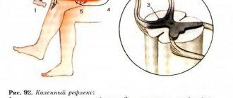
If the spinal cord is cut across, you can see that its tube has an oval, almost round shape. The upper layer of this organ consists of white matter, under which lies the gray spinal substance. In cross section, it has the shape of a butterfly, from which horns of gray matter extend forward, backward and sideways. In the central part of the spinal substance is the intermediate nucleus of the gray matter. This part is the most important. She is responsible for collecting all the information and making decisions about the activation of one or another reaction of the body.
White matter is made up of bundles of nerve fibers called axons. They allow different levels of the central nervous system to exchange impulses. It is known that each axon bundle differs in the specificity of the signals transmitted. So, some of them (ascending) transmit information to the brain, while the rest (descending) are responsible for transmitting impulses from the brain to neurons in different muscles and glands located throughout the body. White matter axons are covered with an insulating myelin sheath, which facilitates the rapid and free transmission of impulses. Myelin is whitish in color, hence the name white matter.
The gray substance of the spinal cord is formed from nerve cells and their branched processes - dendrites. This substance is responsible for the perception and processing of information. For example, if we talk about the brain, it is the gray matter in it that gives a person the ability to think. If we talk about the spinal cord, the horns of the gray matter also process various information messages from the body. Moreover, each horn has its own function.
How does the spinal cord control the body?
Each segment of the spinal cord controls the functionality of a specific part of the body. Thus, the segment of the central nervous system located along the cervical vertebrae sends impulses to the back of the head, neck and upper limbs. The thoracic segments are responsible for the functionality of the torso (chest, abdomen). The lumbar zone of the central nervous system controls the lower extremities, and the sacrococcygeal segments control the pelvis and internal organs located in this area.
The relationship between the segments of the spinal cord and the parts of the body is always strictly observed. At each “floor” of the body, nerve endings extend from the brain and are distributed throughout the area controlled by these segments.
The spinal cord is a tube that runs inside the spine, and spinal nerve processes emerge from the intervertebral spaces. Understanding the structure of this part of the central nervous system, it becomes clear how dangerous the slightest violation of the natural positions of the vertebrae is for a person. Any displacement of even one vertebra can lead to compression of the spinal nerves and disruption of interaction between the parts of the central nervous system and the part of the body for which the segment is responsible.
Many people know that spinal injuries can lead to paralysis. But this is not the only danger that can arise from a spinal cord injury. By and large, most health problems can arise due to misalignment of the vertebrae - even gastritis or ulcers. Just imagine, the vertebra is displaced, pinching the nerve ending, which is why the brain sends incorrect signals to the digestive system and gastric juice begins to be produced in excess, corroding the gastric mucosa. The result is gastritis, and then an ulcer.
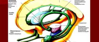
Structure and functions of the brain stem
Let us consider the characteristic features of the structure of the brain, starting with its “lowest” section - the trunk, directly bordering the spinal cord.
The brain stem is covered from above and from the sides by the cerebral hemispheres and the cerebellum. Its structure has features similar to the spinal cord; cranial nerves (from III to XII pairs) depart from it, innervating the muscles and scalp, as well as internal organs (respiratory and digestive systems, heart). The brain stem communicates with the spinal cord through special pathways. The brain stem contains centers that are important for the whole organism and are associated with the regulation of breathing, blood circulation, muscle tone, and others. The brainstem combines 3 sections: the medulla oblongata, the pons and the midbrain.
Medulla oblongata The medulla oblongata is a continuation of the spinal cord. Since it is in the medulla oblongata that the vital centers of respiration and blood circulation lie, damage to this section leads to the cessation of respiratory movements, disruption of the heart, a sharp decrease in blood pressure, resulting in rapid death. The centers of vomiting, sneezing and coughing are also located here.
The pons The pons plays an important role in connecting the cerebral cortex with the cerebellum and conducting auditory information.
Midbrain The importance of the midbrain is great for the regulation of skeletal muscle tone, the implementation of protective reflexes in response to strong visual and auditory stimuli, as well as indicative reactions (synchronous rotation of the head and eyes towards the light source).
Functions of the spinal cord
So, the gray matter of the spinal cord has 3 horns, which actually perform the main functions of this organ:
- analyze sensory signals;
- govern internal organs (vegetative function);
- control muscles (motor function).
The collection of sensory signals occurs separately on each “floor” of the body. That is, each segment of the spinal cord accumulates information about what is happening in the territory “controlled” by it. This information is also strictly structured. It concerns three aspects:
- pain, that is, damage to cells and tissues on the “floor”;
- skin sensitivity, that is, heat, cold, vibrations, etc.;
- muscle sensitivity, that is, about the condition of muscles, tendons and joint position.
The spinal cord receives all information through clusters of nerve cells known as the spinal ganglia (spinal ganglia). The number of these nodes exactly corresponds to the number of spinal segments - 31. The cells included in the ganglion have a special structure: 1 long dendrite and 1 axon. The dendrite collects information about what is happening on the controlled “floor”, and the axon connects to the dorsal horn of the gray spinal substance, to which it transmits the received data. In this area, the primary processing of sensory signals occurs and then the transfer of information to the intermediate nucleus of the gray spinal matter, which determines the further reaction of the body. But here it is worth mentioning another interesting point. The neurons of the intermediate nucleus receive signals not only from the horn, but also from the brain, which also retains the right to vote on what the reaction to the impulse received from the horn should be.
To make this information easier to understand, we will use an example. Let's say a person took a glass of boiling water. As a result, sensory fibers send pain information to the spinal cord. Consequently, he is ready to give the signal to throw a red-hot object. But at the same time, another signal comes from the brain that, for example, the glass contains something valuable or the glassware itself is valuable, so the vessel should be brought to the surface. These conflicting signals arrive at the intermediate nucleus of the spinal gray matter, where neurons analyze both options and either trigger or block a response. If the intermediate nucleus approves of the reaction, it transmits the corresponding signal to the anterior and lateral horns of the gray substance.
The anterior horns of the spinal gray matter contain so-called motor neurons, which cause muscle contractions. Autonomic neurons (sympathetic and parasympathetic nervous system) are concentrated in the lateral horns, which regulate the activity of internal organs.
Structure and functions of the diencephalon
Anterior to the brain stem, between the midbrain and the cerebral hemispheres, is the diencephalon. The upper part of the diencephalon is called the thalamus or thalamus optic, the lower part is the hypothalamus.
The meaning of the thalamus The thalamus, a paired ovoid-shaped formation, is a collector of all types of sensitivity from all parts of the body and sensory organs. From here this information is transmitted to the cerebral cortex. Certain areas of the thalamus are important components of the limbic system of the brain, which controls the psycho-emotional behavior of a person, while others are involved in ensuring memory processes. There is evidence that the thalamus is involved in the perception of pain. Destruction of certain areas of the thalamus can lead to a decrease in anxiety, tension, aggressiveness, elimination of obsessive thoughts, as well as a sharp decrease in motor activity.
The significance of the hypothalamus The significance of the hypothalamus is associated primarily with the regulation of the activity of internal organs. The nuclei of the hypothalamus produce special substances - neurohormones, which enter the pituitary gland, and from it into the blood.
The pituitary gland is an endocrine gland, closely related in structure and location to the hypothalamus. The single hypothalamic-pituitary system of the diencephalon controls the work of other endocrine glands and, with their help, regulates the functions of the body. This system controls the state of water-salt balance, metabolism and energy, the functioning of the immune system, thermoregulation, reproductive function of the body, etc. There is evidence that the hypothalamus contains specific pleasure centers that play an important role in the formation of motivations and emotional forms of behavior . In the area of the hypothalamus there are areas of the optic nerves through which information is transmitted from the retina of the eye.
The significance of the pineal gland The diencephalon also includes the pineal gland, or pineal gland, an endocrine gland that influences the work of other endocrine glands and is involved in the regulation of the seasonal rhythms of the body.
Consequences of central nervous system injury
Even partial damage to the spinal cord can cause disruption of the functionality of a wide variety of systems and organs.
Bones and joints
A spinal cord injury can impair the absorption of calcium and some other important minerals into bone tissue. But at the same time, these substances can accumulate in the urinary system, causing the formation of stones. Experts usually explain such processes by a decrease in human motor activity. In addition, decreased mobility can lead to stiffness in joints, including the knees, elbows, and shoulders. To prevent this, there are special sets of exercises for people with disabilities.
Urinary system
The urinary system consists of the kidneys, which filter the blood and produce urine, and the bladder, which collects and then removes it from the body. After a spinal cord injury, the kidneys continue to produce urine, but the bladder may not work as well as before. After an injury, a person may not feel when his bladder is full of fluid, or his urinary system, due to a lack of necessary impulses, may lose the ability to eliminate urine naturally. As a result, a catheter may be needed to empty the bladder. Another option: urine may, on the contrary, come out involuntarily and the person loses the ability to control this process.
Respiratory system
Even after a spinal injury, human lungs continue to function. However, the ability to inhale and exhale air is controlled by the muscles. Depending on the level of injury, a person may lose the ability to cough or take deep breaths. And they are necessary for complete straightening of the lungs and deep air circulation.
The most important muscle for breathing is the diaphragm. It is a large arcuate muscle that is located directly under the lungs. And if the diaphragm is paralyzed due to damage to the spinal cord, special equipment may be needed to maintain respiratory function.
Leather
Damage to the spinal cord can also negatively affect the skin. Skin plays a very important role for humans. It protects the body from germs and other pathogens from the outside world. The spinal cord is an important organ that helps protect the skin from damage. For example, when sitting in one position for a long time, the brain sends signals in the form of a feeling of discomfort, which encourages you to change your position and prevent damage or pressure on the skin. In the same way, the central nervous system protects against burns, cuts and other possible damage to the skin (for example, a person reflexively withdraws his hand from a hot or cutting object). With spinal cord injuries, such signals may not appear, which greatly increases the risk of damage to the skin and, consequently, the penetration of microbes into the body.
Reproductive system
After a spinal cord injury in the lumbar or sacrococcygeal segment, the functionality of the reproductive system may be affected. Men usually experience erectile dysfunction, problems with ejaculation and fertility. In women, after injury, sensitivity in the genital area may be impaired, although fertility itself may not be directly affected.
Intestines
It is well known that the digestive system is responsible for the breakdown of food consumed, the absorption of nutrients from it and the removal of metabolic products. A fairly common consequence of central nervous system injury is impaired intestinal motility. That is, the digestive system continues to absorb and digest food, but the process of eliminating excrement is disrupted. In a healthy body, when the rectum is full, an exchange of impulses occurs between the intestines and the brain. If the spinal cord is damaged, such signals may not be received. As a result, a person loses the ability to control the process of defecation.
Autonomic nervous system
The autonomic nervous system regulates the activity of internal organs, blood and lymphatic vessels, as well as endocrine and exocrine glands. Body temperature, blood pressure, digestion and other important functions depend on it. After a spinal injury, some of its functions may be impaired. Quite often (especially if the damage occurs in the thoracic segment) after an injury the body loses the ability to thermoregulate. What does it mean? If the autonomic nervous system works correctly, then the body temperature remains stable regardless of the weather conditions outside or the indoor microclimate. Due to spinal cord damage, body temperature may rise or fall according to indoor or outdoor temperatures. Disturbances in thermoregulation are most noticeable in parts of the body located below the area of injury.
Structure and functions of the cerebellum
The cerebellum is located above the brain stem and is connected to its parts by 3 pairs of peduncles. The cerebellum has 2 small hemispheres covered by the cerebellar cortex. The main functional significance of the cerebellum is to maintain body balance, regulate and coordinate body movements, giving them smoothness, accuracy and proportionality. The cerebellum programs the automatic execution of movements, which is made possible by its connections with the spinal cord, brainstem and cerebral cortex. For example, when walking and running, the cerebellum controls the position and movement of the torso and arms in accordance with the movements of the legs and the movement of the body's center of gravity. When writing, it is responsible for maintaining optimal posture and coordinating the movements of the head, eyes and hands. The cerebellum plays an important role in performing rapid sequential and simultaneous movements, such as the movements of the hands of a pianist or typist.