A person performs many actions and movements every day, and behind each of the simplest movements there is a huge mechanism of the motor-skeletal system. We get up early in the morning, wash, someone does exercises, have breakfast and go to work, all this happens simply and routinely. But if we could look into what is happening behind the curtain of this performance, we would see that behind all these actions there are neurons of the brain, and, in particular, human motor neurons. What are these physiological mechanisms, where are they located, how do they work, where is the motor neuron located later in this article.
Functions of motor neurons
All physical actions that a person can perform are carried out according to the same principle: through contraction and stretching of muscles and tendons.
These contractions occur due to the existence of communication between all muscles and tendons with a single coordination center - the brain. These messages consist of cells with different tasks - neurons. Accordingly, special motor cells—motoneurons—participate in the implementation of motor functions. Muscle contraction occurs by changing just two commands: relax and tense - that is, straighten and contract. A special motor neuron is responsible for each of these conditions. The motor neuron responsible for contraction is called a flexor, and the motor neuron responsible for relaxation is called an extensor.
Types of motor neurons
Motor neurons are divided into central and peripheral according to their location in the body. Accordingly, central motor cells are located in the spinal cord and brain, and peripheral ones are located directly in the muscles and are connected to them through the axons of neurons.
Central neurons are responsible for conscious and reflex movements; electrochemical impulses with commands diverge from them to the periphery and are transmitted to muscles, organs and other tissues. The main accumulation of groups of motor cells of the somatic nervous system occurs in the region of the anterior horns of the spinal cord. Each group is responsible for contracting its own muscles. For example, a group of motor neurons in the cervical region controls the muscles of the arms.
It is precisely because of the participation of the spinal cord and its motor neurons in the control of the motor system that the spine is dangerous to injure and there is a high risk of injury and disability. And even spinal massage should be trusted only to trusted professionals.
Classification of motor neurons:
- Renshaw cells
- Small alpha motor neurons.
- Large alpha motor neurons.
- Gamma motor neurons.
Large alphas form the trunk of the nerve chain, and small alpha and gamma with their small axons transmit signals to the most inaccessible areas. Renshaw cells perform a special function of signal switching. These are a kind of telephone operators who, in the last century, manually connected different telephone subscribers.
Tips: How to Improve Neuron Functions
As always, healthy habits play an important role in the optimal development of neural function. Our brain thanks us for taking care of our body. As they say, “a healthy mind in a healthy body.” What can we do to improve brain plasticity and neurogenesis?
Sleep while resting: It is not necessary to sleep strictly 8 hours. Each of us has our own sleep rhythm, and there are people for whom sleeping 7 or 7.5 hours is quite enough
However, it is important that sleep is restorative.
Use moderate physical activity and stimulation: neurogenesis occurs to adapt to the environment. It involves overcoming challenges to achieve our goals, which in turn engages our decision-making skills.
Avoid excessive stress: A little stress is good, but we always need to know when we are “crossing the line.”
Having sex: This is a great way to stimulate and relieve stress, as well as exercise.
Do brain exercises: CogniFit is the leader among cognitive stimulation programs, all exercises can be performed online using any device - computer, phone, tablet
Neuropsychologists and neuroscientists have developed fun exercises in the form of simple games that can be used to professionally “train” the basic functions of the brain. This program has been highly acclaimed by the scientific community and is currently being used in various medical institutions, schools, colleges and universities around the world. Discover this simple tool that anyone can use to test and train their brain like a pro.
From the editor: Causes of numbness on the left side of the face, their diagnosis and treatment
Lack of sleep, monotony, constant routine and high levels of stress lead to a slowdown in neurogenesis.
How do motor neurons work?
The entire nervous system, central and peripheral nerves, is a large and complex mechanism in which many elements work in concert. In fact, human upright posture is a unique and very costly function for the body, which requires a special kind of motor mechanism, and humans have it.
Any physical action comes down to the fact that a certain group of muscles flexes and extends, and for this there are special “flexor and extensor” cells.
A motor signal is formed in the corresponding part of the cerebral cortex. Another specialized cells are involved in this, which are called pyramidal for their shape. Pyramidal cells make up the pyramidal motor pathway through which the signal reaches the spinal cord.
Different areas of the cerebral cortex are responsible for the work of flexors and extensors, as a result of which muscle contraction occurs: a signal is formed in the area of the precentral gyrus, and the posterior regions of both hemispheres are already responsible for the work of flexors and extensors.
Reflexes
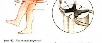
Reflexes are quick, involuntary reactions in response to stimuli. The most well-known reflex is the patellar reflex, which is tested when a doctor taps a patient's knee during a physical examination. Reflexes are integrated in the gray matter of the spinal cord or brain stem. Reflexes allow the body to respond to stimuli very quickly, sending responses to effectors before nerve signals reach the conscious part of the brain. This explains why people often pull their hands away from a hot object before they realize they are in danger.
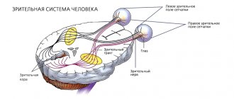
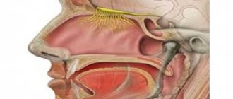
Functions of Cranial Nerves Each of the 12 cranial nerves has a specific function within the nervous system. The olfactory nerve (I) carries odor information to the brain from the olfactory epithelium in the roof of the nasal cavity. The optic nerve (II) transmits visual information from the eyes to the brain. The oculomotor, trochlear, and abducens nerves (III, IV, and VI) all work together to allow the brain to control eye movement and focusing. The trigeminal nerve (V) carries sensations from the face and innervates the muscles of mastication. The facial nerve (VII) innervates the facial muscles to make facial expressions and carries taste information from the anterior 2/3 of the tongue. The vestibulocochlear nerve (VIII) carries auditory information from the ears to the brain.
Editor's Note: Ask an Expert
The glossopharyngeal nerve (IX) carries taste information from the posterior 1/3 of the tongue and aids in swallowing.
The vagus nerve (X), called the vagus nerve because it supplies many different areas, travels through the head, neck, and torso. It carries information about the state of vital organs in the brain, provides motor signals for speech control, and provides parasympathetic signals to many organs.
The accessory nerve (XI) controls movements of the shoulders and neck.
The hypoglossal nerve (XII) moves the tongue for speech and swallowing.
Types of motor neurons
Motor neurons and cells are functionally divided into the following groups:
- Sensitive (afferent). Receive and process signals from the brain and spinal cord.
- Motor (efferent). Directly attached to muscle fibers. Each muscle has its own motor nerve.
- Intercalated (associative). They are a kind of distribution transformer booths in the body. They receive the signal and, depending on the instructions received, can amplify, weaken and transmit it further along the chain.
What muscles are motor neurons connected to?
All muscle fibers have their own motor neurons attached. Together, the motor cell and the muscle fiber to which it is attached are called a “motor unit.” Each such unit functions independently of other similar units. And each motor unit contains only one type of muscle fiber.
Types of muscle fibers:
- Slow oxidative fibers.
- Fast oxidative fibers.
- Fast glycolytic fibers.
Features of nerve cells
Neurons are somewhat reminiscent of a colony of ants - there are just as many of them and they are divided into various groups according to specialization. It is in the differences between these specializations that their specific features and differences lie.
Types of motor neurons, their characteristics and localization in the cerebral cortex:
- Central innervating flexors: localized in the area of the precentral gyrus and are responsible for compression (contraction) of skeletal muscles.
- Central innervating extensors: localized in the hindbrain region and are responsible for relaxing skeletal muscles.
- Peripheral alpha: cells that transmit commands to muscle fibers to contract. Localized in the anterior horns of the spinal cord.
- Peripheral gamma: cells responsible for muscle tone. They are localized in the same place, in the anterior horns of the spinal cord.
- Intercalary: present in all parts of the central nervous system, and carry out the role of communication of all signals in the central nervous system.
Structure of neurons
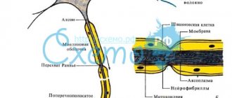
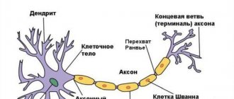
A motor nerve cell consists of three conventional parts: the body of the motor neuron, one axon and many dendrites. Dendrites are active nerve endings of cells through which communication between neurons is established and electrochemical impulses pass. Nerves form connections with each other of varying degrees of stability. And the axons are already connected to other cells and transmit command signals to them, forming the entire nervous system.
Some of the connections form a fully automated system for controlling many physiological processes that a person does not need to consciously control. These connections are called conditioned and unconditioned reflexes. Also, stable neural circuits are formed in the process of any activity, including thinking.
The more often a person performs the same action, thinks the same thoughts, reacts in the same way to the same stimuli, the more stable the connections that these events form become. This is how acquired reflexes, good and bad habits, physical and psychological dependencies are formed. Each person’s return to habitual behavior only strengthens the associated neural circuits, and any attempt to further change one’s character or behavior will encounter more and more resistance from the psyche (where the root of any addiction is located) and a feeling of discomfort.
Reflex arc
The very majority of automated neural circuits that are responsible for the unconscious regulation of all processes in the body are, in fact, a reflex arc. A “reflex arc” is a stable neural connection that is guaranteed to fire under certain identical conditions. For example, withdrawing your hand from a hot object is a reflex that makes a connection. The reflex is triggered by an irritant - in this example, any hot object.
The general mechanism of reflexive activity is as follows:
- The signal about the presence of the stimulus is transmitted to the sensitive nerve endings and, through communication from the dendrites, is redirected for analysis to the brain. Each area of the cerebral cortex is responsible for a specific specialization. Accordingly, nerve endings throughout the body are tied to different areas of the brain, and each neuron sends signals exclusively to its own command center.
- After the dendrites are the first to react to the stimulus, this reaction passes to the cell.
- Information about the event is transformed into an electrochemical impulse, which is immediately transmitted throughout the nervous system to the corresponding parts of the cerebral cortex.
- The brain analyzes the received information and transmits the response impulse back along the entire chain with a set of mandatory instructions for the cells, how they should behave in the response phase and whether this phase is needed.
- The phase of physical response to a stimulus in which cells carry out instructions received.
Structure and functions
The intercalary cell consists of a body from which a single axon and dendrites extend. The dendrites of intercalary cells are often short. Their axons variably pass within the boundaries of the spinal cord from the posterior horns to the anterior ones (close the arc at the level of the spinal cord segment) or spread to other levels of brain structures - spinal, brain.
One of the functions of interneurons is inhibition of the intensity of certain signals. For example, interneurons of the neocortex (the new cortex responsible for higher mental functions - sensory perception, conscious thinking, voluntary motor activity, speech) selectively reduce the intensity of some signals coming from the thalamus to prevent the need to be distracted by extraneous, unimportant stimuli. If the impulse provoked by an external stimulus is not strong enough, it may fade before reaching the cortex of the brain.
The area of influence of intercalary cells is limited by individual structural features - the length of axonal processes, the number of collateral branches. Typically, intercalary ones are equipped with axons with terminals (the terminal section represented by the synaptic ending - the place of contact with other cells) ending within the same center, which determines integration within the group.
Interneurons close reflex arcs, they perceive excitation from afferent nerve structures, process data and transmit them to motor neurons. Associative cells play a leading role in the formation of neural networks, where the storage period of incoming and processed information is extended.