Structure and classification of neurons
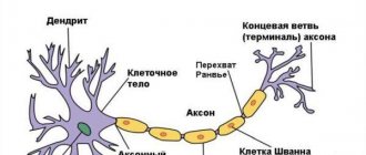
The brain is made up of billions of nerve cells, or neurons. A neuron consists of three main parts: the body of the neuron (soma); dendrites - short processes that receive messages from other neurons; an axon is a long single fiber that transmits messages from the soma to the dendrites of other neurons or body tissues, muscles. The transfer of excitation from the axon of one neuron to the dendrites of another is called neurotransmission or neurotransmission. There is a wide variety of CNS neurons. Most often, neurons are classified according to three characteristics - morphological, functional and biochemical.
The morphological classification of neurons takes into account the number of processes in neurons and divides all neurons into three types - unipolar, bipolar and multipolar.
Unipolar neurons have one process. In the nervous system of humans and other mammals, neurons of this type are rare. Bipolar neurons have two processes - an axon and a dendrite, usually extending from opposite poles of the cell. In the human nervous system, bipolar neurons themselves are found mainly in the peripheral parts of the visual, auditory and olfactory systems. There is a type of bipolar neurons - the so-called pseudounipolar, or false-unipolar neurons.
In them, both cell processes (axon and dendrite) extend from the cell body in the form of a single process, which is further divided in a T-shape into dendrite and axon. Multipolar neurons have one axon and many (2 or more) dendrites. They are most common in the human nervous system. Up to 60–80 varieties of spindle-shaped, stellate, basket-shaped, pear-shaped and pyramidal cells are described by shape.
Classification of neurons
From the point of view of the localization of neurons, they are divided into central (in the spinal cord and brain) and peripheral (located outside the central nervous system, neurons of the autonomic ganglia and the metasympathetic division of the autonomic nervous system).
The functional classification of neurons divides them according to the nature of the function they perform (in accordance with their place in the reflex arc) into three types: afferent (sensitive), efferent (motor) and associative.
1. Afferent neurons (synonyms - sensitive, receptor, centripetal), as a rule, are false unipolar nerve cells. The bodies of these neurons are not located in the central nervous system, but in the spinal or sensory ganglia of the cranial nerves. One of the processes extending from the body of the nerve cell follows to the periphery, to one or another organ and ends there with a sensory receptor that is capable of transforming the energy of an external stimulus (irritation) into a nerve impulse. The second process is directed to the central nervous system (spinal cord) as part of the dorsal roots of the spinal nerves or the corresponding sensory fibers of the cranial nerves. As a rule, afferent neurons are small in size and have a well-branched dendrite at the periphery. The functions of afferent neurons are closely related to the functions of sensory receptors. Thus, afferent neurons generate nerve impulses under the influence of changes in the external or internal environment
Some of the neurons involved in the processing of sensory information, which can be considered as afferent neurons of the higher parts of the brain, are usually divided depending on the sensitivity to the action of stimuli into monosensory, bisensory and polysensory.
Monosensory neurons are located more often in the primary projection zones of the cortex and respond only to signals of their sensory function. Monosensory neurons are divided functionally according to their sensitivity to different qualities of a single stimulus into monomodal, bimodal and polymodal.
Bisensory neurons are more often located in the secondary zones of the cortex of any analyzer and can respond to signals from both their own and other sensory systems. For example, neurons in the secondary visual area of the cerebral cortex respond to visual and auditory stimuli. Polysensory neurons are most often neurons of the associative areas of the brain; they are able to respond to stimulation of different sensory systems.
2. Efferent neurons (motor, motor, secretory, centrifugal, cardiac, vasomotor, etc.) are designed to transmit information from the central nervous system to the periphery, to the working organs. By their structure, efferent neurons are multipolar neurons, the axons of which continue in the form of somatic or autonomic nerve fibers (peripheral nerves) to the corresponding working organs, including skeletal and smooth muscles, as well as to numerous glands. The main feature of efferent neurons is the presence of a long axon with a high speed of excitation.
3. Interneurons (interneurons, associative, transmit the nerve impulse of the afferent (sensitive) neuron to the efferent (motor) neuron. Interneurons are located within the gray matter of the central nervous system. In their structure, these are multipolar neurons. It is believed that functionally these are the most important neurons of the central nervous system, since they account for 97%, and according to some data, even 99.98% of the total number of neurons of the central nervous system. The area of influence of interneurons is determined by their structure, including the length of the axon and the number of collaterals. According to their function, they can be excitatory or inhibitory.In this case, excitatory neurons can not only transmit information from one neuron to another, but also modify the transmission of excitation, in particular, enhance its effectiveness.
The biochemical classification of neurons is based on the chemical characteristics of the neurotransmitters used by neurons in the synaptic transmission of nerve impulses. There are many different groups of neurons, in particular, cholinergic (transmitter - acetylcholine), adrenergic (transmitter - norepinephrine), serotonergic (transmitter - serotonin), dopaminergic (transmitter - dopamine), GABAergic (transmitter - gamma-aminobutyric acid - GABA) , purinergic (mediator - ATP and its derivatives), peptidergic (mediators - substance P, enkephalins, endorphins and other neuropeptides). In some neurons, the terminals simultaneously contain two types of neurotransmitter, as well as neuromodulators.
Other types of neuron classifications. Nerve cells of different parts of the nervous system can be active without influence, that is, they have the property of automaticity. They are called background active neurons. Other neurons exhibit impulse activity only in response to some kind of stimulation, that is, they do not have background activity.
Some neurons, due to their special significance in brain activity, received additional names after the name of the researcher who first described them. Among them are Betz pyramidal cells, localized in the neocortex; pyriform Purkinje cells, Golgi cells, Lugano cells (as part of the cerebellar cortex); inhibitory Renshaw cells (spinal cord) and a number of other neurons.
Among sensory neurons, a special group is distinguished, which are called detector neurons. Detector neurons are highly specialized neurons of the cortex and subcortical formations that are capable of selectively responding to a specific feature of a sensory signal that has behavioral significance. Such cells highlight individual features of a complex stimulus, which is a necessary step for pattern recognition. In this case, information about individual parameters of the stimulus is encoded by the detector neuron in the form of action potentials.
Currently, detector neurons have been identified in many sensory systems of humans and animals. The initial stages of their study date back to the 60s, when orientation and directional neurons were first identified in the retina of the frog, in the visual cortex of the cat, as well as in the human visual system (for the discovery of the phenomenon of orientation selectivity of neurons in the visual cortex of the cat, D. Hubel and T. Wiesel were awarded the Nobel Prize in 1981). The phenomenon of orientation sensitivity lies in the fact that the detector neuron produces a maximum discharge in frequency and number of impulses only at a certain position in the receptive field of a light strip or grating; with a different orientation of the strip or lattice, the cell does not respond or responds weakly. This means that there is an acute tuning of the detector neuron to action potentials that reflect the corresponding feature of the object.
Directional neurons respond only to a certain direction of movement of the stimulus (at a certain speed of movement). In addition to orientation and directional neurons, detectors of complex physical phenomena encountered in life (a moving human shadow, cyclic hand movements), and detectors of approaching and moving away objects have been found in the visual system. In the neocortex, in the basal ganglia, in the thalamus, neurons were found that are particularly sensitive to stimuli similar to the human face or some parts of it. The responses of these neurons are recorded at any location, size, or color of the “facial stimulus.” In the visual system, neurons have been identified with an increasing ability to generalize individual features of objects, as well as multimodal neurons that have the ability to respond to stimuli of different sensory modalities (visual-auditory, visual-somatosensory, etc.).
Human neuron[edit | edit wiki text]
Main source of the section:
Cytology and general histology
[2]
Neurons
(
neurocytes
,
nerve cells
) are cells of various sizes (which vary from the smallest in the body - in neurons with a body diameter of 4-5 microns - to the largest with a body diameter of about 140 microns). Their total number in the human nervous system exceeds 100 billion (1011), and according to some estimates reaches one trillion (1012).
To date, there is indisputable evidence of neurogenesis (the process of regeneration in the nervous system) in the subventricular zone and subgranular zone (part of the dentate gyrus of the hippocampus) in mammals, including humans.[3]
Death of neurons under physiological conditions in an adult
is relatively small and is carried out by the mechanism of apoptosis. Excessive loss of neurons is prevented by their relatively high resistance to the development of apoptosis. Neuronal death accelerates significantly in old age, leading to the loss of 20-40% of cells in some areas of the brain.
Death of neurons in degenerative diseases of the nervous system
(Alzheimer's, Huntington's, Creutzfeldt-Jakob diseases, parkinsonism, amyotrophic lateral sclerosis, etc.) is carried out due to abnormally high apoptosis activity, which leads to a sharp decrease in their content in certain areas of the central nervous system (CNS). The development of neurological disorders, which are detected in 90% of AIDS patients, is associated with the loss of 40-50% of neurons in the cerebral cortex, which also die by apoptosis.
Functional morphology of a neuron[edit | edit wiki text]
The structure of a multipolar neuron (according to Rohen JW, Lutjen-Drecoll E. 1982).
PC - perikaryon, I - nucleus with nucleolus, CS - chromatophilic substance, NF - neurofibrils (aggregates of cytoskeletal elements), D - dendrites. A - axon, NSA - initial segment of axon, AH - axon hillock, CA - axon collaterals, MO - myelin sheath, UP - nodal interceptions, MB - motor plaque (motor nerve ending on a striated muscle fiber). Synapses (C): ADS - axo-dendritic, ACC - axo-somatic, AAS - axo-axonal. Ultrastructural organization of a neuron. I - nucleus (nucleolus shown by arrow), CS - chromatophilic substance, ECS - cytoskeletal elements (neurotubes, neurofilaments), MTX - mitochondria, CG - Golgi complex, L - lysosomes, D - dendrites, A - axon, AH - axon hillock. A neuron consists of a cell body (perikaryon) and processes that ensure the conduction of nerve impulses - dendrites, which carry impulses to the body of the neuron, and an axon (neurite), which carries impulses from the body of the neuron.
Neuron body (perikaryon)[edit | edit wiki text]
The body of the neuron (perikaryon) includes the nucleus and the surrounding cytoplasm (except for that which is part of the processes). The perikaryon contains the synthetic apparatus of the neuron, and its plasmalemma performs receptor functions, since it contains numerous nerve endings (synapses) carrying excitatory and inhibitory signals from other neurons.
Neuron nucleus[edit | edit wiki text]
The nucleus of a neuron is usually one, large, round, light, with finely dispersed chromatin (predominance of euchromatin), one, sometimes 2-3 large nucleoli. These features reflect the high activity of transcription processes in the neuron nucleus. Near the nucleolus in neurons in females, a Barr body is often detected - a large clump of chromatin containing a condensed X chromosome (especially noticeable in the cells of the cerebral cortex and sympathetic nerve ganglia).
Cytoplasm of a neuron[edit | edit wiki text]
The cytoplasm of a neuron is rich in organelles and is surrounded by a plasma membrane, which has the ability to conduct a nerve impulse (propagate depolarization) due to a local current of Na+ into the cytoplasm and K+ from it through voltage-dependent membrane ion channels. The plasmalemma contains Na+-K+ pumps that maintain the necessary ion gradients.
Granular endoplasmic reticulum[edit | edit wiki text]
The granular endoplasmic reticulum is well developed, its cisterns often form separate complexes of parallel lying flattened anastomosing elements, which at the light-optical level, when stained with aniline dyes, have the appearance of basophilic clumps, collectively called chromatophilic substance (substance, or Nissl bodies, tigroid substance, tigroid) . The nature of the distribution and size of the cisterna complexes of the granular endoplasmic reticulum (chromatophilic substance) vary in individual types of neurons (the largest are found in motor neurons) and depend on their functional state. With prolonged irritation or damage to a neuron, complexes of cisterns of the granular endoplasmic reticulum disintegrate into individual elements, which at the light-optical level is manifested by the disappearance of Nissl bodies (chromatolysis, tigrolysis).
Agranular endoplasmic reticulum[edit | edit wiki text]
The agranular endoplasmic reticulum is formed by a three-dimensional network of anastomosing cisterns and tubes involved in synthetic processes and intracellular transport of substances.
Golgi complex[edit | edit wiki text]
The Golgi complex is well developed (it was first described in neurons) and consists of multiple dictyosomes, usually located around the nucleus.
Mitochondria[edit | edit wiki text]
Mitochondria are very numerous and provide the high energy needs of the neuron, associated with significant activity of synthetic processes, the conduction of nerve impulses, and the activity of ion pumps. They are usually rod-shaped and characterized by rapid wear and renewal (short life cycle).
Lysosomal apparatus[edit | edit wiki text]
The lysosomal apparatus (intracellular digestion apparatus) is highly active and is represented by endosomes and numerous lysosomes of various sizes. Intense processes of autophagy ensure constant renewal of the components of the neuron cytoplasm. When certain lysosomal enzymes are defective, undigested products accumulate in the cytoplasm of neurons, which disrupts their functions and causes storage diseases, for example, gangliosidosis (Tay-Sachs disease).
Cytoskeleton of neurons[edit | edit wiki text]
The cytoskeleton of neurons is well developed and is represented by all elements - microtubules (neurotubes), microfilaments and intermediate filaments (neurofilaments). They form a three-dimensional supporting contractile network, which plays an important role in maintaining the shape of these cells and, in particular, their long process, the axon. Numerous intermediate filaments (neurofilaments) are connected to each other and to neurotubules by cross bridges; when fixed, they are glued together into bundles, which are colored with silver salts. Such formations (which are actually artifacts) are described at the light-optical level under the name neurofibrils - filaments 0.5-3 microns thick, forming a network in the perikaryon. Microtubules (neurotubes) and microfilaments have the same structure as in other cells. The cell center is present in all neurons, its main function is the assembly of microtubules.
Inclusions in the cytoplasm of a neuron[edit | edit wiki text]
Inclusions in the cytoplasm of a neuron are represented by lipid droplets, granules of lipofuscin (a pigment of aging, or wear, which, however, is detected even in neurons of fetuses), (neuro)melanin - in neurons of the substantia nigra and locus coeruleus.
Dendrites[edit | edit wiki text]
Dendrites conduct impulses to the neuron body, receiving signals from other neurons through numerous interneuron contacts (axodendritic synapses), located on them in the area of special cytoplasmic protrusions - dendritic spines. Many spines have a special spine apparatus, consisting of 3-4 flattened cisterns, separated by areas of dense substance. Spines are labile structures that break down and form again; their number drops sharply with aging, as well as with a decrease in the functional activity of neurons.
Transport processes in a neuron. AAT - anterograde axonal transport (from the neuron body along the axon) is divided into slow (speed - 1-5 mm/day) and fast (100-500 mm/day). PAT - retrograde axonal transport (from the axon to the neuron body) occurs at a speed of 100-200 mm/day. DT - dendritic transport (from the cell body along dendrites) occurs at a speed of about 70 mm/day. In most cases, dendrites are numerous, relatively short in length, and highly branched near the neuron body. Large stem dendrites contain all types of organelles; as their diameter decreases, elements of the Golgi complex disappear in them, and the cisterns of the granular endoplasmic reticulum are preserved. Neurotubules and neurofilaments are numerous and arranged in parallel bundles; they provide dendritic transport, which occurs from the cell body along the dendrites at a speed of about 3 mm/h.
Axon[edit | edit wiki text]
An axon (neurite) is a long (in humans from 1 mm to 1.5 m) process through which nerve impulses are transmitted to other neurons or cells of working organs (muscles, glands). In large neurons, the axon can contain up to 99% of the volume of cytoplasm. The axon extends from a thickened part of the neuron body that does not contain a chromatophilic substance - the axon hillock, in which nerve impulses are generated; Almost along its entire length it is covered with a glial membrane. The central part of the axon cytoplasm (axoplasm) contains bundles of neurofilaments oriented along its length; closer to the periphery there are bundles of microtubules, cisterns of the agranular endoplasmic reticulum, elements of the Golgi complex, mitochondria, membrane vesicles, and a complex network of microfilaments. There are no Nissl bodies in the axon. An axon can, in its course, produce branches (collaterals), which usually extend from it at right angles. In the final section, the axon often breaks up into thin branches (telodendria). The axon ends in specialized terminals (nerve endings) on other neurons or cells of working organs.
Axon transport[edit | edit wiki text]
Axon transport (current) - movement along the axon of various substances and organelles; is divided into anterograde (direct - from the neuron body to the axon) and retrograde (reverse - from the axon to the neuron body). Substances are transported in cisterns of the agranular endoplasmic reticulum and vesicles, which move along the axon due to interaction with cytoskeletal elements (mainly with microtubules through associated contractile proteins - kinesin and dynein); the transport process is Ca2+-dependent.
Anterograde axonal transport includes slow (speed - 1-5 mm/day), providing axoplasmic flow (carrying enzymes and cytoskeletal elements), and fast (100-500 mm/day), carrying out the transport of various substances, cisterns of the granular endoplasmic reticulum, mitochondria, vesicles containing neurotransmitters.
Retrograde axonal transport (100-200 mm/day) promotes the removal of substances from the terminal area and the return of vesicles and mitochondria.
It is assumed that due to axonal transport, neurotropic viruses (herpes, rabies, polio) that have penetrated into a neuron can spread along neural circuits. The transport phenomenon is used to study interneuronal connections by introducing a marker into the area where terminals or cell bodies are located and identifying areas of its subsequent distribution by the described mechanisms.
Classification of neurons[edit | edit wiki text]
Classification of neurons is carried out according to three characteristics: morphological, functional and biochemical.
Morphological classification of neurons[edit | edit wiki text]
The morphological classification of neurons takes into account the number of their processes and divides all neurons into three types: unipolar, bipolar and multipolar.
Morphological classification of neurons. UN - unipolar neuron, BN - bipolar neuron PUN - pseudounipolar neuron, MN - multipolar neuron. PC - perikaryon, A - axon, D - dendrite(s).
Unipolar neurons[edit | edit wiki text]
Unipolar neurons have one process. According to most researchers, they are not found in the nervous system of humans and other mammals. Some authors still include amacrine neurons of the retina and interglomerular neurons of the olfactory bulb as such cells.
Bipolar neurons[edit | edit wiki text]
Bipolar neurons have two processes - an axon and a dendrite, usually extending from opposite poles of the cell. They are rare in the human nervous system. These include bipolar cells of the retina, spiral and vestibular ganglia.
Pseudounipolar neurons are a type of bipolar neurons, in which both cell processes (axon and dendrite) extend from the cell body in the form of a single process, which is then divided in a T-shape. These cells are found in the spinal and cranial ganglia.
Multipolar neurons[edit | edit wiki text]
Multipolar neurons have three or more processes: an axon and several dendrites. They are most common in the human nervous system. Up to 80 variants of these cells have been described: spindle-shaped, stellate, pear-shaped, pyramidal, basket-shaped, etc. Based on the length of the axon, type I Golgi cells (with a long axon) and type II Golgi cells (with a short axon) are distinguished.
Functional classification of neurons[edit | edit wiki text]
Sensitive (afferent) neurons[edit | edit wiki text]
Sensitive (afferent) neurons generate nerve impulses under the influence of changes in the external or internal environment.
Motor (efferent) neurons[edit | edit wiki text]
Motor (efferent) neurons transmit signals to working organs (skeletal muscles, glands, blood vessels).
Associative (intercalary) neurons (interneurons)[edit | edit wiki text]
Associative (interneurons) neurons carry out connections between neurons and quantitatively predominate over neurons of other types, making up about 99.98% of the total number of these cells in the nervous system.
Biochemical classification of neurons[edit | edit wiki text]
The biochemical classification of neurons is based on the chemical characteristics of the neurotransmitters used by neurons in the synaptic transmission of nerve impulses. There are many different groups of neurons, in particular, cholinergic (mediator - acetylcholine), adrenergic (mediator - norepinephrine), serotonergic (mediator - serotonin), dopaminergic (mediator - dopamine), GABAergic (mediator - gamma-aminobutyric acid, GABA) , purinergic (mediator - ATP and its derivatives), peptidergic (mediators - substance P, enkephalins, endorphins, vasoactive intestinal peptide, cholecystokinin, neurotensin, bombesin and other neuropeptides). In some neurons, the terminals contain two types of neurotransmitter simultaneously.
The distribution of neurons using different transmitters in the nervous system is uneven. Impaired production of certain mediators in certain brain structures is associated with the pathogenesis of a number of neuropsychiatric diseases. Thus, the content of dopamine is reduced in parkinsonism and increased in schizophrenia, a decrease in the levels of norepinephrine and serotonin is typical for depressive states, and their increase is typical for manic states.
Efferent neurons
These are neurons that transmit information from the nerve center to the executive organs.
Pyramidal cells of the motor cortex of the cerebral hemispheres, sending impulses to the motor neurons of the anterior horns of the spinal cord.
Motor neurons - axons extend beyond the CNS and end with synapses on effector structures.
The terminal part of the axon branches, but there are also branches at the beginning of the axon - axon collaterals.
The junction of the motor neuron body and the axon, the axon hillock, is the most excitable area. Here the AP is generated and then propagates along the axon.
There are a huge number of synapses on the neuron body. If a synapse is formed by the axon of an excitatory interneuron, then when a transmitter acts on the postsynaptic membrane, depolarization or EPSP (excitatory postsynaptic potential) occurs.
If the synapse is formed by the axon of the inhibitory cell, then when the mediator acts on the postsynaptic membrane, hyperpolarization or IPSP occurs. The algebraic sum of EPSPs and IPSPs on the nerve cell body is manifested in the occurrence of APs in the axon hillock.
The rhythmic activity of motor neurons under normal conditions is 10 impulses per second, but can increase several times.
Carrying out stimulation.
AP propagates due to local ion currents arising between the excited and unexcited sections of the membrane.
Since AP is generated without energy expenditure, the nerve has the lowest fatigue.
Neuron Unions
There are different terms for associations of neurons.
Nerve center is a complex of neurons in one or different places of the central nervous system (for example, the respiratory center).
Neural circuits are sequentially connected neurons that perform a specific task (from this point of view, a reflex arc is also a neural circuit).
Neural networks are a broader concept, because In addition to serial circuits, there are parallel circuits of neurons, as well as connections between them. Neural networks are structures that perform complex tasks (for example, information processing tasks).
Classification of neurons
I – Morphological classification – according to the number of processes and the shape of the perikaryon:
A). pseudounipolar (with one process) neurocytes, present, for example, in the sensory nucleus of the trigeminal nerve in the midbrain; pseudounipolar cells grouped near the spinal cord in the intervertebral ganglia;
B). bipolar (have one axon and one dendrite), located in specialized sensory organs - the retina, olfactory epithelium and bulb, auditory and vestibular ganglia;
IN). multipolar (have one axon and several dendrites), predominant in the central nervous system.
II – Functional – depending on the nature of the function performed by the cell (by position in the reflex arc):
A). Afferent neurons
(sensitive, sensory, receptor or centripetal).
Neurons of this type include primary cells of the sensory organs and pseudounipolar cells, whose dendrites have free endings.
B).
Efferent neurons (effector, motor, motor or centrifugal). Neurons of this type include the final neurons - ultimatum and penultimate - non-ultimatum.
IN). Association neurons
(intercalary or interneurons) - a group of neurons communicates between efferent and afferent ones; they are divided into intrusive, commissural and projection.
Morphofunctional zones of a neuron.
Microscopic and ultramicroscopic structure of the perikaryon zones, dendrites and axon. Organelles of general and special importance (chromatophilic substance and neurofibrils).
Transport processes in the cytoplasm of neurons.
Morpho-functional characteristics of a neuron (according to Bodian):
1 – The dendritic zone is the receptor zone of a nerve cell; it is represented by a system of cytoplasmic processes tapering towards the periphery, carrying on their surface the synaptic endings of other neurons.
2 – The perikaryon zone is the body of the neuron or the accumulation of neuroplasm around the nucleus; the organelles of the neuron are located here: mitochondria, CG, aEPS, gEPS, elements of the cytoskeleton.
3 – Axon zone – a single process structurally and functionally adapted for conducting a nerve impulse from the nerve cell body.
4 – Axon telodendria – branched and variously differentiated axon endings, where it breaks up into thin branches, which end on other neurons or cells of working organs.
Neuron morphology:
The study of the nerve cell at the optical level led to the discovery of specialized cellular organelles in its composition, which were described as a Nissl substance
and
neurofibrils
.
Nissl's substance at the light-optical level, when using basic dyes, has the appearance of basophilic colored lumps of various sizes and shapes; collectively they are called chromatophilic substance or tigroid substance.
On electrograms, the analogue of this substance is the GEPS; the distribution characteristics and sizes of the complexes of its cisterns are determined by the functional status and type of neurons.
The identified analogy between the clumps of the basophilic substance and the elements of the hEDS led to the conclusion that, according to the KTP, the Nissl substance is a well-developed substance in the neurons of the hEDS.
Neurofibrils are a system of filaments identified in a neuron when stained with silver nitrate.
The filaments are from 0.5 to 3 microns thick, run unoriented in the perikaryon and are quite orderly in the zone of processes.
EM revealed that the filaments are elements of the neuron cytoskeleton, represented by microtubules, microfilaments and intermediate filaments.
Consequently, neurofibrils detected under SM conditions are an artifact (the result of gluing of fibrillar structures during fixation of the material with subsequent deposition of dye on such complexes).
Axon transport (current) – movement along the axon of various things and organelles; divided into anterograde (direct) and retrograde (reverse).
Substances are transported in aEPS cisterns and vesicles, CTP move along the axon due to interaction with cytoskeleton elements (with microtubules through socrates protein kinesin and dynein); the transport process is Ca2+-dependent.
Anterograde axon transport
includes a slow (V=1-5 mm/day), which carries the current of ascoplasma (carrying enzymes and elements of the cytoskeleton), and a fast (100-500 mm/day), which carries out the flow of various substances, hepatocellular reticulum cisterns, mitochondria, vesicles containing neurotransmitters.
Retrograde axon transport
(100-200 mm/day) promotes the removal of substances from the terminal area, the return of vesicles and mitochondria.
3.3. Neurons, classification and age characteristics
Neurons.
The nervous system is formed by nervous tissue, which includes specialized nerve cells -
neurons
and
neuroglial cells.
The structural and functional unit of the nervous system is the neuron
(Fig. 3.3.1).
Rice. 3.3.1 A – structure of a neuron, B – structure of a nerve fiber (axon)
It consists of a body
(soma) and processes extending from it:
axon and dendrites.
Each of these parts of the neuron performs a specific function.
Body
The neuron is covered with a plasma membrane and contains in the neuroplasm a nucleus and all the organelles characteristic of any animal cell. In addition, it also contains specific formations - neurofibrils.
Neurofibrils –
thin supporting structures that pass through the body in different directions and continue into processes, located in them parallel to the membrane.
They maintain a specific neuron shape. In addition, they perform a transport function, carrying various chemical substances synthesized in the neuron body (transmitters, amino acids, cellular proteins, etc.) to the processes. Body
the neuron performs
a trophic
(nutritional) function in relation to the processes.
When the shoot is separated from the body (by cutting), the separated part dies after 2–3 days. The death of neuron bodies (for example, during paralysis) leads to degeneration of processes.
Axon
- a thin long process covered with a myelin sheath.
The place where the axon originates from the body is called axon hillock
; for 50–100 microns it does not have a myelin sheath.
This section of the axon is called the initial segment
; it has higher excitability compared to other sections of the neuron.
Function
axon - conduction of nerve impulses
from the body of a neuron
to other neurons or working organs. The axon, approaching them, branches, its final branches - terminals - form contacts - synapses with the body or dendrites of other neurons, or cells of working organs.
Dendrites
are
short, thick branching processes extending in large numbers from the body of the neuron (similar to the branches of a tree).
Thin branches of dendrites have spines on their surface, on which the axon terminals of hundreds and thousands of neurons end. Function
dendrites - receiving stimuli or nerve impulses from other neurons and conducting them
to the body of the neuron.
The size of axons and dendrites and the degree of their branching in different parts of the central nervous system are different; the most complex structure is that of the neurons of the cerebellum and cerebral cortex.
Neurons performing the same function are grouped together to form nuclei
(nuclei of the cerebellum, medulla oblongata, diencephalon, etc.).
Each nucleus contains thousands of neurons, closely interconnected by a common function. Some neurons contain pigments in the neuroplasm that give them a certain color (red nucleus and substantia nigra in the midbrain, blue spot of the pons).
Classification of neurons.
Neurons are classified according to several criteria:
1) according to body shape
– stellate, fusiform, pyramidal, etc.;
2) by localization –
central (located in the central nervous system) and peripheral (located outside the central nervous system, but in the spinal, cranial and autonomic ganglia, plexuses, inside organs);
3) by number of shoots
– unipolar, bipolar and multipolar (Fig. 3.3.2);
4) according to functionality
– receptor, efferent, intercalary.
Rice.
Receptor
(afferent, sensory) neurons conduct excitation (nerve impulses) from receptors to the central nervous system.
The bodies of these neurons are located in the spinal ganglia; one process extends from the body, which is T-shaped and divided into two branches: the axon and the dendrite.
How does a neuron work?
Neurons exchange information with each other at axonal and dendritic terminals. These terminals form a special structure known as a synapse (Greek "syn" = together + "haptein" = "to interlock"). Communication between two neurons begins with a stream of electrical impulse, known as an action potential, in one of the neurons. This action potential travels down the axon and reaches the synaptic terminal. Here it triggers the release of neurotransmitters at the synaptic cleft, the tiny space between the terminals of two interacting neurons. The released neurotransmitter then binds to receptors on the synaptic terminal of another neuron and also induces an action potential in that neuron. Now the electrical impulse will travel through the neuron.