What is the BBB?
One of the most interesting and mysterious histohematic barriers is the blood-brain barrier, or the barrier between capillary blood and neurons of the central nervous system. In modern, informational language, there is a completely “secure connection” between the capillaries and the substance of the brain.
The meaning of the blood-brain barrier (abbreviation - BBB) is that neurons do not come into direct contact with the capillary network, but interact with the supplying capillaries through “intermediaries”. These mediators are astrocytes, or neuroglial cells.
Neuroglia is an auxiliary tissue of the central nervous system that performs many functions, such as supporting, supporting neurons, and trophic, nourishing them. In this case, astrocytes directly take from the capillary everything that the neurons need and transfer it to them. At the same time, they control that harmful and foreign substances do not enter the brain.
Thus, not only various toxins, but also many drugs do not pass through the blood-brain barrier, and this is the subject of research in modern medicine, since every day the number of drugs that are registered for the treatment of brain diseases, as well as antibacterial and antiviral drugs, is increasing .
A little history
The famous physician and microbiologist, Paul Ehrlich, became a world celebrity thanks to the invention of salvarsan, or drug No. 606, which became the first, albeit toxic, but effective drug for the treatment of chronic syphilis. This medicine contained arsenic.
But Ehrlich also experimented a lot with dyes. He was sure that just as the dye sticks tightly to fabric (indigo, purple, carmine), it will stick to a pathogenic microorganism, as soon as such a substance is found. Of course, it must not only be firmly fixed to the microbial cell, but also be lethal to microbes. Undoubtedly, the fact that he married the daughter of a famous and wealthy textile manufacturer “added fuel to the fire.”
And Ehrlich began experimenting with various and very poisonous dyes: aniline and trypan.
By dissecting laboratory animals, he was convinced that the dye penetrated into all organs and tissues, but was not able to diffuse (penetrate) into the brain, which remained pale.
At first, his conclusions were incorrect: he assumed that the dye simply did not stain the brain because it contained a lot of fat, and it repelled the dye.
And then the discoveries preceding the discovery of the blood-brain barrier rained down as if from a cornucopia, and the idea itself gradually began to take shape in the minds of scientists. The following experiments were of greatest importance :
- if the dye is injected intravenously, the maximum that it can stain is the choroidal choroid plexuses of the ventricles of the brain. Then “the path is closed” to him;
- if the dye was forcibly injected into the cerebrospinal fluid by performing a lumbar puncture, the brain was stained. However, the dye did not get “out” from the cerebrospinal fluid, and the remaining tissues remained colorless.
After this, it was completely logical to assume that cerebrospinal fluid is a liquid that is located “on the other side” of the barrier, the main task of which is to protect the central nervous system.
The term BBB first appeared in 1900, one hundred and sixteen years ago. In the English-language medical literature it is called the “blood-brain barrier”, and in Russian the name has taken root in the form of the “blood-brain barrier”.
Subsequently, this phenomenon was studied in sufficient detail. Before the Second World War, evidence appeared that there is a blood-brain and blood-CSF barrier, and there is also a hematoneural variant, which is not in the central nervous system, but is located in the peripheral nerves.
Structure and functions of the barrier
Our life depends on the uninterrupted operation of the blood-brain barrier. After all, our brain consumes a fifth of the total amount of oxygen and glucose, and at the same time its weight is not 20% of the total body weight, but about 2%, that is, the brain’s consumption of nutrients and oxygen is 10 times higher than the arithmetic average.
Unlike, for example, liver cells, the brain works only “on oxygen,” and aerobic glycolysis is the only possible option for the existence of all neurons without exception . If the supply of neurons stops within 10-12 seconds, the person loses consciousness, and after blood circulation stops, being in a state of clinical death, the chances of complete restoration of brain function exist only for 5-6 minutes.
This time increases with strong cooling of the body, but at normal body temperature, the final death of the brain occurs after 8-10 minutes, so only intense activity of the BBB allows us to be “in shape.”
It is known that many neurological diseases develop only due to the fact that the permeability of the blood-brain barrier is impaired, in the direction of its increase.
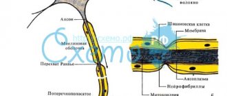
We will not go into detail about the histology and biochemistry of the structures that make up the barrier. Let us only note that the structure of the blood-brain barrier includes a special structure of capillaries. The following features are known that lead to the appearance of a barrier:
- tight junctions between endothelial cells lining the capillaries from the inside.
In other organs and tissues, the capillary endothelium is made “carelessly”, and there are large gaps between the cells through which there is a free exchange of tissue fluid with the perivascular space. Where the capillaries form the blood-brain barrier, the endothelial cells are located very tightly, and the tightness is not broken;
- energy stations - mitochondria in capillaries exceed the physiological need for those in other places, since the blood-brain barrier requires large amounts of energy;
- the height of endothelial cells is significantly lower than in vessels of other localizations, and the number of transport enzymes in the cell cytoplasm is much higher. This allows us to assign a large role to transmembrane cytoplasmic transport;
- the vascular endothelium in its depth contains a dense, skeletal-forming basement membrane, to which the processes of astrocytes are externally adjacent;
In addition to the characteristics of the endothelium, outside the capillaries there are special auxiliary cells - pericytes. What is pericyte? This is a cell that can regulate the lumen of the capillary from the outside, and, if necessary, can have the functions of a macrophage to capture and destroy harmful cells.
Therefore, before reaching neurons, we can note two lines of defense of the blood-brain barrier : the first is tight junctions of endothelial cells and active transport, and the second is macrophage activity of pericytes.
Further, the blood-brain barrier includes a large number of astrocytes, which make up the largest mass of this histohematic barrier. These are small cells that surround neurons and, by definition of their role, can do “almost everything.”
They constantly exchange substances with the endothelium, control the safety of tight junctions, the activity of pericytes and the lumen of capillaries. In addition, the brain needs cholesterol, but it cannot penetrate from the blood into the cerebrospinal fluid or pass through the blood-brain barrier. Therefore, astrocytes take over its synthesis, in addition to the main functions.
By the way, one of the factors in the pathogenesis of multiple sclerosis is impaired myelination of dendrites and axons. And for the formation of myelin, cholesterol is needed. Therefore, the role of BBB dysfunction in the development of demyelinating diseases is established and has recently been studied.
Where there are no barriers
Are there places in the central nervous system where the blood-brain barrier does not exist? It would seem impossible: so much effort has been put into creating several levels of protection from external harmful substances. But it turns out that in some places the BBB does not constitute a single “wall” of protection, but there are holes in it. They are needed for those substances that are produced by the brain and sent to the periphery as commands: these are pituitary hormones. Therefore, there are free areas, just in the zone of the pituitary gland and epiphysis. They exist to allow hormones and neurotransmitters to pass freely into the blood.
There is another zone free from the BBB, which is located in the area of the rhomboid fossa or the bottom of the 4th ventricle of the brain. The vomiting center is located there. It is known that vomiting can occur not only due to mechanical irritation of the back wall of the pharynx, but also in the presence of toxins that have entered the blood . Therefore, it is in this area that there are special neurons that constantly “monitor” the quality of the blood for the presence of harmful substances.
As soon as their concentration reaches a certain value, these neurons are activated, causing a feeling of nausea and then vomiting. To be fair, it must be said that vomiting is not always associated with the concentration of harmful substances. Sometimes, with a significant increase in intracranial pressure (with hydrocephalus, meningitis), the vomiting center is activated due to direct excess pressure with the development of intracranial hypertension syndrome. Therefore, so-called central or cerebral vomiting develops, which can occur suddenly, and without any signs of nausea.
The BBB and the EphR/Ephrin system: links between neurovascular and neuropsychiatric disorders
The structural integrity of the blood-brain barrier (BBB) is essential for establishing and maintaining brain homeostasis. Any disruption of the structural components of the BBB can have a devastating impact on mental health. A string of important studies on neurovascular units (NVUs) have been published over the past couple of years. They consider the BBB as a potential new target for the development of alternative treatments for brain and vascular pathologies.
Structural components and functions of the BBB
The BBB is a cellular vascular structure that separates the central nervous system from the peripheral circulation (Obermeier et al., 2013). It is composed of cerebrovascular endothelial cells (ECs) that form the brain vessels, parts of astrocytes, and extracellular matrix (EM) that provide structural support (Abbott et al., 2006). Pericytes are also involved in the formation of the BBB (Cabezas et al., 2014). All of these elements perform their functions by forming a selective physical (Abbott et al., 2006), transport (Begley and Brightman, 2003) and metabolic (Pardridge, 2003, 2016) barrier that tightly controls the passage of molecules into and out of the brain parenchyma and prevents toxins or pathogens from entering the brain (Obermeier et al., 2013).
Figure 1. Cellular and signaling components of the blood-brain barrier (BBB) are normal. Signaling molecules from astrocytes influence the creation and/or maintenance of the properties of the BBB. In addition, various members of the A and B class A and B family of ephrins (Eph receptor ligands) located on astrocytes and/or pericytes activate EphA and EphB receptors on endothelial cells (ECs) to influence EC differentiation during the time of angiogenesis and the development of tight junctions during barrier formation (barriergenesis).
Endothelial cells
Cerebral ECs have unique characteristics compared to peripheral ECs: cells are connected to each other by continuous intracellular multiprotein complexes called tight junctions (Fig. 1). They limit the flow of intercellular substances and direct molecules across the BBB along a controlled path (Abbott et al., 2006). This physical barrier allows only small gaseous and lipophilic molecules to pass freely into and out of the brain, while large molecules must be actively transported through specialized transport systems such as glucose transporter type 1 (GLUT-1) or large neutral amino acid transporter type 1 (LAT). -1) (Borst and Schinkel, 2013). Potentially harmful compounds such as glutamate are actively released from the brain even against a concentration gradient, requiring ATP as an energy source (Hawkins and Viña, 2016). In general, large hydrophilic molecules cannot be transported across the BBB except by specific receptor or adsorption-mediated transcytosis (Pardridge, 2003, 2021, Strazielle and Ghersi-Egea, 2016).
Tight junctions are key regulators of intercellular permeability. The main components of tight junctions are intermembrane molecules such as occludins (Yu et al., 2005), which bind to the cytoskeleton through accessory proteins, claudins (Piehl et al., 2010) and junctional adhesion molecules (JAM-A, -B, -C, Mandel et al., 2012). During early embryogenesis, vascular sprouting already occurs, and angiogenesis is actively occurring (Obermeier et al., 2013). Mature dense channels are fixed and must be maintained throughout life.
Astrocytes
Astrocytes, through their processes with which they contact the brain vessels, regulate the properties of the BBB (Kettenmann and Verrratsky, 2008). Other functions include regulation of the ion balance around the BBB and the secretion and processing of neurotrophic factors necessary for the control of tight junctions (Gee and Keller, 2005). In particular, astrocytes secrete molecules such as glial-derived neurotrophic factor (GDNF; Igarashi et al., 1999), transforming growth factor β (TGF-β; Dobolyi et al., 2012), angiopoietin 1 (ANG1, Easton, 2012), fibroblast growth factor 2 (FGF2; Reuss et al., 2003) and vascular endothelial growth factor (VEGF; Rosenstein et al., 2010), which acts on ECs to either promote tight junction formation or regulate BBB permeability (Figure 1) .
Meningeal blood vessels, which do not have contacts with astrocytes, exhibit higher vascular permeability than is the case with BBB endothelial cells, supporting the requirement of astrocytes to induce BBB properties (Lécuyer et al., 2016).
basement membrane
The noncellular component of the NSE is a basic membrane composed of structural proteins such as collagen-IV, laminin, and fibronectin ( Cardoso et al., 2010 ). The main function of the basement membrane is to provide stability to other NCE structures and regulate their interactions (Baeten and Akassoglou, 2011).
Erythropoietin-producing hepatocellular carcinoma receptors (EphR) and ligands that interact with Eph receptors (ephrin).
In 1990, the erythropoietin-producing hepatocellular carcinoma (EphR)/ephrin system was first discovered. EphrinA1 has been characterized as a tumor necrosis factor (TNF)-inducible protein in human umbilical vein endothelial cells (HUVEC; Holzman et al., 1990). EphRs/ephrins typically regulate contact dependence between cells, determining their interactions. During organismal development, this system plays an important role in spatial organization, axon guidance, formation of synaptic connections, and blood vessel remodeling. In adulthood, the system mainly regulates synaptic remodeling, epithelial differentiation, bone remodeling, immune function, insulin secretion, and stem cell self-renewal (Kullander and Klein, 2002; Yamaguchi and Pasquale, 2004; Pasquale, 2005, 2008).
Eph receptors contain the largest family of receptor tyrosine kinases (RTKs). Eph receptors and ephrins can be divided into subclasses A and B. Nine EphA and five EphB receptors are known in humans. They consist of an extracellular part, including a globular ligand-binding domain and a cytoplasmic domain. Ephrins can also be distinguished by their membrane attachment: ephrins A are anchored by glycosylphosphatidylinositol (GPI), whereas ephrins B are attached through a single transmembrane domain containing a short cytoplasmic PDZ-binding portion (Lisabeth et al., 2013).
EphR/Ephrin system: signaling mechanisms
In addition to the known bidirectional signaling activated by cell-cell interactions (Pasquale, 2008; Murai and Pasquale, 2011; Klein, 2012; Lisabeth et al., 2013), there are several alternative signaling mechanisms through the EphR/ephrin system.
Upon receptor/ligand interaction, several downstream signaling cascades are activated mediating cellular attraction-adhesion or repulsion depending on the type and abundance of ligands and receptors present on cell surfaces (Janes et al., 2012). Among others, these signaling pathways include Src family of kinases, mitogen-activated protein kinase, and integrin-mediated pathways (Lackmann and Boyd, 2008; Pasquale, 2008; Pitulescu and Adams, 2010). After the initial receptor/ligand interaction, intact EphR/Ephrin complexes, along with potentially associated cytoplasmic proteins and the surrounding membrane, are implanted into cells. This mechanism is called transendocytosis. It provides a mechanism for switching between cellular attraction-adhesion/repulsion and termination of receptor signaling activity (Lisabeth et al., 2013).
In addition to transendocytosis, another alternative signaling mechanism is the activation of enzymes that initiate proteolytic cleavage (Atapattu et al., 2014). These cleavage enzymes include A disintegrin and metalloproteases (ADAMs), as well as matrix metalloproteases (MMPs) (Atapattu et al., 2014).
Several EphR/ephrins of both subclasses A and B can interact with ADAM, leading to their own cleavage. Cleavage of the ligand-bound receptor results in the destruction of molecular bonds between interacting cells.
MMPs degrade proteins located either on membranes or in the extracellular space (Miller et al., 2008). Their main function is to disrupt structural components of endothelial cells to facilitate cell migration (Streuli, 1999), especially during angiogenesis and inflammatory processes (Kessenbrock et al., 2010; Palmisano and Itoh, 2010). Recently, MMPs were shown to release ephrinA1 and ephrinA2 from their GPI moiety, resulting in the release of functional soluble monomers that can act on distant Eph receptors ( Beauchamp and Debinski, 2012 ). Following the initial proliferation step mediated by ADAMs or MMPs, EphRs/ephrins can be further processed by intramembrane cleavage proteases such as γ-secretase (Bergmans and De Strooper, 2010) or neuropsin (Attwood et al., 2011; Morohashi and Tomita, 2013 ). These interactions generate cytoplasmic active moieties (Litterst et al., 2007; Xu and Henkemeyer, 2009), which can, for example, regulate behavioral responses such as anxiety (Attwood et al., 2011).
Such Eph/ephrin signaling mechanisms may play a role in the development of brain pathology, providing an opportunity to identify alternative targets for therapy.
The role of the EphR/Ephrin system in the development and functioning of the vasculature and the BBB
The interaction of specific cell types in the proper development of the vascular system and the functioning of the BBB is an important process that requires appropriate coordination in time and space. The EphR/ephrin system also plays a role in this coordination.
During angiogenesis, VEGF induces the expression of ephrinA1, which activates EphA2 on adjacent ECs, thereby exerting angiogenic effects in vitro and in vivo (Cheng et al., 2002; Brantley-Sieders et al., 2004). Astrocytes release VEGF during embryonic development and may therefore contribute to the early formation of tight junctions. With further differentiation of ECs and proper formation of the BBB, inhibition of EphA2 activity in human brain microvascular endothelial cells is observed, which plays an important role in further strengthening of tight junctions (Zhou et al., 2011;). Thus, regulation of EphA2 dosages may underlie the “switch” between the early/angiogenic and late/barrier effects of EphA2 on endothelial cells. Moreover, it has been suggested that interactions between EphA2-expressing endothelial cells with ephrinA1-expressing perivascular astrocytes or pericytes may also control the formation of tight junctions under physiological conditions or their dysfunction during pathogenic processes (Lécuyer et al., 2016). It should be noted that overexpression of certain EphR/ephrin interactions may affect the integrity of the BBB, ultimately affecting brain homeostasis. Astrocytes express several members of the EphR/ephrin system (Nestor et al., 2007), which may be important during angiogenesis and/or barrigenesis. For example, proper interaction between EphA4/ephrinA5 on ECs and astrocyte processes, respectively, is required for the development of normal vasculature in the adult mouse hippocampus (Hara et al., 2010). EphA4 signals have also been shown to direct the growth of new small vessels in response to glial-dependent stimulation (Goldshmit et al., 2006).
Regarding B types, ephrinB2 controls the incorporation of VEGF receptor (VEGFR)-2, which is necessary for activation of this receptor and proper development of endothelial cells during angiogenesis (Bochenek et al., 2010; Sawamiphak et al., 2010; Pitulescu and Adams, 2014). At the same time, the EphB2/ephrinB2 interaction is required for proper blood vessel formation (Foo et al., 2006). For the development of the cardiovascular system, EphB4/ephrinB2-mediated signals are also important for the correct determination of arterial or venous identity (Wang et al., 1998; Adams et al., 1999; Gerety et al., 1999; Gale et al., 2001; Augustin and Reiss, 2003).
BBB dysfunction and the Eph/Ephrin system. Is there a connection between neurovascular and neuropsychiatric pathology?
BBB leakage is a hallmark of neurovascular pathologies associated with neuroinflammatory processes (Lee et al., 2009; Abbott and Friedman, 2012). Signs of BBB dysfunction have also been shown in neuropsychiatric disorders, which are accompanied by increased levels of circulating proinflammatory cytokines and TNF (Miller et al., 2009; Janelidze et al., 2011; Liu et al., 2012; Salim et al., 2012; Najjar et al., 2012). al., 2013). In addition, preclinical and clinical studies indicate a wide range of neurovascular and neuropsychiatric disorders in which neuroinflammation is detected (Dantzer et al., 2008; Wood, 2014; Hodes et al., 2015; Patel and Frey, 2015; Seligman and Nemeroff, 2015 Miller and Raison, 2021, Barnes et al., 2021, Menard et al., 2017). It can be assumed that common neurobiological substrates may underlie such diseases. In view of the regulatory functions of the EphR/ephrin system during the formation and maturation of proper BBB sealing properties, it seems clear that this system can be considered the center of brain disorders associated with BBB dysfunction.
Preclinical studies have shown that members of the group A EphR/ephrin system (notably the EphA2 receptor) mediate inflammation during injury, ischemia, and other chronic inflammatory conditions in mice with neurovascular diseases (Jellinghaus et al., 2013; Thundyil et al. , 2013; Ende et al., 2014). In particular, activation of the EphA2 receptor occurs after traumatic brain injury and promotes inflammation by increasing BBB permeability (Thundyil et al., 2013). Interestingly, the ephrinA1 promoter, the most highly active ligand for EphA2, is a target of the proinflammatory marker TNF (Ende et al., 2014). In addition, while TNF has angiogenic properties during early embryogenesis (Cheng and Chen, 2001; Munthe and Aasheim, 2002), it also causes BBB hyperpermeability in adults through the activation of both EphA2 and EphA4 in endothelial cells, which is accompanied by development or exacerbation of neurovascular disorders (Jellinghaus et al., 2013; Thundyil et al., 2013; Ende et al., 2014). Thus, the study of EphA2/ephrinA1-mediated signaling pathways may reveal new molecular triggers of pathology with inflammatory/neurovascular/neuropsychiatric disorders, which may point us to alternative targets for therapeutic interventions. The association of ephrinA5, expressed on astrocytes, with its corresponding receptor EphA4 on endotheliocytes has also been shown to be increased in the hippocampus of mice with temporal lobe epilepsy, suggesting increased small vessel development that is detrimental to brain homeostasis (Shu et al. , 2016). Interestingly, selective blockade of the EphA4/epHRin interaction leads to attenuation of disease manifestations, further supporting the therapeutic value of selectively targeting the EphR/ephrin system in neurovascular/neuropsychiatric disorders.
Figure 2. Cellular and signaling components of the BBB under pathological conditions. In brain pathology, excessive activation of astrocyte- or pericyte-dependent ephrin signaling can influence tight junctions by increasing the activity of Eph receptors with a subsequent increase in the permeability of the BBB to inflammatory factors circulating in the blood, such as tumor necrosis factor (TNF). It has been proven that severe stress exposure correlates with dysfunction of the BBB and disruption of tight junctions, which can cause more pronounced manifestations of neurological and neuropsychiatric disorders. However, the specific molecular mediators of these effects have not yet been identified.
Regarding members of the B group, it has been suggested that the relationship between TNF and EphB2 is relevant to the induction of inflammatory pathways (Pozniak et al., 2014). EphB2 has also been shown to regulate cognitive functions and resistance/vulnerability to stress (Yuferov et al., 2013; Zhang et al., 2016). Among the triggers of neuropsychiatric disorders, stress is one of the most harmful (Charney and Manji, 2004). It has been shown that just 2 days of stress provoke morphological changes in endothelial cells, which are accompanied by dysregulation of the expression of claudin-5 and occludin (Fig. 2). These changes are accompanied by a decrease in glial fibrillary acidic protein expression, indicating additional negative effects on astrocytes (Sántha et al., 2016). A study last year clearly demonstrated the detrimental effects of stress on BBB permeability through increased BBB permeability (Menard et al., 2017).
Evidence that the EphR/ephrin system may represent an important therapeutic target for the treatment of neurovascular and neuropsychiatric disorders is the controlled reactivation of EphB4/ephrinB2 in cardiovascular disorders, which improves BBB repair mechanisms (Ghori et al., 2017).
Conclusion
Although several studies have shown that BBB dysfunction occurs in a wide range of psychopathologies, it is still unknown whether BBB dysfunction is a cause or a consequence of the disease. It has already been determined that the permeability of the BBB increases during inflammation, which has a positive side, as it ensures the passage of growth factors or antibodies that prevent the inflammatory process. On the other hand, improving the sealing properties of the BBB will help during periods of stress or hypoxia (Abbott et al., 2006).
Studying the influence of the EphR/ephrin system in individual components of the BBB during angiogenesis and barrier genesis, as well as their action in physiological and pathological processes, may open new ways for understanding the neurobiological basis of neurological diseases. This could help identify new therapeutic targets for the development of new types of effective drugs.
Source.
Other articles in the issue
| Plasma β-amyloid, cerebral atrophy and risk of dementia |
| Autoantibodies against neuronal surface proteins in hemorrhagic stroke |