Laws of axon work
When conducting a nerve impulse along axons, four main laws apply:
- Law of anatomical and physiological integrity. Conduction is possible only through intact neuron processes. This rule also applies to damage resulting from changes in membrane permeability (under the influence of drugs or poisons).
- Excitation isolation law. One axon – conduction of one excitation. Axons do not share nerve impulses with each other.
- Law of unilateral conduct. The axon conducts impulses either centrifugally or centripetally.
- The law of no loss. This property is non-decremental - when an impulse is carried out, it does not subside or change.
The location of these nerve cells
Pyramidal neurons can be found at various points in the nervous system, but they are much more common in some specific areas. Among them the following stand out.
Cortex
Pyramidal neurons are found primarily in the cerebral cortex, forming part of most of it, and are found in five of the six layers that make up this area of the brain. In particular, they can be observed in the granular and pyramidal layers, both external and internal.
They are especially prominent in the third and fifth layers (which are actually called the outer pyramidal and inner pyramidal), and they are larger the deeper into the cortex. There are also areas within the cortex where its existence has been discovered more frequently.
Motor cortex
In the motor cortex we can find a large number of pyramidal neurons, especially those associated with motor control. In this area of the cortex there are many giant pyramidal neurons known as Betz cells that transmit motor information from the brain to areas of the spinal cord, where they synapse with motor neurons that activate movement.
Prefrontal cortex
Pyramidal neurons can also be found in the prefrontal cortex, influencing higher mental processes. It is believed that these cells are the main primary excitations of prefrontal neurons, participating in numerous functions and considering themselves primordial for the existence of behavioral control.
Corticospinal tract
Pyramidal neurons are especially visible along the corticospinal tract, which sends motor information from different brain nuclei responsible for motor control to motor neurons that will cause muscle contraction as they pass through the spinal cord.
Hippocampus
Not only in the cortex we can find pyramidal neurons, but we can also find in subcortical structures. One of them is the hippocampus, associated with aspects such as memory and orientation.
Article on the topic: “Hippocampus: functions and structure of the memory organ”
Amygdala
Another structure in which these neurons are found is in the amygdala, an area of the limbic system associated with emotional memory.
Structure of a nerve cell
Neurons in the brain have an irregular shape, they can look like a leaf or a flower, and have various grooves and convolutions. The color palette is also varied. Scientists believe that there is a relationship between the color and shape of a cell and its purpose.
For example, the receptive fields of cells in the projection area of the visual cortex have an elongated shape, this helps them selectively respond to individual fragments of lines with different orientations in space.
Each cell has a body and processes. Brain tissue is usually divided into gray and white matter. The cell bodies of neurons, together with glial cells that provide protection, insulation and preservation of the structure of nervous tissue, make up the gray matter. The processes, organized into bundles in accordance with their functional purpose, are white matter.
Types of processes:
- axons - have an elongated appearance, at the end they branch into terminals - nerve endings that are necessary for transmitting impulses to other cells;
- dendrites - shorter than axons, also have a branched structure; through them the neuron receives information.
Thanks to this structure, neurons in the brain “communicate” with each other and are combined into neural networks, which form brain tissue. Both dendrites and axons are constantly growing. This plasticity of the nervous system underlies the development of intelligence.
Classification
Structural classification
Based on the number and arrangement of dendrites and axons, neurons are divided into axonless neurons, unipolar neurons, pseudounipolar neurons, bipolar neurons, and multipolar (many dendritic arbors, usually efferent) neurons.
Axonless neurons
- small cells, grouped near the spinal cord in the intervertebral ganglia, which do not have anatomical signs of division of processes into dendrites and axons. All processes of the cell are very similar. The functional purpose of axonless neurons is poorly understood.
Unipolar neurons
- neurons with a single process, present, for example, in the sensory nucleus of the trigeminal nerve in the midbrain. Many morphologists believe that unipolar neurons do not occur in the body of humans and higher vertebrates.
Bipolar neurons
- neurons with one axon and one dendrite, located in specialized sensory organs - the retina, olfactory epithelium and bulb, auditory and vestibular ganglia.
Multipolar neurons
- neurons with one axon and several dendrites. This type of nerve cells predominates in the central nervous system.
Pseudounipolar neurons
- are unique in their kind. One process extends from the body, which immediately divides in a T-shape. This entire single tract is covered with a myelin sheath and is structurally an axon, although along one of the branches the excitation goes not from, but to the body of the neuron. Structurally, dendrites are branches at the end of this (peripheral) process. The trigger zone is the beginning of this branching (that is, it is located outside the cell body). Such neurons are found in the spinal ganglia.
Functional classification
Based on their position in the reflex arc, afferent neurons (sensitive neurons), efferent neurons (some of them are called motor neurons, sometimes this not very accurate name applies to the entire group of efferents) and interneurons (interneurons) are distinguished.
Afferent neurons
(sensitive, sensory, receptor or centripetal). Neurons of this type include primary cells of the sensory organs and pseudounipolar cells, whose dendrites have free endings.
Efferent neurons
(effector, motor, motor or centrifugal). Neurons of this type include the final neurons - ultimatum and penultimate - non-ultimatum.
Association neurons
(interneurons or interneurons) - a group of neurons communicates between efferent and afferent ones.
Secretory neurons
- neurons that secrete highly active substances (neurohormones). They have a well-developed Golgi complex, the axon ends at axovasal synapses.
Morphological classification
The morphological structure of neurons is diverse. Several principles are used to classify neurons:
- take into account the size and shape of the neuron body;
- number and nature of branching of processes;
- axon length and the presence of specialized sheaths.
According to the shape of the cell, neurons can be spherical, granular, stellate, pyramidal, pear-shaped, fusiform, irregular, etc. The size of the neuron body varies from 5 μm in small granular cells to 120-150 μm in giant pyramidal neurons.
Based on the number of processes, the following morphological types of neurons are distinguished:
- unipolar (with one process) neurocytes, present, for example, in the sensory nucleus of the trigeminal nerve in the midbrain;
- pseudounipolar cells grouped near the spinal cord in the intervertebral ganglia;
- bipolar neurons (have one axon and one dendrite), located in specialized sensory organs - the retina, olfactory epithelium and bulb, auditory and vestibular ganglia;
- multipolar neurons (have one axon and several dendrites), predominant in the central nervous system.
From the editor: Alzheimer's disease?measure
Classifications of neurons
Lecture 2 Anatomy of the central nervous system 2009
Lecture outline
- Microstructure of nervous tissue
- Neuron as the basic morpho-functional unit
- nervous system
- Classifications of neurons
- Morpho-functional characteristics of the synapse
- General characteristics of nerve fibers
- Structural and functional characteristics of glial cells
Microstructure of nervous tissue
Cell. According to cellular theory, a cell is an elementary unit of structure, functioning and development of a living organism.
Tissue is a historically established collection of cells and intercellular substance that have a common origin, structure and functions. There are 4 main types of tissues in the human body: epithelial, muscle, connective and nervous. Epithelial tissue covers the outside of the body and lines the cavities of the internal organs. Muscle tissue forms the skeletal muscles of the body, the heart muscle and the muscles of internal organs and blood vessels. Connective tissue forms bones, cartilage, dense membranes around internal organs, blood, etc. Nervous tissue forms the nervous system.
There are 4 main types of tissues in the animal and human body: muscle, connective (blood, bones, ligaments, cartilage, subcutaneous fat, etc.), epithelial (skin and mucous membranes of organ cavities) and nervous.
Nervous tissue is formed by two types of cells: neurons, which perform specific functions of the nervous system, and glial cells, which are auxiliary and perform the functions of support, insulation, and nutrition (trophism) of neurons. Neurons in nervous tissue, due to their processes, are connected into very complex systems; interaction between neurons is carried out through specific contacts called synapses.
Neuron as the basic morpho-functional unit
Nervous system
According to the “neural doctrine” formulated by the leading Spanish neuroanatomist Santiago Ramon y Cajal (1852-1934), the neuron is the basic structural, functional and genetic unit of the nervous system. There are up to 1011 neurons in the nervous system, varying in shape and function.
A neuron is a highly specialized cell capable of perceiving irritation, converting it into a nerve impulse (excitability property) and conducting it along the cell surface (conductivity property) for transmission at the synapse to other nerve cells or cells of effector (working) organs (muscle or glandular).
A neuron consists of a soma (body or perikaryon) and processes. The dimensions of the neuron body range from 5 to 150 microns. The short processes branch like a tree and are therefore called dendrites
(from Greek
dendron -
tree).
Their number in different neurons ranges from one to thousands. Small membranous outgrowths, microspines up to 2-3 µm long, are formed on the dendrites. Spines are sites of synaptic contacts. They do not occur at the site where dendrites extend from the soma. Nervous excitation always passes in the direction from the dendrite to the soma. The soma and dendrites of the neuron are covered only with a cell membrane and outwardly look like the gray matter of the nervous system.
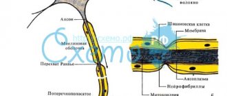
One long process, the axon, extends from the soma. It is the basis of the nerve fiber. The length of such a process in humans can reach 120 cm. The axon serves to conduct nerve impulses from the cell body to other neurons (or effector organs). The initial part of the axon, extended in the form of a funnel from the cell body, is called the axon hillock.
And the xonic hillock is the most excitable and is the most frequent site of generation of nerve impulses. The axon itself, or axial cylinder, is gray in color. But its main part is covered like a cover with a white, fat-like myelin sheath. Therefore, outwardly, clusters of axons look like the white matter of the nervous system. The myelin sheath periodically thins, forming nodes of Ranvier. Due to the myelin sheath, the nerve impulse travels along the axon tens of times faster than along the dendrites or soma.
At a relatively large distance from the soma, the axon can branch. Such lateral processes are called collaterals. Each of the collaterals at the very end also, as a rule, branches; these branches are called terminals (from the Latin terminalis - final, final). The terminals are no longer covered with myelin. At the end of each terminal there is a swelling, which is an integral part of the synapse (presynapse).
A neuron, like a typical animal cell, has in its internal structure a plasma membrane, a nucleus, cytoplasm and organelles.
A structural feature of the neuron is a large number of ribosomes on the endoplasmic reticulum in the soma, which, with special staining methods, looks like a tigroid substance (Nissl substance). The cytoplasm of a neuron contains special-purpose organelles, microtubules.
and
microfilaments
, which vary in size and structure. Microfilaments represent the internal skeleton of the cytoplasm and are located in the soma. Microtubules stretch along the axon along the internal cavities from the soma to the end of the axon. Biologically active substances spread through them.
After maturation, neurons are not capable of division due to their high specialization. It is this feature of neurons that ensures the preservation of all the information that the body assimilates throughout life. Accordingly, dead neurons are not replaced; however, when an axon is cut off in the peripheral nervous system, it can re-grow into the innervated organ.
Classifications of neurons
When classifying neurons, various bases are used to separate them: by the shape of the soma, the number of processes, by functions and by the effects that the neuron has on other cells.
1. Depending on the shape of the soma , granular (ganglionic) neurons are distinguished, in which the soma has a rounded shape, pyramidal neurons of different sizes - large and small pyramids, stellate neurons, fusiform neurons, etc. (Fig. 6).
2. Based on the number of processes in a neuron, they are distinguished:
a) multipolar neurons –
consist of a body, several dendrites extending from it and one axon (found most often in the human central nervous system):
b) bipolar neurons
– consist of a body, an axon and one dendrite (for example, peripheral sensory nerves).
c) unipolar neurons
– have only one axon process, perceive excitation due to synapses located on the cell body (in humans, such neurons are found only in the sensory nucleus of the trigeminal nerve at the level of the midbrain).
d) pseudounipolar neurons
. Such cells are formed from bipolar neurons when two processes merge into one. This process then branches T-shaped into two fibers: afferent and efferent (they are located in the spinal ganglia of the dorsal roots and in the sensory ganglia of the cranial nerves). This structure of sensory neurons ensures rapid transmission of signals to the central nervous system, since the process that performs the functions of a dendrite is also myelinated.
3. Depending on the functions , neurons are usually distinguished:
a) sensory
(sensitive, afferent);
b) effector
(efferent, motor and autonomic);
c) insertion
(interneurons, combinational, associative).
Among them, modulator neurons
, which do not themselves participate in reactions, but can change the level of activity of other nerve cells.
d) secretory neurons.
Secretory neurons produce various hormones that are released into the blood and carry out humoral regulation of the work of various organs and systems (for example, neurosecretory cells of the hypothalamus).
4. Based on the effect that neurons have on other cells, excitatory neurons and inhibitory neurons are distinguished. Excitatory neurons increase the activity of the cells with which they are connected until they generate excitation. Inhibitory neurons, on the contrary, reduce the excitability of cells, causing an inhibitory effect and making it difficult for excitation to occur in them.
Neuroglia
Neurons are not able to divide, which is why the statement appeared that nerve cells do not regenerate. That is why they should be protected with special care. Neuroglia cope with the main function of the “nanny”. It is located between the nerve fibers.
These small cells separate neurons from each other and hold them in place. They have a long list of features. Thanks to neuroglia, a constant system of established connections is maintained, the location, nutrition and restoration of neurons is ensured, individual mediators are released, and genetically foreign substances are phagocytosed.
Thus, neuroglia perform a number of functions:
- supporting;
- delimiting;
- regenerative;
- trophic;
- secretory;
- protective, etc.
In the central nervous system, neurons make up the gray matter, and outside the brain they accumulate in special connections, nodes - ganglia. Dendrites and axons create white matter. At the periphery, it is thanks to these processes that the fibers that make up the nerves are built.
Functions of neurons
What are the functions of nerve tissue? The nervous system, in the order of the endocrine system, coordinates the activities of the entire organism. The cutaneous neuron is partly involved in the coordination of one or another physiological (or mental) process.
To put it simply, the main function of a neuron is to receive and transmit information. It is true that whatever cells the system is looking at, what it does is that it removes some cells and transmits information to others in the form of nerve impulses.
Read also: Human blood groups - description, type, like wines, like madness, Rh blood factors
However, for different neurons, more specific functions are also seen.
Types of neurons by function:
- Afferent (sensitive): to receive information directly from the receptors interacting between the external light and our nervous system.
- Efferents (rukhov): represent the performance of specific actions – shortened meat, seen in the secretion of the plant.
- Associative (inserted): all the “middle” neurons in the lanyard, they may not be at all or they may be part of the same reflex node. The smells are concentrated in the central nervous system, indicating the processing of information and, seemingly rudely, the nervous system making a decision about the body.
Integumentary tissues
Integumentary tissues are also called epithelial tissues
Integumentary tissues line not only the surfaces of the body, but also the cavities of internal organs. So the stomach, intestines, oral cavity, bladder, etc. are lined with integumentary tissues from the inside.
There is almost no intercellular substance in epithelial tissues. Their cells adhere tightly to each other and form from one to several layers.
The main functions of the epithelium are protection, secretion production, gas exchange, absorption, and excretion.
is expressed in protecting the deeper tissues of the animal from damage, temperature changes, and the entry of harmful microorganisms. This function is performed by the skin.
epithelium is characteristic of the intestine. Here nutrients are absorbed into the blood using the intestinal villi.
of the animal's integumentary tissue is observed in the stomach, where its cells secrete mucus. There are also various glands in the skin.
carried out by the epithelium of the lungs; in some animals the skin also takes part in gas exchange.
performs the epithelium of the excretory organs.
Structure of neurons
File:Neuron-rus.svg Neuron diagram
Cell body
The body of a nerve cell consists of protoplasm (cytoplasm and nucleus), and is externally bounded by a membrane of a double layer of lipids (bilipid layer). Lipids consist of hydrophilic heads and hydrophobic tails, arranged with hydrophobic tails facing each other, forming a hydrophobic layer that allows only fat-soluble substances (eg oxygen and carbon dioxide) to pass through. There are proteins on the membrane: on the surface (in the form of globules), on which growths of polysaccharides (glycocalyx) can be observed, thanks to which the cell perceives external irritation, and integral proteins that penetrate the membrane through, in which ion channels are located.
A neuron consists of a body with a diameter of 3 to 130 µm, containing a nucleus (with a large number of nuclear pores) and organelles (including a highly developed rough ER with active ribosomes, the Golgi apparatus), as well as processes. There are two types of processes: dendrites and axons. The neuron has a developed and complex cytoskeleton that penetrates its processes. The cytoskeleton maintains the shape of the cell; its threads serve as “rails” for the transport of organelles and substances packaged in membrane vesicles (for example, neurotransmitters). The cytoskeleton of a neuron consists of fibrils of different diameters: Microtubules (D = 20-30 nm) - consist of the protein tubulin and stretch from the neuron along the axon, right up to the nerve endings. Neurofilaments (D = 10 nm) - together with microtubules, provide intracellular transport of substances. Microfilaments (D = 5 nm) - consist of actin and myosin proteins, especially expressed in growing nerve processes and in neuroglia. A developed synthetic apparatus is revealed in the body of the neuron; the granular ER of the neuron is stained basophilically and is known as the “tigroid”. The tigroid penetrates the initial sections of the dendrites, but is located at a noticeable distance from the beginning of the axon, which serves as a histological sign of the axon. Neurons vary in shape, number of processes, and functions. Depending on the function, sensitive, effector (motor, secretory) and intercalary are distinguished. Sensory neurons perceive stimuli, convert them into nerve impulses and transmit them to the brain. Effector (from the Latin effectus - action) - generate and send commands to the working organs. Intercalary neurons - communicate between sensory and motor neurons, participate in information processing and the generation of commands.
There is a distinction between anterograde (away from the body) and retrograde (toward the body) axon transport.
Dendrites and axon
Main articles: Dendrite
,
Axon
File:Complete neuron cell diagram ru.svg Diagram of the structure of a neuron
An axon is usually a long extension of a neuron that is adapted to conduct excitation and information from the body of the neuron or from the neuron to the effector. Dendrites are, as a rule, short and highly branched processes of a neuron, serving as the main site of formation of excitatory and inhibitory synapses influencing the neuron (different neurons have different ratios of axon and dendrites lengths), and which transmit excitation to the body of the neuron. A neuron may have several dendrites and usually only one axon. One neuron can have connections with many (up to 20 thousand) other neurons.
Dendrites divide dichotomously, while axons give off collaterals. Mitochondria are usually concentrated at branching nodes.
Dendrites do not have a myelin sheath, but axons may have one. The place of generation of excitation in most neurons is the axon hillock - a formation at the point where the axon departs from the body. In all neurons, this zone is called the trigger zone.
Synapse
Main article: Synapse
Synapse
(Greek σύναψις, from συνάπτειν - hug, clasp, shake hands) - the place of contact between two neurons or between a neuron and the effector cell receiving the signal. It serves to transmit a nerve impulse between two cells, and during synaptic transmission the amplitude and frequency of the signal can be adjusted. Some synapses cause depolarization of the neuron, others cause hyperpolarization; the former are excitatory, the latter are inhibitory. Typically, stimulation from several excitatory synapses is necessary to excite a neuron.
Editorial: How to determine that the brain is dead
The term was introduced in 1897 by the English physiologist Charles Sherrington.
The structure of nerve cells at the cellular and subcellular levels
The size of neurons ranges from 4 to 80 microns, their bodies are located in the gray matter of the brain and in the ganglia (nodes) of the peripheral nervous system.
At the cellular level (Fig. 4), each neuron consists of a body
,
processes
(
dendrites and axon
) and
nerve endings
, or
synapses
(Greek “
synapsis
” - contact, connection), through which nerve cells interact with each other and with working organs.
In addition, an axonal hillock
- a part of the cell body, elongated in the form of a funnel, directly passing into the axon.
The structure of nerve cells at the subcellular level is fundamentally similar to the structure of other types of cells, although the specialization of neurons has determined some features. The outer surface of the neuron, like that of any other cell, is formed by the bilipid layer of the plasma membrane
(Fig. 5, A).
The intracellular space is filled with the nucleus
and
cytoplasm
. The nucleus contains chromosomes, which are strands of deoxyribonucleic acid (DNA). The sequence of nucleotides in DNA encodes all the information necessary for the development and subsequent functioning of a nerve cell.
Cytoplasm is a chemically complex liquid that forms the intracellular environment of the cell in which cytoplasmic organelles
. The most important of them are:
1) mitochondria , within which, during the aerobic oxidation of glucose, ATP molecules are synthesized - the universal carrier of energy in the body. Mitochondria are a kind of energy stations that supply energy to all cellular structures.
2) lysosomes
Nerve cells under electron microscopy look like densely packed plates of the endoplasmic membrane, hence their other name -
dense body
. These structures contain various enzymes that are necessary for the normal functioning of metabolism in the cell.
3) Inside the neuron there is a system of membrane tubules through which various substances are transported in the cell. This network of tubules is called the endoplasmic reticulum
(EPR).
There are two types of endoplasmic reticulum. On the inner surface of the membrane of the “rough” or granular reticulum there are ribosomes , which determine this “roughness” of the membranes. Ribosomes synthesize various protein substances intended for secretion. The same ribosomes located in the cytoplasm independently, separately from the endoplasmic reticulum, are called free ribosomes
(4). The substances that are synthesized in them are not secreted, but are used inside the cell. The second type of endoplasmic reticulum is called “smooth”, which is explained by the absence of ribosomes. In the smooth ER, also called the Golgi apparatus, the substances intended for secretion are packaged into membrane membranes in the form of granules. Subsequently, these granules are transported along special microtubules to the cell surface, where they are brought out.
When neurons are stained with hematoxylin and eosin, the rough reticulum stains as clumps of basophilic material ( Nissl substance
). Noteworthy is the uneven distribution of the Nissl substance in the neuron: it is found in the dendrites and body, but it is not in the axon and in the axonal hillock. This reflects the functional role of different parts of the neuron and makes it possible to distinguish axons from dendrites in histograms.
5) Contractive elements
nerve cells (Fig. 5, B).
Inside neurons, especially near the cytoplasmic membrane, there are a large number of microfilaments
(neurofibrils) and
microtubules
(neurotubes). Microfilaments are thread-like polymer formations 5–7 nm thick, formed from monomers of the F-actin protein dissolved in the cytoplasm. Microtubules are similarly formed from monomers of the protein tubulin and are about 10 nm thick.
Microfilaments and microtubules form a dense network under the outer membrane of the cell, connecting with membrane proteins and with each other; some fibers penetrate the cytoplasm that fills the body and processes of the nerve cell. Thus, microfilaments and microtubules form the contractile skeleton of the cell ( cytoskeleton
).
Contractile proteins ensure the movement of sections of the cell cytoplasm relative to each other, the movement of substances on the inner and outer surfaces of the cell membrane, inside the cell, the elongation of axons and dendrites, changes in their diameter, as well as the formation (protrusion) of small membrane outgrowths on the axons and dendrites - microspikes
(see Fig. 4, 5B).
Microspikes located on dendrites and axons carry synapses on their surface designed to transmit excitation from one nerve cell to another. With frequent use of synapses connecting two neurons, the number of microspikes and synapses on the contacting processes increases. This process, called neosynaptogenesis
, goes in parallel with the disintegration of unused synapses, ensuring the plasticity of the functions of the nervous system.
Neurons are excitable cells, meaning they are capable of changing the charge of the cell membrane and generating nerve impulses under the influence of electrical impulses transmitted from other nerve cells. When excitatory synapses are activated, excitation from the presynaptic neuron spreads along the dendrites to the body of the postsynaptic neuron, resulting in depolarization of its entire membrane. As soon as a critical level of depolarization is reached for the axonal hillock, from which the axon directly departs, centrifugal nerve impulses are formed, traveling along the axon to the periphery. Thus, the nervous system, in the form of nerve impulses, encodes, transmits and processes information about the state of the external and internal environment; the impulse code is also used to transmit commands to working organs.
Evolution of fabric
The main property of a living organism is irritability or sensitivity. The type of nervous tissue is determined by the phylogenetic position of the animal and is characterized by wide variability, becoming more complex in the process of evolution. All organisms require certain parameters of internal coordination and regulation, proper interaction between stimulus for homeostasis and physiological state. The nervous tissue of animals, especially multicellular ones, the structure and functions of which have undergone aromorphoses, contributes to survival in the struggle for existence. In primitive hydroids, it is represented by stellate, nerve cells scattered throughout the body and connected by thin processes intertwined with each other. This type of nerve tissue is called diffuse.
The nervous system of flat and roundworms is stem, scalene type (orthogonal) consists of paired cerebral ganglia - clusters of nerve cells and longitudinal trunks extending from them (connectives), interconnected by transverse cords-commissures. In the rings, from the peripharyngeal ganglion, connected by cords, the abdominal nerve chain departs, in each segment of which there are two close nerve ganglia connected by nerve fibers. In some soft-bodied animals, nerve ganglia are concentrated to form the brain. Instincts and spatial orientation in arthropods are determined by the cephalization of the ganglia of the paired brain, the peripharyngeal nerve ring and the ventral nerve cord.
In chordates, the nervous tissue, the types of tissues of which are strongly expressed, is complex, but such a structure is evolutionarily justified. Different layers arise and are located on the dorsal side of the body in the form of a neural tube, the cavity is the neurocoel. In vertebrates, it differentiates into the brain and spinal cord. As the brain forms, swellings form at the anterior end of the tube. If in lower multicellular organisms the nervous system plays a purely connecting role, then in highly organized animals it stores information, retrieves it when necessary, and also ensures processing and integration.
In mammals, these cerebral swellings give rise to the main parts of the brain. And the rest of the tube forms the spinal cord. Nervous tissue, the structure and functions of which are unique in higher mammals, has undergone significant changes. This is the progressive development of the cerebral cortex and all parts of the nervous system, which determine complex adaptation to environmental conditions and the regulation of homeostasis.
Structure and functions of a neuron[edit | edit code]
A. Structure and functions of a nerve cell
Excitable cells respond to stimuli by changing the state of their membranes. There are two types of excitable cells: nerve cells, which conduct and transduce impulses in the nervous system, and muscle cells, which contract either in response to nerve impulses or autonomously.
Nervous system
The human body consists of more than 1010 nerve cells, or neurons.
A neuron
is a structural and functional unit of the nervous system. A typical neuron (motoneuron, A1) consists of a soma, or cell body, and two types of processes - axon and dendrites. In addition to the usual cellular organelles, such as the nucleus and mitochondria (A2), the neuron contains neurofibrils and neurotubules. A neuron receives afferent signals (excitatory and inhibitory) from several and sometimes several thousand neighboring neurons through dendrites (usually tree-like), and the signals are summed along the neuron body on the cell membrane (summation).The axon starts from the axon hillock of the neuron body: it carries out transmission of efferent nerve signals to nearby or distant effectors (muscle and secretory cells) and nearby neurons. Axons often have branches (collaterals), which branch further and end in swellings - synaptic vesicles or synaptic endings. If the total potential at the axon hillock exceeds a certain threshold, an action potential is generated and transmitted down the axon, where it reaches the next synapse through the synaptic terminal (A1, 3), described below.
Vesicles containing various substances (proteins, lipids, sugars and neurotransmitter molecules) are transported from the Golgi complex in the soma to the synaptic terminal and to the tips of dendrites by rapid axonal transport (40 cm/day). This type of anterograde (forward-directed) transport along neurotubules is carried out by kinesin (myosin-like protein), and the energy required for this is supplied by ATP. Endogenous and exogenous substances, such as nerve growth factor (NGF), herpes virus, polio virus, and tetanus toxin, are transported retrogradely from peripheral sites to the soma at a rate of ~25 cm/day. Slow axonal transport (~1 mm/day) plays an important role in the treatment of severe neuritis.
The plasma membrane of the soma continues along the axon and is called the axolemma (A1, 2).
In the central nervous system (CNS), the axolemma is surrounded by oligodendrocytes, and in the peripheral nervous system, by Schwann cells (A1, 2). A nerve fiber consists of an axon and its sheath. In some neurons, Schwann cells form a multilayered myelin sheath of double phospholipid layers (A1, 2) around the axon, which insulates the axon from ionic currents. The myelin sheath is interrupted approximately every 1.5 mm at the nodes of Ranvier (A1). The conductivity of myelinated nerve fibers is much higher than that of unmyelinated nerve fibers and increases with the diameter of the nerve fiber.
A synapse (A3) is the site where a neuron's axon interacts with effectors or other neurons. Synaptic transmission in almost all mammals is carried out by chemical compounds rather than by electrical signals. In response to an electrical signal in the axon, neurotransmitters are released from vesicles on the presynaptic membrane by exocytosis. The transmitter diffuses through the synaptic cleft (10-40 nm) to the postsynaptic membrane, where it connects with receptors that create new electrical signals (AZ). Depending on the type of neurotransmitter and receptor involved in the process, the neurotransmitter has either an excitatory (for example, acetylcholine in skeletal muscle) or an inhibitory effect (for example, glycine in the central nervous system) on the postsynaptic membrane. Because the postsynaptic membrane does not normally release neurotransmitters (there are only a few exceptions), nerve impulses can only pass through the synapse in one direction. Thus, the synapse acts as a valve that ensures orderly signal transmission. Synapses are also sites at which the transmission of a nerve impulse can be converted by other (excitatory or inhibitory) neurons.
From the editor: Routes of transmission, symptoms and treatment of viral meningitis
Types and characteristics of neurons
Nerve cells called neurons receive, send, and conduct bioelectrical signals. There are efferent (motor) neurons - these are components of the central nervous system that redirect signals to executive organs, for example, skeletal muscles. Afferent (sensitive) neurons are cells that perceive external and internal stimuli, which ensures the body’s connection with the external environment and reactions to changes in the functional activity of internal organs.
Intercalary cells provide interconnections within the overall neural network. Neurons of all types (sensitive, efferent, associative) are functional units that support the activity of the nervous system; they are found in all tissues of the body, where they play the role of connecting links between receptor (perceiving irritating stimuli) and effector organs that respond to irritating stimuli.
Effector organs include muscles and glands, and receptor organs include sensory organs. The significance of the signals transmitted varies significantly depending on the type of cell and its role in the functioning of the central nervous system. For example, sensory neurons that perceive impulses from the external environment transmit signals from skin receptors and sensory organs in the direction of the brain; motor neurons redirect commands generated in the brain, causing contraction of skeletal muscles and initiating movement.
Despite the different meanings of bioelectric impulses, their nature is the same and consists in changing the electrical potential in the area of the plasma membrane of the nerve cell. The mechanism of propagation of nerve impulses is based on the ability of an electrical disturbance that appears in one place of the cell to be transmitted to other areas. In the absence of factors that enhance the signal, the pulses fade away as they move away from the source of excitation.
Sensory, also known as sensitive, is an afferent neuron that conducts impulses from distal parts of the body to the central parts of the central nervous system. For example, sensory fibers form fibers that extend from the light-sensitive cells of the organs of vision. Signals leave the retina, traveling along millions of axons belonging to the structures of the basal ganglia, towards a region of the visual cortex.
The sensory neuron, together with the executive (motor) neurons, forms a simple reflex arc.
For example, the knee reflex is an unconditioned stretch reflex reaction that occurs as a result of the activity of a similar reflex arc. A reaction in the form of uncontrolled extension of the lower leg occurs when there is a mechanical impact on the tendon of the femoral muscle, which lies under the patella. Reaction mechanism:
- Mechanical effects on the neuromuscular spindles located in the hip extensor muscle.
- An increase in the intensity of nerve signals in the endings entwining the neuromuscular spindles due to their stretching.
- Transmission of impulses to sensory neurons located in the spinal ganglia through dendrites extending from the femoral nerve.
- Transmission of impulses from sensory cells to alpha motor neurons located in the anterior horns within the boundaries of the spinal cord.
- Signal transmission from alpha motor neurons to contractile muscle fibers of the femoral muscle.
The knee reflex mechanism involves interneurons that transmit inhibitory impulses to motor neurons of the flexor muscles, and other interneurons, for example, Renshaw cells. The knee reflex mechanism also involves gamma motor neurons, which regulate the intensity of spindle stretch.
In the spinal cord, formed by gray matter, there are three types of neurons - motor, intercalary, and autonomic. Moreover, the vegetative ones are located in the visceral (related to the internal organs) nuclei. These cells interact with afferent (ascending pathways that transmit impulses from peripheral receptors to the central zones of the central nervous system) fibers responsible for general visceral sensitivity.
Visceral afferents conduct nerve signals (usually painful or reflex sensations) from internal organs, elements of the circulatory system, glands to the corresponding zones of the central nervous system. Visceral afferents are part of the autonomic nervous system. Reflex arcs within the autonomic department of the central nervous system differ in structure from the arcs of the somatic department.
Efferent components (descending pathways that transmit impulses from the cortical and subcortical zones of the brain to peripheral areas) are formed by two types of neurons - intercalary and effector (motor). The intercalary ones are located in the nuclei belonging to the vegetative part of the central nervous system. The name “intercalary” is due to its location between the sensory and motor neurons.
Sensitive
A sensory neuron is a component of the nervous system that transmits information to the brain about stimuli affecting a specific area of the body. Examples of irritants include factors: sunlight, mechanical impact (impact, touch), the action of a chemical substance. Sensory neurons are located in the ganglia of the brain - the spinal cord and brain.
The connection formed with a sensitive neuron can provoke excitation or inhibition, which is sent along nerve fibers to the cortical parts of the brain. As the level of sensory pathways increases, the transmitted information is processed to identify important features. Sensitive neurons belong to pseudounipolar neurons - their axon and dendrites extend from the body together, subsequently separate and are located in the spinal cord, brain (axon) and in the peripheral parts of the body (dendrites).
Insert
Interneurons transmit converted nerve impulses obtained as a result of processing sensory information received from various sources, for example, from the organs of vision and skin receptors. As a result, the processed information becomes the initial data for the formation of adequate motor commands.
Motor
Motor nerve cells are of two types - large and small. In the first case we are talking about α-motoneurons, in the second – about γ-motoneurons. Alpha motor neurons are present in the basal ganglia of lateral (closer to the lateral plane) and medial (closer to the median plane) localization. These are the largest cells present in nervous tissue.
Their axons interact with striated fibers contained in skeletal muscles. As a result, synapses (sites where nerve signals are transmitted) are formed. The axons of alpha motor neurons interconnect with intercalary analogues, also known as Renshaw cells, leading to the formation of collateral pathways and inhibitory synapses in the spinal cord.
Gamma motor neurons are part of the neuromuscular spindle, which is a complex receptor consisting of nerve endings (afferent, efferent). The main function of neuromuscular spindles is to regulate the force and rate of contraction or stretching of skeletal muscles.
Neuron and its structure
You can often hear that a person’s mental abilities are guaranteed by the presence of gray matter. What is this substance and why is it gray? This is the color of the cerebral cortex, which consists of microscopic cells. These are neurons or nerve cells that ensure the functioning of our brain and control of the entire human body.
How does a nerve cell work?
A neuron, like any living cell, consists of a nucleus and a cell body called the soma. The size of the cell itself is microscopic - from 3 to 100 microns. However, this does not prevent the neuron from being a real repository of various information. Each nerve cell contains a complete set of genes - instructions for producing proteins. Some of the proteins are involved in the transmission of information, others create a protective shell around the cell itself, others are involved in memory processes, others provide mood changes, etc.
Even a small malfunction in one of the programs for the production of a certain protein can lead to serious consequences, illness, mental impairment, dementia, etc.
Each neuron is surrounded by a protective sheath of glial cells; they literally fill the entire intercellular space and make up 40% of the brain substance. Glia or a collection of glial cells performs very important functions: it protects neurons from unfavorable external influences, supplies nerve cells with nutrients and removes their waste products.
Glial cells guard the health and integrity of neurons, and therefore prevent many foreign chemicals from entering nerve cells. Including medications. Therefore, the effectiveness of various drugs designed to enhance brain activity is completely unpredictable, and they affect each person differently.
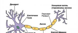
Dendrites and axons
Despite the complexity of the neuron, it itself does not play a significant role in the functioning of the brain. Our nervous activity, including mental activity, is the result of the interaction of many neurons exchanging signals. The reception and transmission of these signals, more precisely, weak electrical impulses, occurs with the help of nerve fibers.
The neuron has several short (about 1 mm) branched nerve fibers - dendrites, so named because of their resemblance to a tree. Dendrites are responsible for receiving signals from other nerve cells. And the axon acts as a signal transmitter. A neuron has only one fiber, but it can reach a length of up to 1.5 meters. Connecting with the help of axons and dendrites, nerve cells form entire neural networks. And the more complex the system of relationships, the more complex our mental activity.
Neuron operation
The most complex activity of our nervous system is based on the exchange of weak electrical impulses between neurons. But the problem is that initially the axon of one nerve cell and the dendrites of another are not connected; between them there is a space filled with intercellular substance. This is the so-called synaptic cleft, and the signal cannot cross it. Imagine that two people reach out to each other and just barely reach each other.
This problem is easily solved by a neuron. Under the influence of a weak electric current, an electrochemical reaction occurs and a protein molecule, a neurotransmitter, is formed. This molecule blocks the synaptic cleft, becoming a kind of bridge for the passage of the signal. Neurotransmitters also perform another function - they connect neurons, and the more often a signal passes along this nerve chain, the stronger this connection. Imagine a ford across a river. Walking along it, a person throws a stone into the water, and then each subsequent traveler does the same. The result is a strong, reliable transition.
This connection between neurons is called a synapse, and it plays an important role in brain activity. It is believed that even our memory is the result of the work of synapses. These connections provide a high speed of passage of nerve impulses - the signal along the chain of neurons moves at a speed of 360 km/h or 100 m/sec. You can calculate how long it takes for a signal from a finger that you accidentally pricked with a needle to reach your brain. There is an old riddle: “What is the fastest thing in the world?” Answer: "Thought." And this was very accurately noted.
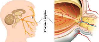
Let's sum it up
All our automatic and reflex actions occur under the supervision of the spinal cord. The only exceptions are those that are controlled by the brain itself. For example, when we perceive what we see using the optic nerve, which goes directly to the brain, we change the angle of vision using the muscles of the eyeball, which are already controlled by the spinal cord. By the way, we also cry on the orders of the spinal cord - it is he who “commands” the lacrimal glands. Our conscious actions begin in the brain, but as soon as they become automatic, their control passes to the spinal cord. You could say that our inquisitive brains love to learn. And when he has already learned, he becomes bored and gives the “reins of power” to his brother, who is more ancient in evolutionary terms.