Neuron connections
Nerves, as individual objects, consist of processes of nerve cells - neurons. Neurons, like electrical wiring, run throughout the human body.
- The main components of a nerve are axons. They are sharp, thin processes of neurons that conduct electrical impulses from neuron to neuron, as well as to other internal organs;
- Dendrites are short processes of neurons that receive signals coming from other neurons;
- With the help of synapses, individual neurons communicate with each other in a special way;
- Thanks to biological substances - a mediator, as well as through chemical compounds and with the help of electrical signals, a signal passes through the synapse;
- Dendrites receive the signal and transmit it further.
Definition
The medulla is a highly organized structure formed by nerve cells from which axons extend. Brain tissue is made up of nerve cells. Axon, translated from Greek, means “axis” - this is a process, an element of the medulla, which ensures interaction between cells of different types (neurons, cells of innervated organs), which is associated with subtle, precise control of the functioning of organs and systems. Functions of central nervous system tissue:
- Perceives irritations, converting them into impulses.
- Supports the transmission of impulses from the control parts of the brain to the executive organs.
- Forms a response to irritating influence.
- Provides interaction in the functioning of systems and organs, supports the integration of structural units of the body.
- Provides interaction between the body and the external environment.
According to the definition in biology, an axon is an elongated process along which impulses travel from the body of a neuron to other nerve cells and structural elements of all tissues of the body. During fetal development, brain tissue is formed from the neural plate. The edges of the plate bend, which leads to the formation of ridges and grooves. As a result of the closure of the edges of the ridges, a neural tube is formed - the basis of the central nervous system.
Differentiation of the cells forming the tube leads to the appearance of neuroblasts and spongioblasts. The former serve as the basis for the formation of neurons, the latter – for the formation of neuroglia. Neurons (anat.) are the main structural elements of the brain matter. They are characterized by the absence of a division function, which leads to a gradual decrease in their numbers. The body of a neuron consists of a nucleus and cytoplasm. Depending on the type of neurons, the geometric shape of the body changes, which can be round, oval, pyramidal and others.
The cytoskeleton, consisting of microtubules and neurofibrils, provides support and trophic function. The cytoskeleton maintains the shape of the neuron and ensures the transport of substances and organelles. Branches from the body include a single axon and multiple dendrites. The neuron axon has almost no branches and sometimes forms collateral (bypass) segments. The end segments (terminations) branch out and are called terminals.
The terminals are interconnected with the endings of other neurons and with cells that form the parenchyma (tissue) of working organs - muscles, glands. The number of dendrites varies from 1 to several. Thin branches of dendrites end in small spines, where the terminals of the axonal processes of many thousands of other cells are concentrated. Dendrites receive stimuli or action potentials from other cells and transmit them along fibers to the body of their neuron.
Axon growth depends on the structural features and vital activity of the neuron, which supports the function of feeding the process. For example, if an axonal trunk is cut, the segment connected to the body remains viable and continues to function, while the section that has lost connection with the body dies. Axons form nerves, which suggests a complex structural and morphological organization of the central nervous system.
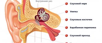
Peripheral nervous system
There are about 90-100 billion neurons in the entire human body. More than 80 percent of neurons are located in the spinal cord and brain, while the rest belong to the peripheral nervous system.
Structure of the spinal nerve:
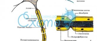
- The central part of the nerve is the axons;
- The insulating covering of axons is the myelin sheath;
- bundle of axons (nerve bundle);
- The sheath surrounding the bundles of nerve fibers is the perineurium;
- Blood vessels.
This system includes 31 pairs of spinal nerves and cranial nerves. The peripheral nervous system (PNS) has a major role in communicating between the central nervous system and the sensory organs and muscles.
Properties
A very active lateral transport of small and large molecules is carried out throughout the neutrite body. Macromolecules and organelles formed in the neuron itself continuously move along this process to its sections. The activation of this movement is a forward propagating current (transport). This electric current is realized by three transports of different speeds:
- A very weak current (at a rate of a few ml per day) transports proteins and filaments from actin monomers.
- A current at an average speed moves the main energy stations of the body, and a fast current (the speed of which is 100 times greater) moves the molecules that are contained in the bubbles required for the site of communication with other cells at the time of signal retranslation.
- In parallel with the forward-moving current, a retrograde current (transport) operates, which moves certain molecules in the opposite direction (towards the neuron itself), including material captured by endocytosis (including viruses and toxic compounds).
This phenomenon is used to study the projections of neurons; for this purpose, the oxidation of substances is used in the presence of peroxide or other constant substance, which is introduced into the area where synapses are located and after a certain time its distribution is monitored. Motor proteins associated with axonal flow contain molecular motors (dynein) that move various “cargos” from the outer boundaries of the cell to the nucleus, characterized by ATPase action, located in microtubules, and molecular motors (kinesin) that move various “cargos” from the nucleus to the periphery cells, forming a forward propagating current in the neurite.
The affiliation of nutrition and elongation of the axon to the body of the neutron is undoubted: when the axon is excised, its peripheral section dies, but the beginning remains viable.
With a circumference of a small number of microns, the total length of the process in large animals can be 100 cm or more (for example, branches directed from spinal neurons to the arms or legs).
Most representatives of invertebrate species have very large neural processes with a circumference of hundreds of microns (in squids - up to 2-3 mm). As a rule, such neutrites are responsible for transmitting impulses to muscle tissue, which provides a “signal to escape” (getting into a hole, quickly swimming away, etc.). With other similar factors, as the circumference of the appendix increases, the speed of transmission of nerve signals throughout its body increases.
Structure
The contents of the material substrate of the axon - axoplasm - contain very thin fibers - neurofibrils, and in addition microtubules, energy organelles in the form of granules, cytoplasmic reticulum, which ensures the production and transport of lipids and carbohydrates. There are pulpy and non-pulpous brain structures:
- The pulpy (also known as myelin or mislin) sheath of neutrites is found exclusively in representatives of the vertebrate species. It is formed by special lemmocytes (additional cells formed along the neutrites of the nervous structures of the periphery) “wound” around the process, in the middle of which the places unoccupied by the mislin membrane, the belt of Ranvier, are preserved. Only in these areas are voltage-gated sodium channels located and the activity potential appears again. In this case, the brain signal moves through the mislin structure in steps, which significantly increases the speed of its transmission. The speed of movement of the pulse along the neutrons with the pulpy layer is 100 meters per second.
- The pulpless shoots are smaller in size than the neutrites provided by the pulpy shell, which makes up for the losses in the speed of signal transmission in comparison with the pulpy branches.
At the site of union of the axon with the body of the neuron itself, an axonal eminence is located in the largest cells in the form of pyramids of the 5th shell of the cortex. Not long ago, there was a hypothesis that it is in this place that the post-connective capabilities of a neuron are converted into nerve signals, but this fact has not been proven through experiments. Fixation of electrical capabilities determined that the nerve signal is concentrated in the body of the neurite, or more precisely in the starting zone, at a distance of ~50 μm from the nerve cell itself. In order to maintain the strength of activity in the starting zone, a high content of sodium passages is necessary (up to a hundred times, with regard to the neuron itself).
Axon growth and development[edit]
Neuron
Axon growth occurs through its environment, in the form of a growth cone, which is located at the axon tip. The growth cone has a broad leaf-like extension called lamellipodia, which contains protrusions called filopodia. Filopodia is a mechanism representing the process of holding surfaces. It analyzes the immediate environment. Actin plays a major role in the motility of this system. Environments with high levels of cell adhesion molecules or "CAM" create an ideal environment for axonal growth. This appears to provide a "sticky" surface for axons to grow forward. Examples of CAM specific to nervous systems include: N-CAM, neuroglial CAM or NgCAM, TAG 1, MEG, and DCC, all of which are part of the immunoglobulin superfamily. Another set of molecules called extracellular matrix adhesion molecules also provide a sticky base for axons to grow forward. Examples of these molecules include laminin, fibronectin, tenascin, and perlecan. Some of them are surface bound to cells and thus act as short range attractants or repellents. Others are difusible ligands and thus can long-last range effects.
Bell cells and pointer cells help in guiding neuronal axon growth. These cells are typically other, sometimes immature, neurons.
Classification
Neurons are divided into types depending on the type of mediator (mediator of the conductive impulse) released at the axon terminals. This can be choline, adrenaline, etc. Depending on their location in the parts of the central nervous system, they can relate to somatic neurons or autonomic ones. There are receptive cells (afferent) and transmitting feedback signals (efferent) in response to irritation. Between them there may be interneurons responsible for the exchange of information within the central nervous system. Depending on the type of response, cells can inhibit excitation or, conversely, increase it.
According to their state of readiness, they are distinguished: “silent”, which begin to act (transmit an impulse) only in the presence of a certain type of irritation, and background, which constantly monitor (continuous generation of signals). Depending on the type of information perceived from the sensors, the structure of the neuron also changes. In this regard, they are classified into bimodal, with a relatively simple response to irritation (two interrelated types of sensation: a prick and, as a result, pain, and polymodal. This is a more complex structure - polymodal neurons (specific and ambiguous reaction).