Prefrontal cortex
Dorsal lateral prefrontal cortex, external view.
Inferiorbital prefrontal cortex, external view.
Dorsal-lateral prefrontal region (3D surface image, lateral view).
Inferior-orbital prefrontal cortex (internal structure).
Inferior-orbital prefrontal cortex (internal structure). 3D image - bottom surface.
Prefrontal region. Three-dimensional image of the surface, top view.
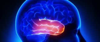
The prefrontal cortex (PC) is the most developed part of the brain. This area is located in the anterior third of the hemispheres, just behind the frontal bone. Often, three zones are distinguished in the prefrontal region: the dorsolateral (outer surface of the PC), the cortex of the inferior surface of the frontal lobes, and the cingulate gyrus (located in the middle part of the frontal lobes).
We will talk about the cingulate gyrus, which is often considered part of the limbic system, in a separate chapter. The dorsolateral and internal ocular gyri are often referred to as the main control center of the brain. We'll look at both of them in this chapter. If necessary, I will clarify what exactly is known about their functions.
In general, the PC is the part of the brain that monitors, controls, directs, manages and focuses your actions. It is responsible for “executive functions”—the ability to manage time, judgment, impulses, planning, organization, and critical thinking. The ability of humans as a species to think, plan for the future, use time wisely, and communicate with others largely depends on this part of the brain. The PC is responsible for activities that help you stay goal-oriented, socially responsible, and efficient.
Thomas Galtieri, a psychiatrist at North Carolina State, succinctly described the functions of the human PC as “the ability to formulate goals, formulate plans to achieve them, implement them effectively, and change course or improvise in the face of obstacles or failure, and do so successfully in the absence of external guidance.” or structures. The ability of an individual to set goals and achieve them is considered the main characteristic of a mature, effective personality. Moreover, this ability is not determined by social conventions or cultural baggage. This ability is embedded in the structure of the prefrontal cortex and its connections.”2
2 John Ratey, M.D., ed., The Neuropsychiatry of Personality Disorders (Cambridge, Mass.: Blackwell Science, 1995), p. 153.
The prefrontal cortex (especially the inferior frontal cortex) helps us think about what we say or do before we say or do it. Let's say, if you are arguing with your husband (wife) and your PC is working well, most likely you will react thoughtfully, and your answers will be able to correct the situation. If your PC isn't working well, you'll probably say something that will make everyone feel worse. The PC helps you solve problems, calculate how the situation will develop, and, using experience, choose the best one from several options. A good PC function is essential in a game like chess.
This same part of the brain is responsible for how you learn from your mistakes. A good PC performance doesn't mean you don't make mistakes. However, it will help you avoid making the same mistake over and over again. You are able to learn from experience and apply the lessons of the past. Let's say a student with good PC performance is able to learn that by starting work on a large project as early as possible, he will thereby ensure himself more time for research work and will have less reason to worry about not being able to turn it in on time. Meanwhile, a student with reduced PC function does not take past worries and failures into account and will constantly put everything off until the last minute. Those who are unable to learn from past failures tend to have impaired PC function. They constantly make the same mistakes. Their actions are not based on their experience, but on what they want at the moment.
The PC (especially the dorsolateral zone) is also involved in maintaining sustained attention. It helps you focus on important information and filter out less significant thoughts and sensations. Sustained attention plays an important role in short-term memory and learning. Through its many connections to other areas of the brain, the PC allows you to focus on one project for a long time and not be distracted from it until it is completed. The PC sends calming signals to the limbic and sensory systems of the brain when you need to focus, and reduces distracting impulses coming from other areas of the brain. When PC function is reduced, a person becomes more easily distracted from the main activity (we will talk about this in more detail when we begin to discuss attention deficit disorder).
The PC, and especially the dorsolateral area, allows us to experience and express emotions; feel happy, sad, feel joy and love. The way this happens in the PC is different from the more primitive limbic system. Although the limbic system controls mood and sex drive, the PC can translate processes in the limbic system into recognizable feelings, emotions, and words, such as love, passion, or hate. Reduced activity or injury in this area of the brain often results in a decreased ability to express thoughts and feelings.
PC has a major impact on the ability to reflect and control impulsive behavior. The ability to think through the consequences of your actions (choosing a partner, working with clients, dealing with difficult children, spending money, driving on the highway) is critical to living a fulfilling life in almost every aspect of it. Without normal PC operation, it is difficult to act thoughtfully and consistently, and then impulsiveness begins to play a decisive role in our behavior.
The PC has many connections with the limbic system. She sends inhibitory signals that help her keep the limbic system under control. It gives her the opportunity to “think with her head, and not just with her emotions.” When this area of the brain, especially the left side, becomes underactive or damaged, the PC is no longer able to properly influence the limbic system, which can cause an increased susceptibility to depression if the limbic system becomes overactive. . A classic illustration of this would be patients who have suffered a hemorrhage in the left frontal lobe of the brain. Sixty percent of these patients develop severe depression within the first year after a stroke.
Patience found in the orbitofrontal and prefrontal cortex
Giphy.com
Japanese scientists conducted an experiment on mice and found that for deciding whether to wait for a reward (that is, patience), in the brain, in addition to the raphe nuclei, two more areas associated with them by the serotonergic system are responsible: the orbitofrontal and the medial part of the prefrontal bark. At the same time, these areas process information about a possible reward differently: the orbitofrontal cortex evaluates the time spent waiting, and the prefrontal cortex evaluates the situation as a whole. The results of the study were published in the journal Science Advances
.
Patience, that is, the ability to wait a long time for reward, is a quality that helps animals and humans adapt to a changing environment. In the body, patience is regulated by serotonin, a neurotransmitter that is also responsible for mood, sleep-wake cycles, appetite, and impulsive behavior, that is, behavior designed to obtain an immediate reward.
A group of neuroscientists led by Katsuhiko Miyazaki from the Okinawa University Institute of Science and Technology has long been studying how exactly serotonin regulates patience. Scientists have already found that serotonin is most effective in this role when confidence in receiving a reward is high. Previous studies in mice have also shown that anticipation of future rewards is associated with activation of serotonergic neurons in the dorsal raphe nucleus (a region in the medulla oblongata). Neurons from this area extend to other areas—the nucleus accumbens, orbitofrontal cortex, and medial frontal cortex—and their damage increases impulsive behavior. But exactly how these regions regulate patience remained unknown.
Researchers led by Miyazaki decided to find out in a new study. To do this, they used optogenetics: they took 27 genetically modified mice in which serotonergic neurons expressed a light-sensitive protein. By stimulating axon terminals with light through an optical fiber implanted in the heads of animals, it is possible to induce the release of a neurotransmitter and increase neural activity. The scientists also took 15 mice, which were also implanted with optical fiber, but there was no light-sensitive protein in their neurons.
The mice took part in a series of experiments in which they were placed in cages with a hole in the wall. If the mouse poked its nose into the hole, a sound was heard and a piece of food rolled into the cage, but not immediately: sometimes the food arrived six seconds after the action, sometimes ten seconds later. In some experiments, food was delivered after a random period of time ranging from two to ten seconds. At the same time, mice received food only in 75 percent of cases. During the experiment, scientists assessed how long mice poked their nose and waited for a reward, depending on whether they stimulated serotonergic neurons or not.
A - design of the experimental cell, B - experimental diagram, C - location of the studied brain areas: orbitofrontal cortex (OFC) and dorsal raphe nuclei (DRN)
Katsuhiko Miyazaki et al. / Science Advances, 2020
Share
It turned out that stimulating the projections of serotonergic neurons to the nucleus accumbens did not increase latency. This means that serotonin in this area of the brain probably does not play a role in regulating patience. The activity of serotonergic neurons in the orbitofrontal cortex, on the contrary, increased the time during which subjects were willing to wait (p = 0.033), and was as effective as stimulation of the neurotransmitter in the raphe nuclei. The result did not depend on the time period.
When serotonergic neurons were active in the medial prefrontal cortex, mice waited patiently longer, but only when they did not know when they would receive a reward (p = 0.023), that is, in experiments with a random time interval. At a fixed time (two, six, or ten seconds each time, depending on the experiment), no such effect was observed.
When scientists compared the results of experiments for normal and genetically modified animals, it turned out that the latter, on average, waited much longer: from this, scientists concluded that it was all about the stimulation of serotonergic neurons. To explain how this is related to the behavior of animals, scientists built a mathematical model based on the experimental results: as the main parameter, they took the ratio of the time of waiting for a reward with the activation of serotonergic neurons to the time of waiting without the activation of neurons.
The model took into account that the brain of mice predicts the time of food delivery and estimates the likelihood that they will receive food in principle: the more time passes, the lower the likelihood that a reward will follow. Stimulating the release of serotonin increased this probability: for example, if a reward came only 75 percent of the time, then the probability was 75 percent, but stimulating serotonergic neurons increased this probability in mice to 94 percent. The effect of serotonin increased the animals' confidence that they were participating in a reward trial, so they waited longer.
The model also suggests that the orbitofrontal cortex and the medial prefrontal cortex use different reward anticipation strategies. Information processing in the first area affects the assessment of time spent, and the second area is responsible for the general assessment of the situation of delayed reward, since it specializes to a lesser extent in the analysis of time intervals. The brain then likely combines the results of calculations across different regions to decide whether to be patient or not.
Scientists have previously shown that serotonin stimulates the repair of severed motor neuron axons—at least in zebrafish.
Evgenia Shcherbina
Stroke in the left frontal lobe
Three-dimensional image of the surface, side view.
3D image, bottom surface.
Notice the large hole in brain activity located in the left frontal lobe.
When scanning the PC with methods such as SPECT, scientists often conduct two studies: one on the brain at rest and another during a concentration exercise. When assessing brain function, it is important for them to see a picture of an active brain. When a normal brain is presented with a task that requires concentration—a math problem or card sorting—PC activity increases. In some brain disorders, such as schizophrenia or attention deficit disorder, PC activity decreases when it is necessary to solve an intellectual problem.