MRI of the pituitary gland: the essence of the procedure, preparation and indications for the procedure
MRI of the pituitary gland is a modern and highly accurate diagnostic method, the purpose of which is to identify changes in the structure of the lower medullary appendage due to the development of diseases and pathological processes. This type of diagnosis is considered the most highly informative, allowing one to visualize even minor pathological conditions in which other examination methods will be ineffective.
What is diagnostics using a tomograph?
MRI uses a powerful magnetic field. Through its effect on the body, radio frequency impulses are formed, the analysis of which is carried out by specialists on a computer. Three-dimensional images can be printed, saved or sent, if necessary, to the subject’s e-mail address.
MRI of the pituitary gland provides an opportunity to fully evaluate existing changes in the functioning of the pituitary gland, as well as examine nearby tissues. Scanning allows you to analyze the state of operation of a separate part of the brain, which ultimately can be used to assess the functioning of many organs and systems.
In the photographs after the diagnosis, you can see in detail the sinuses with the eye orbits. If it becomes necessary to examine other parts of the brain, a full scan is performed to clarify or identify the causes of the pathological condition.
How to properly prepare for an MRI of the pituitary gland
Magnetic resonance imaging is a type of pre-diagnosis for which patients do not need to comply with strict restrictions and extensive preparatory measures. True, there is one exception - if the scanning is planned to be carried out using contrast, in this case some preparation will still have to be completed. So, it is unacceptable to eat on the day of the study, and if the diagnosis is carried out in the evening, you need to wait a 6-hour interval. Otherwise, nausea and vomiting may occur, which will be completely inappropriate.
If an MRI of the pituitary gland is performed without contrast, such restrictions do not apply to the procedure.
Before scanning, all patients must:
- Remove any clothing that has metal inserts. You will have to leave all decorations and gadgets outside the office. Any metal objects can not only affect the operation of the equipment, but also have a negative impact on the quality of the images, which will complicate diagnostic measures.
- The radiologist must be informed about the implants present in the body. Some of them may act as strict contraindications to scanning.
- Women expecting the birth of a baby should not undergo an MRI in the first trimester of pregnancy. Despite the fact that tomography is considered one of the safest examination methods, there is still a potential risk of negative effects on the developing organism.
- As a rule, young children are allowed to be scanned after 5-6 years. MRI is indicated for children only in the most extreme cases and, most often, in a state of medicated sleep.
Preparation and performance of MRI
A little preparation will be required before administering the MRI contrast agent and undergoing the procedure to ensure the results of the examination are as detailed as possible and the patient does not encounter problems
Therefore, it is important not only to familiarize yourself with the features of the study, but also to prepare for it in advance.
Preparation
You should start preparing for an MRI a few days before the procedure.
This is very important to obtain the most accurate results. The first element of preparation is a conversation with the doctor, during which all important information about the patient is clarified, which could become a contraindication for MRI.
Therefore, it is very important to remember whether you have previously had surgery with the installation of implants or other metal elements. This is especially true for dental procedures.
After this, it will be very easy to prepare for the procedure, because... there are no special requirements. You only need to follow these rules:
Prepare psychologically
It is important to set yourself up for the absence of fear, the rapid completion of the procedure, and a positive result. This should be done a few days before the MRI. Stop using makeup
From the editor: What to do if you have a headache and a fever
It is recommended not to use cosmetics before undergoing brain imaging, because this may harm the accuracy of the result. Remove all metal items and electronic devices. It is best not to take anything metal or electronic with you, so as not to accidentally forget about these things. If you have them with you, be sure to remove them before performing the diagnostics.
It is strictly forbidden to drink alcoholic beverages before the tomography. It is also not recommended to smoke, be nervous, or influence your brain or blood vessels in any way.
These rules apply to any type of MRI with contrast.
However, when examining the abdominal cavity, it is important to adhere to a strict diet, avoiding junk food, and also not to eat a day before the procedure, while refusing to drink liquids 5 hours before the MRI
Indications for MRI of the pituitary gland
In most cases, the doctor writes a referral for examination if there is compelling evidence for it. First of all, he conducts a comprehensive examination and issues a referral for preliminary tests.
If the cause of the complaints cannot be identified, the specialist decides on the need for additional examination. The following pathological conditions may be indications for tomography:
- severe periodic pain in the head;
- metabolic disorders in the body;
- existing vision problems;
- menstrual irregularities;
- impotence;
- elevated TSH levels in tests.
Contraindications
Contraindications to tomography are divided into relative and absolute. If a patient has absolute contraindications, then MRI will be prohibited for him, regardless of whether there are compelling reasons for this examination or not. These include:
- metal implants, insulin pumps, pacemakers present in the body.
- the presence in the body of the subject of metal objects received as a result of injury.
Relative or temporary bans on scanning include:
- first trimester of pregnancy. In exceptional cases, diagnostics can still be carried out on expectant mothers if the expected benefit from the procedure for the mother exceeds the potential risk to the fetus;
- the patient is in extremely serious condition;
- epilepsy;
- contrast intolerance;
- disorders of the kidneys;
- fear of closed spaces;
- involuntary movements of the limbs.
If the patient has any doubts or concerns before undergoing a tomography, he can always ask all the questions that interest him to the radiologist.
Preparation for MRI of the pituitary gland (with contrast)
Preparation for an MRI of the pituitary gland is necessary if the study is performed with contrast. Then you must go for the procedure on an empty stomach, or at least 5 hours must have passed since your last meal. Otherwise (non-contrast MRI of the sella turcica is performed less frequently), you can go for the study without preparation.
You need to take with you, if you have:
- doctor's referral;
- an extract from the medical history or outpatient card;
- photographs and descriptions of the results of previous studies (not only MRI, but also others);
- other documents related to your disease.
MRI of the pituitary gland for young children
This type of diagnosis is prescribed to children, most often after 5 years. To obtain clear images, the patient should not make any movements during the examination, which is very difficult to achieve from young patients. Children simply cannot be in a calm state inside a confined space due to overwhelming fear. To examine children, doctors use open-type devices. However, even in this case, the baby must remain still throughout the scan. Parents are allowed to be with their child during the examination. If the baby shows excessive anxiety, cries a lot or refuses to go into the office, the doctor may decide on the need to use sedatives or conduct diagnostics in a state of medicated sleep.
MRI of the pituitary gland without contrast is most often performed on children.
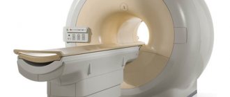
How does the scan work?
MRI is a diagnostic method that is quite simple, and the absence of the need to follow preparatory measures is another big advantage of tomography. The device on which the examination will be carried out is located in a separate room.
Medical staff monitors the examination from the next room, and communication with the subject is carried out using a special speaker. Therefore, you should not worry about the fact that if your health worsens, you will not have the opportunity to inform your doctor about it.
Before being examined, the patient must change into hospital clothes. It is more spacious, comfortable and does not have any metal parts. If the scan is to be performed on a child, he or she enters the office accompanied by an adult. When performing an MRI of the pituitary gland with contrast, the patient is given an intravenous injection of a contrast agent.
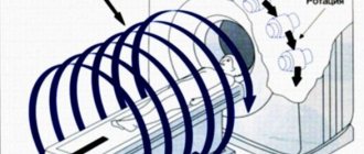
Regardless of whether an MRI of the pituitary gland is performed with or without contrast, as soon as the patient is ready to begin the procedure, he will be asked to take a horizontal position on a special extendable table of the tomograph.
The patient's body will be fastened with special straps. There is no need to be afraid of this; this is done to ensure complete immobility and eliminate involuntary movement of the limbs during the procedure.
Actually, to carry out scanning, the table is rolled into the apparatus, which looks like a large tunnel. The tomograph makes specific noises during operation - this is quite normal and does not pose any danger to the patient. The duration of the scan varies from 25 to 55 minutes and depends directly on whether an MRI of the pituitary gland is performed with or without contrast, as well as on the required number of images.
Upon completion of the procedure, the doctor can immediately analyze the resulting images. The evaluation of the obtained images is carried out on a computer monitor using a special program. The doctor evaluates the results obtained and writes a conclusion based on them.
At the time of diagnosis, the patient can await results in the clinic. The results can be sent to him by email. We must not forget that once the results are ready, in order to prescribe treatment, it will be necessary to contact the specialist who issued the referral for examination.
There is no need to observe any restrictions in food or lifestyle after an MRI. After the patient leaves the clinic, he can return to his usual activities without any restrictions.
Contraindications for MRI of the pituitary gland (with contrast)
The MRI procedure is strictly contraindicated if the patient’s body contains any metal parts that are attracted by a magnet. Under the influence of the field, they can heat up and move, causing tissue damage and burns. For this reason, MRI is not performed for patients with wire braces, metal vascular staples, and other “add-ons.”
The study is prohibited for patients with a pacemaker, hearing aid, insulin pump, implanted cardioverter-defibrillator and other medical devices, since radio waves can affect and disrupt the operation of these devices.
MRI is not recommended for claustrophobia, severe pain and hyperkinesis (involuntary movements), when the patient cannot remain motionless for a long time.
Contrast-enhanced MRI diagnostics are contraindicated in pregnant and lactating women, since the contrast is toxic to the fetus and passes into breast milk.
Also, contrast studies are prohibited for patients with renal failure: if its excretion is impaired (the drug is excreted in the urine), side effects and poisoning of the body are possible.
MRI of the pituitary gland with contrast
In order to obtain more informative images, a specialist may decide on the need for an MRI with contrast. A contrast agent is administered intravenously to the patient before the procedure begins. Once in the bloodstream, contrast when assessing the resulting images allows the doctor to examine the vascular network in the required area. Almost always, contrast makes it possible to assess the size and location of the pathological process and determine the intensity of blood flow. Scanning using a contrast agent, in most cases, is prescribed to patients before surgery to remove pituitary tumors. The contrast is concentrated in those areas where there is increased blood supply, in particular, in those tissues in which the tumor is located. As a result, it is possible to diagnose even small tumors.
MRI of the pituitary gland without contrast is performed much more often, because pituitary tumors are a rather rare pathological process.
MRI with and without contrast - what's the difference?
Having received an appointment for an examination, the patient is usually interested in what an MRI without contrast shows and why in some cases doctors decide to use this very contrast.
Contrast or contrast agent is a solution that is administered intravenously to the patient before starting the magnetic resonance diagnostic method. It is usually administered when a clearer three-dimensional image of an organ is needed. An image is created from a large number of photographs, which are superimposed on each other in a special way. And if any picture is blurry, the whole picture will change, which will make it difficult to make a diagnosis.
Contrast is especially often used for MRI of the brain or when it is necessary to detect a cancerous tumor. In the latter case, contrast allows you to visually assess not only the location of the tumor, but also its quality and size, as well as determine whether there are the first signs of metastasis.
Gadolinium salts are most often used for contrast. They are characterized by low toxicity and practically do not cause an allergic reaction; moreover, these substances are quickly eliminated from the body without accumulating in it. However, salts in their pure form are still not recommended for use, so during MRI the patient is injected with a chelate complex containing gadolinium ions.
From the editor: Foci of glial changes in the brain
How it works?
Contrast plays the role of a special indicator of disorders in the brain. It actively reacts to pathologies, accumulating exclusively in the area of the disease. If we clarify what MRI with contrast shows, then, basically, this is:
- Benign and malignant tumors
- Cysts
- Metastases
- Brain injury
- Meningioma
- Genetic and infectious diseases
- Alzheimer's disease
- Neurodegenerative disorders of the central nervous system.
The contrast can determine not only the location where the pathological process develops, but also the nature of the tumor. Therefore, resonance with contrast is considered a more effective method than research without contrast.
The use of contrast does not harm a person and is absolutely painless. The only contraindication is considered to be individual intolerance to the substance, which should be notified to the doctor in advance.
It is necessary to take into account that if you do an MRI for a fee, the price of a study with contrast will increase significantly compared to conventional diagnostics. However, your health and life are worth more than any money, so you should not skimp on such diagnostic methods.
Magnetic resonance allows in most cases to obtain a very accurate result in the form of a three-dimensional image of the organ. That is why this non-invasive diagnostic method, which has been used for almost half a century, is becoming increasingly popular these days.
The magnetic field created by the tomograph is completely safe. The only absolute indication is the presence of a pacemaker, insulin pump and other electronic devices or metal structures in the patient’s body (wires, endoprostheses, dental implants).
The absence of harm to health and high efficiency make MRI virtually no alternative to diagnosing various diseases.
However, sometimes before the procedure the doctor may announce that this time the tomography will be done using a contrast agent. In this regard, the patient may wonder whether contrast is needed in MRI and how it works.
Also, tomography with contrast is often performed after removal of intervertebral hernias to monitor the condition of scar tissue and prevent relapse. Other indications are:
- sinusitis, hearing impairment;
- headache;
- pituitary adenoma;
- paroxysmal state;
- suspicion of an infectious disease of the nervous system;
- stroke;
- cerebrovascular diseases.
Most often, a contrasting solution is used in diagnosing the brain, since in other cases natural contrast - blood circulation - is sufficient.
If you are unsure whether you can have an MRI with contrast, talk to your doctor or get additional tests from an allergist.
This is what a photo looks like with and without contrast
A brain tumor. Left without contrast, right with contrast.
Which is better: MRI of the pituitary gland with or without contrast?
The specialist may prescribe the patient to undergo an MRI of the pituitary gland without contrast, or magnetic resonance imaging using a contrast agent. Most often, paramagnetic substances are used for this purpose, administered intravenously a few minutes before the start of the scan. The dosage of the drug is calculated individually, based on the patient’s body weight. An MRI of the pituitary gland with or without contrast is always decided based on the patient’s medical history. If it is necessary to identify clear boundaries of a neoplasm, its shape, and the condition of nearby tissues, it makes sense to use a contrast agent. In any case, only the doctor decides whether to conduct an MRI of the pituitary gland with or without contrast; the advisability of using contrast is considered on an individual basis.
MRI indications of the pituitary gland (with contrast)?
- if pituitary tumors are suspected;
- if you suspect empty sella syndrome.
Why is an MRI of the pituitary gland done (with contrast)?
MRI diagnosis of the pituitary gland in both cases, the basis for prescribing the procedure are certain symptoms - headaches, obesity, signs of dysfunction of the thyroid glands and adrenal glands, heart pain, vegetative manifestations and others, as well as corresponding laboratory changes. However, symptoms and hormone levels can be completely different and change over time, so if you suspect these diseases, consult an endocrinologist. Only a specialist can determine how legitimate your assumption is and correctly prescribe an examination. Otherwise, you may confuse the signs of one disease with another.
As a rule, tomography, regardless of the type of device and magnetic field strength, allows one to accurately determine a particular disorder. But in complex cases, diagnosis requires high accuracy, so open MRI of the pituitary gland, as it is somewhat less sensitive, is not recommended. Our clinic uses the most modern tomographs. This can be seen in the enlarged photo on the right.
What will the examination show?
MRI of the pituitary gland with or without contrast allows you to visualize any changes in the pituitary gland. Thanks to scanning, it is possible to diagnose any tumors, as well as identify their type and exact location. A radiologist will be able to identify abnormal development of the pituitary gland, its structural changes and changes in size. In addition, MRI provides the opportunity to identify the very cause of the pathological process. It is important to remember that timely diagnosis will be the key to a complete recovery with well-chosen treatment tactics.
MRI of the pituitary gland (with contrast)
- Immediately before the procedure, you must remove all metal objects from yourself: dentures, jewelry, watches, piercings, etc.
- During the examination, you will lie on a mobile table, while your body position will be fixed with special belts and bolsters. When the procedure begins, the table will move inside the annular part of the tomograph, where you will remain until the end of the procedure.
- It is not recommended to make any movements during the scan, so be sure to make sure you are comfortable before starting.
- Lighting and a fan work inside the ring, and if there are any problems, you can always contact the technologist via two-way communication.
- If necessary, MRI of the pituitary gland is performed in the presence of a relative or loved one.
Get specialist advice or make an appointment
+7 (499) 400-47-33
MRI photo of the pituitary gland with and without contrast
Below you can see a photo of a magnetic resonance imaging scan. To better see the difference between an MRI image with and without contrast, hover over the photo and click on it. In the window that opens, you can better see the differences between the studies.
Possibility of complications
An absolutely harmless and safe diagnosis by undergoing an MRI of the pituitary gland can still cause unpleasant symptoms in some patients. So, some patients may complain of:
- the appearance of nausea;
- feeling of extreme weakness;
- the appearance of dizziness;
- severe headaches;
- increased heart rate;
- excessive nervous excitability.
As a rule, all such symptoms are caused not because of the tomography, but because of the patient’s suspiciousness and fear of the procedure. MRI of the pituitary gland without contrast should not cause any negative consequences at all, because this is an absolutely harmless procedure, performed even on small children.
But when undergoing an MRI with contrast, it is quite possible that a real complication may arise – an allergic reaction. However, the occurrence of contrast intolerance is extremely rare.
To avoid negative consequences, you need to conduct an allergy test - if its results do not cause concern among specialists, an MRI with contrast can be performed without any fear.
Where to undergo magnetic resonance imaging of the pituitary gland
Every patient who has been scheduled for a scan, regardless of whether an MRI of the pituitary gland will be performed without contrast or with its use, wants to undergo diagnostics quickly, efficiently and without waiting in long lines. It is almost impossible to do this in public clinics. According to the coupon, you will have to wait a very long time for your turn to undergo the examination, and not every government agency has modern equipment that allows you to obtain high-quality images during scanning.
As a result, people are starting to look for a private clinic to undergo examination. The pages of our website present a large number of clinics with detailed information about each of them. All medical institutions provide diagnostics at an affordable cost, which is a key point for patients. In addition, we provide each patient with an additional discount on MRI of the pituitary gland, so contacting us will guarantee a low cost for the examination.