How to take a puncture
For this reason, spinal cord puncture is also called lumbar puncture.
To carry out the puncture, special long Beer needles of reinforced design (thick-walled) with a mandrin (stiletto) are used.
Preparation for puncture
Before collecting cerebrospinal fluid for analysis, it is necessary to conduct an examination:
- pass general and biochemical blood and urine tests;
- do a blood coagulogram;
- change fundus pressure and intracranial pressure;
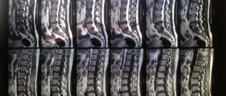
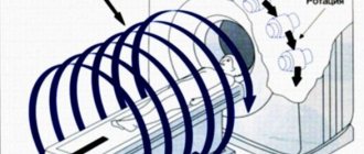
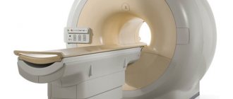
- for neurological disorders, cerebral signs indicating dislocations - CT or MRI of the brain;
- other tests prescribed by your doctor.
How is a spinal cord puncture performed?
The patient lies on his side on a hard couch, bending his knees to his stomach and bending his back as much as possible
A sitting position is also allowed. The surface of the lower back is treated with iodine solution. The needle is inserted into the intervertebral space between the second and third (third and fourth in children) vertebrae, at the level of the spinous processes, slightly at an upward angle. At the beginning of the needle's advancement, an obstacle is soon felt (these are the vertebral ligaments), but when 4 to 7 cm have been passed (about 2 cm in children), the needle falls under the arachnoid membrane and then moves freely. At this level, the progress stops, the mandrin is removed, and by the flow of drops of colorless liquid from it, it is confirmed that the goal has been achieved. If the liquid does not drip, and the needle rests on something hard, it is carefully returned back without completely removing it from the subcutaneous layer, and the injection is repeated, slightly changing the angle. Cerebrospinal fluid is collected into a test tube, the sampling volume is 120 g. If you need to examine the epidural space to see adhesions and tumors, or the condition of the spinal ligaments, a three-channel test is performed (saline is supplied through one channel, a needle with a catheter is supplied through the second, a microchamber is supplied through the third review). Anesthesia or therapy is administered by administering an anesthetic or medicinal drug through a catheter.
Does it hurt when taking a puncture?
Many patients are afraid that it will hurt. You can calm them down: before the analysis itself, local anesthesia is usually performed: layer-by-layer injection of novocaine (1 - 2%) into the area of the future puncture. And even if the doctor decides that local anesthesia is not needed, in general the puncture is no more painful than a regular injection.
Complications and consequences of spinal cord puncture
After the puncture, the following complications are possible:
- On the membranes of the spinal cord, when subcutaneous epithelial cells are introduced with a needle, the development of an epithelial tumor - cholesteatoma - is possible.
- Due to a decrease in the volume of cerebrospinal fluid (daily circulation volume - 0.5 l), intracranial pressure decreases, and a headache may occur for a week.
- If nerves or blood vessels are damaged during a puncture, the consequences can be very unpleasant: pain, loss of sensitivity; formation of hematoma, epidural abscess.
However, such phenomena are extremely rare, since spinal puncture is usually performed by experienced neurosurgeons with experience in numerous operations.
Thank you
The site provides reference information for informational purposes only. Diagnosis and treatment of diseases must be carried out under the supervision of a specialist. All drugs have contraindications. Consultation with a specialist is required!
puncture
When is a lumbar puncture necessary and why not?
lumbar puncture
Lumbar puncture is performed both for diagnostic purposes and for therapy, but always with the consent of the patient, except in cases where the latter, due to his serious condition, cannot contact the staff.
For diagnosis, a spinal puncture is performed if it is necessary to examine the composition of the cerebrospinal fluid, determine the presence of microorganisms, fluid pressure and patency of the subarachnoid space.
Therapeutic puncture is needed to evacuate excess cerebrospinal fluid or introduce antibiotics and chemotherapy drugs into the intrathecal space in case of neuroinfection or oncopathology.
The reasons for lumbar puncture are mandatory and relative, when the decision is made by the doctor based on a specific clinical situation. Absolute indications include:
- Neuroinfections - meningitis, syphilitic lesions, brucellosis, encephalitis, arachnoiditis;
- Malignant tumors of the brain and its membranes, leukemia, when CT or MRI cannot make an accurate diagnosis;
- The need to clarify the causes of liquorrhea with the introduction of contrast or special dyes;
- Subarachnoid hemorrhage in cases where non-invasive diagnosis is not possible;
- Hydrocephalus and intracranial hypertension - to remove excess fluid;
- Diseases requiring the administration of antibiotics and antitumor agents directly under the membranes of the brain.
Among the relative ones are pathology of the nervous system with demyelination (multiple sclerosis, for example), polyneuropathy, sepsis, unidentified fever in young children, rheumatic and autoimmune diseases (lupus erythematosus), paraneoplastic syndrome. A special place is occupied by lumbar puncture in anesthesiology, where it serves as a method of delivering an anesthetic to the nerve roots to provide fairly deep anesthesia while maintaining the patient’s consciousness.
If there is reason to suspect a neuroinfection, then the cerebrospinal fluid obtained by puncture of the intrathecal space will be examined by bacteriologists, who will establish the nature of the microflora and its sensitivity to antibacterial agents. Targeted treatment significantly increases the patient's chances of recovery.
With hydrocephalus, the only way to remove excess fluid from the subarachnoid spaces and the ventricular system is puncture, and often patients feel relief almost immediately as soon as cerebrospinal fluid begins to flow through the needle.
If tumor cells are detected in the resulting liquid, the doctor has the opportunity to accurately determine the nature of the growing tumor, its sensitivity to cytostatics, and subsequent repeated punctures can become a way to administer drugs directly to the area of tumor growth.
Lumbar puncture may not be performed on all patients. If there is a risk of harm to health or danger to life, then the manipulation will have to be abandoned. Thus, the following are considered contraindications for puncture:
- Cerebral edema with risk or signs of herniation of brain stem structures or cerebellum;
- High intracranial hypertension, when removal of fluid can provoke dislocation and wedging of the brain stem;
- Malignant neoplasms and other space-occupying processes in the cranial cavity, intracerebral abscesses;
- Occlusive hydrocephalus;
- Suspicion of dislocation of stem structures.
The conditions listed above are fraught with the descent of the stem structures to the foramen magnum with their wedging, compression of vital nerve centers, coma and death of the patient. The wider the needle and the more fluid removed, the higher the risk of life-threatening complications. If the puncture cannot be delayed, then the minimum possible volume of cerebrospinal fluid is removed, but if wedging occurs, a certain amount of liquid is reintroduced.
If the patient has suffered a severe traumatic brain injury, massive blood loss, has extensive injuries, or is in a state of shock, it is dangerous to perform a lumbar puncture.
Other obstacles to the procedure may include:
- Inflammatory pustular, eczematous skin changes at the point of the planned puncture;
- Pathology of hemostasis with increased bleeding;
- Taking anticoagulants and antiplatelet agents;
- Aneurysm of cerebral vessels with rupture and bleeding;
- Pregnancy.
These contraindications are considered relative, increasing the risk of complications, but in cases where puncture is vitally necessary, they can be neglected if maximum caution is observed.
Indications for manipulation
A pleural puncture can be performed for both diagnostic and therapeutic indications. Firstly, the reason for diagnosis is effusion, an increase in the amount of fluid in the pleural cavity to 3-4 ml, as well as taking a tissue sample for examination if a tumor is suspected.
Symptoms of effusion include:
- The appearance of pain when coughing and taking a deep breath.
- Feeling of fullness.
- The appearance of shortness of breath.
- Constant dry reflex cough.
- Asymmetry of the chest.
- Changes in percussion sound during tapping in specific areas.
- Weak breathing and voice tremors.
- Darkening on an x-ray.
- Changes in the location of the anatomical space in the middle parts of the chest (mediastinum).
Secondly, pleural puncture is indicated for collecting contents from the cavity for bacteriological and cytological analysis in order to identify and confirm pathologies such as:
- Congestive effusion.
- Inflammatory process due to fluid stagnation (inflammatory exudate).
- Accumulation of air and gases in the pleural cavity (spontaneous or traumatic pneumothorax).
- Collection of blood (hemothorax).
- The presence of pus in the pleura (pleural empyema).
- Purulent melting of lung tissue (lung abscess).
- Accumulation of non-inflammatory fluid in the pleura (hydrothorax).
In some cases, diagnostic pleural puncture can simultaneously become therapeutic. The therapeutic indication for pleural puncture is the need to carry out a number of therapeutic manipulations, such as:
- Extraction of contents from the cavity in the form of blood, air, pus, etc.
- Drainage of a lung abscess found in close proximity to the chest wall.
- Introduction of antibacterial or antitumor drugs into the pleural cavity directly into the lesion.
- Lavage (therapeutic bronchoscopy) of the cavity for certain inflammations.
Puncture for diagnostic and treatment purposes
During this procedure, for diagnostic purposes, a biological sample is collected using a thin needle, which is then sent for laboratory testing. If a therapeutic goal is pursued, then a medicinal product is injected into the affected area using a syringe or air or liquid is extracted, for example, from a tumor. The puncture can be performed on different organs of the body. Let's take a closer look at a few of these procedures.
Abdominal puncture
In this case, it is used to remove ascites or to administer special substances. Sterile instruments are used for puncture:
In the morning before the procedure, it is necessary to cleanse the intestines with an enema. The manipulation in this case is carried out in a sitting position. The skin is treated with novocaine in the puncture area. The liquid is taken from the cavity with a trocar, which makes a puncture and subsequent suction. The sample is placed in a test tube, and the puncture site is sealed with an aseptic sticker. The entire procedure is carried out under ultrasound guidance.
Bone marrow puncture
It is performed to obtain a bone marrow sample from the calcaneus or ilium, as well as the sternum and tibial epiphysis. A syringe with a long needle is used to take a sample. Depending on the puncture site, the patient lies on his back or sits down; before this, it is necessary to clear the puncture site from clothing. The doctor uses a needle to puncture the skin and remove a sample from the bone. During its implementation, the patient may feel a slight nagging pain.
Lumbar puncture
In this case we are talking about taking cerebrospinal fluid. Puncture can be performed for both diagnosis and treatment. It is carried out using a regular needle. This procedure is often performed on children. The patient lies on his left or right side, with his legs pressed to his stomach and his head to his chest. The doctor then inserts a needle between the 3rd and 4th lumbar vertebrae. If the procedure is performed for diagnostic purposes, 20 ml of fluid is taken. It is placed in a test tube and sent to the laboratory. There, all its characteristics are studied in detail, including the degree of transparency. After the manipulation, the patient should lie on his stomach for three hours, placing a pillow under his stomach. But it is best to stay in this position for a day. After the puncture, headache or weakness, nausea and back pain are possible. If such symptoms appear, you should consult a doctor. Hexamine, phenacetin and amidopyrine will be prescribed.
Needle biopsy
Used for biopsy of soft tissues of internal organs, such as:
In this case, 3 types of needles can be used: cutting, aspiration and modified. Both diagnostic and therapeutic punctures can be performed; the price in this case will depend on the purpose of the procedure. Depending on which needle will be used, the doctor will select a specific puncture technique. In terms of implementation, it is not much different from the above methods. A puncture is made through which a sample is taken for laboratory testing or a medicinal solution is injected.
Where can a puncture be done?
This procedure can be completed in our center. We are ready to perform it for both therapeutic and diagnostic purposes. Our staff consists of experienced specialists who have been carrying out similar procedures for many years. By contacting our center, you will get quick results. Come to us, we will definitely help you.
Technique for performing lumbar (lumbar, spinal) puncture.
There is no special preparation for lumbar puncture. But it is imperative that the patient first undergo a CT or MRI of the brain, evaluate the results and take into account contraindications.
click on the image to enlarge
Lumbar punctureclick on image to enlarge
For an experienced doctor, the algorithm for performing a lumbar (lumbar, spinal) puncture is not difficult. The patient's position is lying on his side, usually the left. The legs are bent as much as possible at the knee and hip joints, the knees are pressed to the stomach, the spine is bent, the chin is pressed to the chest, the hands are clasped around the knees. Rarely, in some cases, for example, in obese people, it is possible to perform a lumbar puncture in a sitting position; the torso should be tilted as far forward as possible, with the spine bent. The puncture is performed in the lumbar region. A typical point is the space between the spinous processes of the third and fourth lumbar vertebrae (L3-L4); it is possible to perform a lumbar puncture in the spaces L2-L3, L4-L5. It is impossible to penetrate the spinal cord with a needle, since the spinal cord in adults ends at the level of the second lumbar vertebra (L2). The puncture site is treated with antiseptics, after which local anesthesia with novocaine or lidocaine is applied in layers. A lumbar puncture needle (Beer needle) is inserted along the midline between the spinous processes. When the needle passes the interspinous ligament, there is a feeling of failure - this means that the needle has entered the epidural space. The needle is passed a little deeper through the dura mater and arachnoid membrane, after which the mandrel is removed from the needle and cerebrospinal fluid begins to flow. If the needle rests on the bone, then it must be removed, leaving the end in the subcutaneous tissue, then change the direction and insert until the needle passes the interspinous ligament. Having received the required amount of cerebrospinal fluid, the needle is removed and the puncture site is sealed with a sterile napkin. After performing the manipulation, the patient should lie on his stomach for at least two hours, since cerebrospinal fluid may continue to be released into the epidural space for some time through a defect in the dura and arachnoid membranes. After a lumbar puncture, there may be a headache caused by a decrease in intracranial pressure, which usually disappears without treatment after 5-7 days.
Sometimes, when performing a lumbar (lumbar, spinal) puncture, the needle may damage the venous plexus of the spinal canal (epidural venous plexus), which will be accompanied by the release of cerebrospinal fluid mixed with travel blood. Receipt of travel blood may be mistaken for subarachnoid hemorrhage (SAH). To eliminate such errors, there are several techniques to distinguish travel blood from true subarachnoid hemorrhage (SAH).
- When you receive blood-stained cerebrospinal fluid, you need to pull the needle slightly towards yourself. In the presence of travel blood, the cerebrospinal fluid in subsequent samples will become lighter.
- If bloody cerebrospinal fluid gets on a white cloth, such as a gauze pad, in a true hemorrhage, the spot will remain evenly colored, and in the case of travel blood, a rim of clear cerebrospinal fluid will appear around the blood stain - this is called a double spot symptom.
- After centrifugation, the cerebrospinal fluid in SAH will always remain xanthochromic (reddish), and in the presence of travel blood it will become colorless.
Indications and contraindications
Indications for lumbar puncture are:
- encephalitis, meningitis and other damage to the nervous system caused by infections - bacterial, viral and fungal, including syphilis and tuberculosis;
- suspicion of hemorrhage under the arachnoid membrane (subarachnoid space), when blood leaks from a damaged vessel;
- suspicion of a malignant process;
- autoimmune diseases of the nervous system, in particular suspected Guillain-Barré syndrome and multiple sclerosis.
We advise you to study - MRI of the thoracic spine
Contraindications relate to conditions when, with a sharp drop in cerebrospinal fluid pressure, wedging of brain matter into the foramen magnum may occur, or puncture will not improve the person’s condition. A puncture is never performed if there is a suspicion of displacement of brain structures; this has been prohibited since 1938. A puncture is not performed in case of cerebral edema, large tumors, sharply increased cerebrospinal fluid pressure, hydrocephalus or cerebral hydrocephalus. These contraindications are absolute, but there are also relative ones.
Relative are conditions in which puncture is undesirable, but when life is threatened they are neglected. They try to avoid a puncture in case of diseases of the blood coagulation system, pustules on the skin in the lumbar region, pregnancy, taking antiplatelet agents or blood thinning medications, bleeding from an aneurysm. For pregnant women, it is performed only as a last resort if no other way to save life is possible.
How to take a puncture
The sequence of actions when performing a spinal puncture is as follows:
- The patient is placed on his side and asked to press his knees to his stomach and tilt his head. This position allows you to expand the spaces between the vertebrae for unhindered penetration of the needle. In some cases, the procedure is performed in a sitting position with a rounded back.
- The health worker selects the puncture site: this is the space between the 3rd and 4th or 4th and 5th lumbar vertebrae. In this place, the risk of damage to nerve tissue is eliminated, since the spinal cord ends higher.
- The skin in this area is treated with an antiseptic.
- Using a regular syringe with a thin needle, local anesthesia is performed with a solution of novocaine or lidocaine.
- After the anesthetic has taken effect, a puncture needle can be inserted. This is a special needle, 7-10 cm long, with a large clearance of 4-6 mm. The lumen of the needle is closed by a mandrin - this is a metal rod inside the needle, which is removed only when it enters the subarachnoid space of the spinal cord. The mandrin ensures the cleanliness of the needle lumen - it does not become clogged with tissue.
- During the puncture process, the needle is directed almost at a right angle to the body, pointing slightly upward. At a depth of 5-6 cm in adults or 2 cm in children, a “needle drop” is felt - tissue resistance disappears. This means that the needle entered the subarachnoid space where cerebrospinal fluid circulates.
- The mandrin is removed, and a container for collecting cerebrospinal fluid or a syringe is placed at the outer end of the needle. Normally, cerebrospinal fluid drips slowly from the needle. In cases of a strong increase in intracranial pressure, it can flow out in a stream under pressure.
- When enough cerebrospinal fluid has been drawn, the needle is slowly withdrawn. The needle insertion site is once again treated with an antiseptic and a cotton swab with collodion is applied. Collodion is a film-forming preparation (so-called skin glue).
Important. At the end of the procedure, the patient is left in a supine position for several hours.
this helps the body stabilize cerebrospinal fluid pressure and recover from shock.
Some patients (especially those who have problems with the nervous system) may react to the puncture as follows:
- general weakness
- headache,
- back pain,
- nausea (with possible vomiting),
- urinary retention.
If the procedure is carried out for the purpose of anesthesia, then a syringe with novocaine is attached to the needle and slowly, as the needle moves through the tissue, it is injected for pain relief.
The bulk of the anesthetic is injected into the subarachnoid space to temporarily block the sensory nerve fibers that approach the spinal cord.
Laboratory examination of cerebrospinal fluid
Analysis of cerebrospinal fluid begins from the moment it flows out of the needle. The ideal speed is 1 drop per second. If this indicator is elevated, then we can talk about increased intracranial pressure.
For reference. Next, the clarity of the cerebrospinal fluid, the presence of sediment and odor are assessed. Normally, cerebrospinal fluid will be in the form of distilled water. Some diseases of bacterial etiology lead to turbidity of the cerebrospinal fluid and the appearance of a sharp purulent odor (meningitis, encephalitis).
In the presence of pathology, the fluid may acquire a yellowish tint (this color is characteristic of the disease xanthochromia) or become cloudy (this is characteristic of inflammation of the meninges).
Various types of laboratory research are carried out on the selected material:
- biochemical analysis - allows you to evaluate the composition of the fluid and detect pathological components;
- bacteriological culture - makes it possible to detect the presence of microorganisms in the cerebrospinal fluid (normally it should be sterile);
- immunological analysis - checking for the presence of leukocytes in the cerebrospinal fluid (immune cells).
For reference. The data from these studies confirm or refute the proposed diagnosis. With such information, the doctor can prescribe or adjust treatment for the patient.
Evaluation of the result of spinal puncture
As a rule, CSF is collected in 3 containers, which are then sent for general, biochemical and microbiological analysis.
Doctors pay attention to the color of the cerebrospinal fluid:
- Bloody - an admixture of blood in the fluid may indicate blood leaking into the cavity between the arachnoid and pia mater.
- The yellowish color of the CSF indicates long-term development of hemorrhagic processes, for example, subdural hematoma (accumulation of blood between the brain and membranes), metastases in the meninges, blockage of the cerebrospinal fluid pathways.
- Grayish-green – neoplasms in the brain.
- Transparent – the person is healthy.
The ventricular mass is carefully examined, doctors measure pressure, determine the amount of protein, glucose, etc.
Normal results of a cerebrospinal fluid test look like this:
- liquid color – transparent;
- protein level – from 150 to 450 mg/l;
- glucose concentration – from 4 to 60% of the blood level;
- there are no atypical cells;
- leukocytes – up to 5 in 1 mm³ of blood;
- neutrophils and red blood cells are absent;
- pressure – from 150 to 200 mm Hg. Art.
Important. If the cerebrospinal fluid pressure is higher than normal, then decongestant therapy should be performed.
If this indicator is underestimated, then this indicates brain pathologies.
To study First aid for spinal injury
Red blood cells, neutrophils and pus indicate blood diseases. Atypical cells are found in brain tumors, and sugar levels decrease in bacterial meningitis.
This is the very first study that is carried out directly during the collection of cerebrospinal fluid.
The assessment of the indicators is as follows:
- Normal pressure in a sitting position is 300 mm water column.
- Normal pressure in a lying position is 100-200 mm water column.
However, in this case, the pressure is assessed indirectly - by the number of drops flowing out in 1 minute. The normal value of cerebrospinal fluid pressure in the spinal canal in this case is 60 drops/min.
An increase in this indicator indicates the following:
- Hydrocephalus.
- Water stagnation.
- Various tumor formations.
- Inflammation affecting the central nervous system.
Next, the cerebrospinal fluid is collected by the doctor into two 5 ml tubes. The liquid is sent to the laboratory to carry out the necessary research - bacterioscopic, physicochemical, bacteriological, PCF-diagnostic, immunological, etc.
Among other things, when analyzing biomaterial, the laboratory technician is obliged to identify the following:
- Protein concentration in the cerebrospinal fluid sample.
- Concentration of white blood cells in the mass.
- The presence and absence of certain microorganisms.
- The presence of abnormal, deformed, cancer cells in the sample.
- Other indicators characteristic of cerebrospinal fluid.
Do you often face the problem of back or joint pain?
- Do you have a sedentary lifestyle?
- You can’t boast of a royal posture and try to hide your stoop under clothes?
- It seems to you that this will soon go away on its own, but the pain only gets worse...
- Many methods have been tried, but nothing helps...
- And now you are ready to take advantage of any opportunity that will give you the long-awaited well-being!
An effective remedy exists. Doctors recommend >>
!
Spinal cord puncture (lumbar puncture) is one of the most complex and responsible diagnostic methods. Despite the name, the spinal cord is not directly affected, but cerebrospinal fluid (CSF) is collected. The procedure is associated with a certain risk, therefore it is carried out only in case of urgent need, in a hospital and by a specialist.
Why is a spinal cord puncture performed?
Spinal cord puncture is most often used to identify infections (meningitis), clarify the nature of a stroke, diagnose subarachnoid hemorrhage, multiple sclerosis, identify inflammation of the brain and spinal cord, and measure cerebrospinal fluid pressure. Also, a puncture can be performed to administer medications or a contrast agent during an X-ray examination to determine herniated intervertebral discs.
How is a spinal cord puncture taken?
During the procedure, the patient takes a position lying on his side, pressing his knees to his stomach and his chin to his chest. This position allows you to slightly move apart the processes of the vertebrae and facilitate the penetration of the needle. The area around the puncture is disinfected first with iodine and then with alcohol. Then local anesthesia is performed with an anesthetic (most often novocaine). The anesthetic does not provide complete pain relief, so the patient must prepare for some unpleasant sensations in advance in order to remain completely still.
The puncture is carried out with a special sterile needle up to 6 centimeters long. A puncture is made in the lumbar region, usually between the third and fourth vertebrae, but always below the spinal cord.
After inserting a needle into the spinal canal, cerebrospinal fluid begins to flow out of it. Typically, about 10 ml of cerebrospinal fluid is required for the study. Also, when taking a spinal cord puncture, the rate of its flow is assessed. In a healthy person, cerebrospinal fluid is clear and colorless and flows out at a rate of approximately 1 drop per second. In the case of increased pressure, the flow rate of the liquid increases, and it can even flow out in a trickle.
After receiving the required volume of liquid for research, the needle is removed and the puncture site is sealed with a sterile napkin.
Consequences of spinal cord puncture
After the procedure, for the first 2 hours the patient should lie on his back, on a flat surface (without a pillow). In the next 24 hours, it is not recommended to take a sitting or standing position.
Some patients may experience nausea, migraine-like pain, pain in the spine, and lethargy after a spinal tap is performed. For such patients, the attending physician prescribes painkillers and anti-inflammatory drugs.
If the puncture was performed correctly, then it does not have any negative consequences, and the unpleasant symptoms disappear quite quickly.
Why is spinal puncture dangerous?
The spinal cord puncture procedure has been performed for more than 100 years, and patients often have a prejudice against its use. Let us consider in detail whether spinal puncture is dangerous and what complications it can cause.
One of the most common myths is that during a puncture the spinal cord can be damaged and paralysis can occur. But, as mentioned above, a lumbar puncture is performed in the lumbar region, below the spinal cord, and thus cannot touch it.
There is also a concern about the risk of infection, but usually the puncture is carried out under the most sterile conditions. The risk of infection in this case is approximately 1:1000.
Possible complications after a spinal tap include the risk of bleeding (epidural hematoma), the risk of increased intracranial pressure in patients with tumors or other brain pathologies, and the risk of spinal nerve injury.
Thus, if a spinal cord puncture is performed by a qualified doctor, the risk is minimal and does not exceed the risk of performing a biopsy of any internal organ.