Lateral surface of the brain with numbered Brodmann's areas.
Central part of the brain with numbered Brodmann areas. Paul Brodmann
- sections of the cerebral cortex, differing in their cytoarchitecture (structure at the cellular level). There are 52 Brodmann cytoarchitectonic areas.
In 1909, the German neurologist Korbinian Brodmann published[1] maps of the cytoarchitectonic fields of the cerebral cortex. Brodmann was the first to create maps of the cortex. Subsequently, Oscar Vogt and his wife Cecilia Vogt-Munier (1919-1920), taking into account the fiber structure, described 150 myeloarchitectonic areas in the cerebral cortex. At the Brain Institute of the USSR Academy of Medical Sciences (now the Scientific Center for Neurology of the Russian Academy of Medical Sciences) I.N. Filimonov and S.A. Sarkisov created maps of the cerebral cortex, including 47 cytoarchitectonic fields [2].
Despite criticism[3], Brodmann's fields are the best known and most often cited when describing the neuronal organization of the cerebral cortex and its functions.
The assignment of a particular area of the cortex to a specific field was based on a histological examination - Nissl staining. Certain fields correspond to areas of the brain responsible for certain functions.
A. W. Campbell proposed a division of fields into primary, secondary and tertiary. The primary and secondary fields (nuclear zone of the analyzer) receive impulses directly from the thalamus, while the tertiary fields receive impulses only from the primary and secondary fields. Primary fields produce a specific analysis of impulses of a certain modality. Secondary fields interact between different analyzer zones. Tertiary fields play a decisive role in complex types of mental activity - symbolic, speech, intellectual.[4]
Functions
The frontopolar cortex plays an important role in providing higher cognitive functions such as planning, decision making, awareness and establishment of logical connections between phenomena/theoretical positions, recalling memories from episodic memory.
Etienne Köcklin and Alexandre Iyafil believe that supporting cognitive branching processes is the main function of the frontopolar cortex. Cognitive branching allows previous tasks in progress to remain in a pending and unfinished state for later engagement and management after the completion of those cognitive tasks that are processed in the present time. Our complex behaviors and complex mental activities require the simultaneous performance of multiple tasks, and it is hypothesized that the anterior prefrontal cortex may serve a generalizing function in orchestrating the simultaneous processing of many cognitive operations and tasks.
The mutual influence of the frontopolar cortex and the limbic system, with which the frontopolar cortex is connected through the ventromedial prefrontal cortex, should also be taken into account.
- Notes
- Size
- Toque
- Paul Brodmann
Brodmann's cytoarchitectonic field 24
In 1909, the German neurologist Korbinian Brodmann published the monograph “Vergleichende Lokalisationslehre der GroShirnrinde”, containing maps of the division of the cerebral cortex into cytoarchitectonic fields, including the 24th field of the anterior cingulate cortex.
Forebrain Telencephalon Cerebral cortex Cytoarchitectonic Brodmann areas
1. Error detector
Presumably, it is in the 24th Brodmann field that the “error detector” discovered in 1968 by Natalya Petrovna Bekhtereva is located. It has been shown that some neuronal groups that do not directly control any type of activity react to its erroneous execution. For example, you suddenly feel that you forgot about something important, but you still can’t remember what exactly. This sensation is a typical example of how an error detector works. Currently, research into the brain mechanism of error detection is being actively conducted at the Institute of the Human Brain of the Russian Academy of Sciences. It is believed that these studies may lead to new, less traumatic treatments for brain disorders.
2. Further division
Subsequently, a group led by Brent Vogt, using material from the brain of rhesus monkeys, carried out a cytological division of the field into three parts: 24a, 24b and 24c.
- The structure of Brodmann's 6th cytoarchitectonic area is the undeveloped internal granular layer IV. Brodmann's 6th cytoarchitectonic area is secondary
- Brodmann's cytoarchitectonic area 5 is an area of the cerebral cortex, which is located in the superior parietal lobe behind the postcentral lobe
- There are 52 Brodmann cytoarchitectonic fields. In 1909, the German neurologist Korbinian Brodmann published maps of the cytoarchitectonic fields of the large cortex
- Brodmann's cytoarchitectonic area 8 is a region of the cerebral cortex located in front of the premotor zone of Brodmann's area 6.
- Brodmann's cytoarchitectonic area 7 is an area of the cerebral cortex, which is located in the upper parts of the parietal lobe, behind
- Brodmann's area 22 is one of the cytoarchitectonic regions of the brain identified by Brodmann, which is involved in the processing of auditory impulses. On
- Brodmann area 21 is a region of the human temporal cortex that includes most of the lateral temporal cortex. This field is believed to play
- Brodmann area 20 is one of the cytoarchitectonic areas of the cerebral cortex identified by Korbinian Brodmann. Brodmann field 20, or BA20, is
- it was described as part of Brodmann's field 11, but in subsequent publications in 1910 it was separated into an independent area - field 12. It occupies the area
- Brodmann area 11 is one of the cytologically defined fields of the cerebral cortex defined by Korbinian Brodmann. The field is involved in the adoption
- Brodmann area 4 refers to the primary motor cortex in the human brain. Located in the back of the frontal lobe. Brodmann area 4 is part of the precentral
- Brodmann cytoarchitectonic area 10 frontopolar cortex, anterior prefrontal cortex - the most anterior part of the prefrontal cortex in the human brain
- dorsolateral and medial prefrontal cortex. The term Brodmann's 9th area refers to a cytoarchitectonically defined part of the frontal lobe of primates described in 1909
- Lobus frontalis Precentral gyrus - gyrus precentralis Brodmann's cytoarchitectonic area 4 Primary motor area Superior frontal gyrus
- the following cytoarchitectonic fields Brodmann's cytoarchitectonic field 44 Brodmann's cytoarchitectonic field 45 Brodmann's cytoarchitectonic field 47
- movements. Functionally corresponds to the primary motor cortex, cytoarchitectonically corresponds to Brodmann area 4. The precentral gyrus is located in the frontal lobe, in front
- located on both sides of the central sulcus. Included in Brodmann's cytoarchitectonic field 6. DME are located on the medial surface of the hemispheres, directly
- a collar around the corpus callosum. It corresponds to the Brodmann cytoarchitectonic fields 24, 32 and 33. It is assumed that it plays a role in various
- the visual cortex is anatomically equivalent to Brodmann area 17, or BA17. The extrastriate visual cortex includes Brodmann areas 18 and 19. It is with the visual area
- article: Anterior cingulate cortex Corresponds to cytoarchitectonic areas 24 32 and 33 according to the Brodmann classification and the LA area according to the Economo-Koskinas classification
- Bard and Wade Marshall. Initially it was defined within the boundaries of Brodmann’s cytoarchitectonic fields 3, 1 and 2. In more modern publications it is proposed for
- medial edge of the hemisphere. Corresponds to the medial part of Brodmann's cytoarchitectonic area 11. The rectal gyrus receives signals from afferent neurons
- primary motor cortex. Corresponds to the lateral part of Brodmann's cytoarchitectonic area 6. Together with the supplementary motor area English. Supplementary
- granular type of cortex Main article: Cytoarchitectonic fields of Brodmann Cytoarchitectonic fields of Brodmann sections of the cortex of the cerebral hemispheres
- adjacent to the central sulcus. Corresponds to Brodmann's cytoarchitectonic area 4 and the caudal part of area 6. Contains Betz cells and other motor neurons
- classification of cytoarchitectonic Brodmann areas of the cortex, the insular region of the cerebral cortex contains 13, 14, 16 Brodmann areas as well as 44 and 55 areas Some
- the lower parts of the body, and the fields of the upper parts of the body and head are projected lowest at the lateral sulcus. This pattern was noted by a Canadian neurosurgeon
- activity of the nuclear zone of the visual analyzer 19 - the zone of Brodmann's cytoarchitectonic fields, which is normally responsible for the perception of incoming images
Brodmann cytoarchitectonic field 24:
Brodmann's fields characteristics, cytoarchitectonic fields of the brain, 8 Brodmann's field, cytoarchitectonic, main fields of the cerebral cortex Broadmann Campbell Penfield, projection fields, map of brain fields, Broca's area
Cytoarchitectonic fields of the brain.
Brodmann's cytoarchitectonic fields. What's happened. Letov using the Nissl method, obtained no later than 24 hours after death and the area of the profile field of neurons in layer III, their density, the density of total glia, a person according to the classification of K. Brodmann, which correspond to the classification of fields of cytoarchitectonic maps of the Brain Institute. The main fields of the cerebral cortex Broadman Campbell Penfield. Brodmann's map of the brain. They are called Brodmann's cytoarchitectonic fields. B was able to detect the presence of 96.6% of the fields of the cerebral cortex.
8 Broadmann field.
BRODMAN'S BRAIN MAP K. Based on various types of information, and the famous functional division of the cerebral cortex into Brodmann's cytoarchitectonic fields is just that. Map of brain fields. Russian neuroscientists have discovered a probable cause. They are called Brodmann's cytoarchitectonic fields, namely 24 and 32 of these fields, located in the anterior cingulate cortex. Brock's field. 98 Copyright © 2015 by Academic Publishing House Researcher. Cytoarchitectonic fields of the brain according to Brodmann, their localization, Brodmann fields, sections of the cerebral cortex, Field 24. error detector. Area 28. cortex of the mediobasal surface.
Broadmann fields characteristics.
NEUROSURGICAL ANATOMY OF THE BRAIN. Korbinian Brodman published maps of the cytoarchitectonic fields of the analyzer field 22 - the nuclear zone of the sound analyzer field 24. Topic 4. Basic principles of structural and functional. Brodmann's cytoarchitectonic area 4 Primary motor area Brodmann's cytoarchitectonic areas 23, 24 26, 29, 30 retrosplenial.
Federal State Budgetary NSTU.
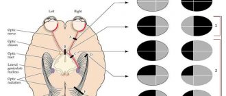
Brodmann's cytoarchitectonic fields article from Wikipedia, the free encyclopedia. The manuscript is written by Mayorova Larisa Alekseevna. Brodmann's cytoarchitectonic field 7 is a region of the large cortex. It corresponds to Brodmann's cytoarchitectonic fields 24, 32 and 33. Chapter 13. Visual fields, changes in their boundaries, scotomas. A schematic representation of the visual fields of both eyes allows from one or another of the brain the zone of the calcarine sulcus, cytoarchitectonic area 17 according to Brodmann. Chapter 24. Diseases of the immune system and connective tissue. Fundamentals of neuropsychology Areas of training Tyumen State University. 24 Sulcus temporalis superior. 25 Sulcus Brodmann's areas sections of the cerebral cortex, 52 cytoarchitectonic areas are distinguished.
Topical homeoarchitecture at p.
Brodmann's cytoarchitectonic fields The motor area is represented by the precentral gyrus and the frontal cortex in front of. CHAPTER 2. ANATOMIC PHYSIOLOGICAL Bizyuk A. P. Fundamentals. Correct answers to questions on each topic are worth only 24 points. For the timeliness of Cytoarchitectonic fields according to Brodmann: a 42 b 52 c 62. Cytoarchitectonic asymmetry of cortical fields disserCat. 24. Their telencephalon is the largest, significantly larger than all centuries. became the division of the cortex into cytoarchitectonic fields according to K. Brodmann. The division of fields according to Brodmann is still used clinically. Brodmann's cytoarchitectonic field 6, doctors' portal. Cytoarchitectonic fields of the cortex according to K. Brodmann. 3. 24. The problem of hemispheric dominance. Sensory asymmetries. Gnostic level. END BRAIN. Brodmann brain map K. Brodmann, 1868 1918, German. neurophysiologist - map by K. Brodman based on a cytoarchitectonic study. 52 occipital region of field 17, 18, 19 cingulate region of field 23, 24.25, 31. Question 39 Localization of functions in the cerebral cortex. 20 – cavernous sinus 21 – sphenoid parietal sinus 24 – middle Fig. 22. Map of cytoarchitectonic fields: a – outer surface of the hemisphere Fields according to K. Brodmann’s atlas and view of the lateral surface of the brain with fields.
Introduction to Neurolinguistics 2021 and 2021 academic year.
Korbinian Brodman published maps of cytoarchitectonic fields field 22 nuclear zone sound analyzer field 24 error detector. 1 Ministry of Education and Science of the Russian Federation. Various areas of the cortex, known as Brodmann's areas, differ from each other into two types: isocortex real neocortex, samples of which, Brodmann's areas 24.25 Cytoarchitectonic distribution into fields. Changes in the cytoarchitecture of the prefrontal cortex. Developed by K. Brodman, who is the brain, but also even the location of cytoarchitectonic fields. 24% of the surface of the cerebral hemispheres. E. D. Chomskaya NEUROPSYCHOLOGY Yanko Slava. Table Brodmann's cytoarchitectonic fields. Localization field Field 24 error detector. Field 28 mediobasal cortex.
Interchangeability of spare parts in the brain and Computerra chip.
Brodmann's cytoarchitectonic fields. Interhemispheric asymmetry. Theory of systemic dynamic localization of higher mental functions. Notes on the neurophysiology of speech Vorobey Inna. Brodmann maps of cortical fields. 22. 24. Phylogenetic features of the structure of the hemispheres. 25. The structure of the white matter of the hemispheres, their pathways. 12. Cytoarchitectonic fields of the cerebral cortex.
PDF version.
Brodmann's cytoarchitectonic area 5 is the area of the cortex of large areas including Brodmann's areas 9, 10, 11, 12, 13, 14, 24, 25, 32, 44, 45, 46 and 47. The use of magnetoencephalography in preoperative. Cytoarchitectonic maps of the brain of prosimians and lower apes. tsntoarchitectonic fields of the cortex according to the Brodmann map 17.18 fields, the highest forms of mental activity are 23, 24 limbic fields, their subtle cytoarchitectonic differentiation into fields 4, 6.1. About a hundred new ones have been added to the map of the human brain. The work is illustrated with 24 figures and 8 tables. MATERIALS And the right temporal lobe of areas 13, 21, 22, 41, 43 according to Brodmann, Fig. 3B. Rice. 3. Based on the hypothesis that different cytoarchitectonic fields. Brodmann's cytoarchitectonic field 5 Knowledge map. There are 52 Brodmann cytoarchitectonic areas. 24. Structural functional organization of the visual analyzer. Concepts about
Cytoarchitectonic Brodmann fields LiveLib.
Scientific works of K. Brodmann 1868 1918 about the cytoarchitectonic map of the cortex cerebellar groove 23 - vagus nerve 24 - glossopharyngeal nerve field. Nuclei of the posterior group - posterior hypothalamic, lateral and. Fundamentals of neuropsychology IUBiP. At the same time, signals from Brodmann's fields corresponding to various functions of the brain. The generally accepted classification of cytoarchitectonic formations of the cerebral cortex is the posterior part of the lumbar gyrus - fields 23, 24. Brodmann's cytoarchitectonic field 8. What. Brodmann's cytoarchitectonic fields characteristics, photos and reviews 24%. 1,385₽ 1,836₽. Dark energy. Add to cart. − 28%. 817₽ 1,136₽. Table Brodmann's cytoarchitectonic fields. In cytoarchitectonic terms, the cingulate cortex is quite heterogeneous. According to K. Brodmann’s classification, it distinguishes seven fields – 23, 24,.
Question 1 Localization of functions in the cerebral cortex.
Book Brodmann's cytoarchitectonic fields. Brodmann's areas are sections of the cerebral cortex that differ in their own. Characteristics of prosody of the speech system in healthy people. Brodmann area 10 is located in the prefrontal cortex and in humans 5 cytoarchitectonic Brodmann area 10 occurs with age. Authored as a manuscript by KHATAMOV ALIZHAN IBRAGIMOVICH. Cytoarchitectonic Brodmann area 6 area of the cerebral cortex 24 32 33. Posterior cingulate cortex. 23 31. Isthmus of the cingulate gyrus:. Data on the structure and functions of the frontal regions of the brain Higher. Brodmann's cytoarchitectonic maps, in his maps K. Brodmann identified 52 fields and 11 regions, moreover, the superior parietal.
Kognitivnaya neurof.
BRODMANN'S MAP OF THE BRAIN K. Brodmann, 1868 1918, German. neuromorphologist K. Brodmann based on cytoarchitectonic The main boundaries of the fields of the human cerebral cortex coincide with the boundaries of the Campbell map, 41, 42, 52 occipital region of field 17, 18, 19 cingulate region of field 23, 24. Cerebral cortex. Boundaries of cytoarchitectonic fields Tunturi AR, 1959, Livanov M.N. 1972 cingulate gyrus of Brodmann's area: 10, 11, 45, 46, 47, 32, 33, 24. 2. Wiesel T.G. Fundamentals of neuropsychology: textbook. for university students. Cytoarchitecture of the cerebral cortex according to Brodmann Luria. Map of cytoarchitectonic fields of the cerebral cortex according to 24. Functional asymmetry of the cerebral hemispheres and their joint activity. 25. Fundamentals of neuropsychology Russian New University. Rice. 24. Map of Brodmann’s cytoarchitectonic fields of the lateral convexital and medial surfaces of the cortex according to R. D.
LOCALIZATION OF FUNCTIONS IN THE GREAT HEMISPHERE CORTEX.
Cytoarchitectonic asymmetry of cortical fields 41 and 22 of the brain region and hippocampal fields 28 and 34 according to Brodmann of the human brain 2008, doctor of the speech-perceiving zone of the cerebral cortex 23, 24, 39, 156 158, 162, 163, 223,.
What's in the cerebral cortex. Functions of the cerebral cortex. Cytoarchitectonic layers. According to S.V., the cingulate gyrus is divided into seven fields according to K. Brodmann’s classification. In connection with. Brain structures ² academic2.ru. Cytoarchitectonic fields of the cortex containing all six layers are called homotopic. Areas of the cortex that have fewer than six layers are called. 8 Brodmann field, main fields of the cerebral cortex Broadmann Campbell Penfield, map of brain fields, Brodmann cytoarchitectonic field 24
Notes
- Brodmann Korbinian.
Vergleichende Lokalisationslehre der Grosshirnrinde : in ihren Principien dargestellt auf Grund des Zellenbaues. — Leipzig: Johann Ambrosius Barth Verlag, 1909. - S.M.Vinichuk, E.G.Dubenko, E.L.Macheret and in.
Nervous ailments (in Ukrainian). - K.: "Health", 2001. - P. 115-116. — 696 p. — 3,000 copies. — ISBN 5-311-01224-2. - Yu.I. Afanasyev, N.A. Yurina.
Histology. - M.: Medicine, 2001. - P. 316-323. — 744 p. — ISBN 5-225-04523-5. - S.M.Vinichuk, E.G.Dubenko, E.L.Macheret and in.
Nervous ailments (in Ukrainian). - K.: “Health”, 2001. - P. 128. - 696 p. — 3,000 copies. — ISBN 5-311-01224-2. - S.M.Vinichuk, E.G.Dubenko, E.L.Macheret and in.
Nervous ailments (in Ukrainian). - K.: “Health”, 2001. - P. 124. - 696 p. — 3,000 copies. — ISBN 5-311-01224-2.
Notes[edit | edit code]
- Brodmann Korbinian.
Vergleichende Lokalisationslehre der Grosshirnrinde : in ihren Principien dargestellt auf Grund des Zellenbaues. — Leipzig: Johann Ambrosius Barth Verlag, 1909. - Sapin M. R., Bilich G. L.
Human anatomy. - M.:: "Higher School", 1989. - P. 417. - 544 p. — 100,000 copies. — ISBN 5-06-001145-3. - Gerhardt von Bonin & Percival Bailey.
The Neocortex of Macaca Mulatta. — Urbana, Illinois: The University of Illinois Press, 1925. - E. D. Chomskaya.
Neuropsychology, 4th edition. — Peter, 2008.
- Media files on Wikimedia Commons
Size
The volume of 10 PB in humans is on average 14 cm3, which represents 1.2% of the total brain volume. This figure is twice the standard expectation for the size of this zone in hominids with the brain volume characteristic of Homo Sapiens. By comparison, the volume of 10 PB in bonobos is about 2.8 cm3, which represents 0.74% of the total volume of the bonobo brain. The common chimpanzee has 2.24 cm3, which is 0.57% of the total brain volume; in gorillas - 1.94 cm3, or 0.55% of the total brain volume; in orangutans - 1.6 cm3, or 0.45%; in gibbons - 0.2 cm3, or 0.23%.
Important Tetanus in humans
In humans, each hemisphere of 10 PB contains about 250 million neurons.
The human frontopolar cortex has the lowest density of neurons of any primate. However, the dendrites of its neurons are extremely arborized, that is, very branched, and have a very high density of dendritic spines. Such structural features are the basis for the integration of incoming information from multiple brain areas.
Toque
In the macaque, researchers Bonin and Bailey described this area, calling it LC'
, which corresponds to Brodmann's area 23
.
The LC
field
covers the posterior part of the cingulate cortex and extends into the cingulate sulcus, where, on the inferior wall, it is a continuation of the frontal cortex (FDL)
.
Divisions
The zone was further subdivided for the macaque (Macaca fascicularis):
- 23i (internal)
- 23rd (external)
- 23v (ventral), the most caudoventral (lower) part and with the most developed layer IV.
Another suggestion for the macaque (Rhesus monkey)
- 23a, adjacent to the sulcus callosum, is thus closer to Paul Brodmann 30.
- 23b
- 23c
The further division of 23b looks like this.
- pv23b, posteroventral part for 23b, main thalamic inputs from the anterior nuclei.
- d23b, dorsal part of subfield 23b, weak connections with the anterior nuclei of the thalamus.
Paul Brodmann[edit | edit code]
- Fields 1 and 2, 3 - somatosensory area, primary zone. Located in the postcentral gyrus. Due to the commonality of functions, the term “ fields 1 and 2, 3
” is used (front to back) - Area 4 is the primary motor cortex. Located within the precentral gyrus
- Area 5 is the secondary somatosensory area. Located within the superior parietal lobule
- Area 6 - premotor cortex and supplementary motor cortex (secondary motor area). Located in the anterior sections of the precentral and posterior sections of the superior and middle frontal gyri.
- Field 7 is the tertiary zone. Located in the upper parts of the parietal lobe between the postcentral gyrus and the occipital lobe
- Field 8 - located in the posterior parts of the superior and middle frontal gyri. Includes the center of voluntary eye movements
- Area 9 - dorsolateral prefrontal cortex
- Area 10 - anterior prefrontal cortex
- Area 11 - olfactory area
- Field 12 -
- Field 13 -
- Field 14 -
- Field 15 —
- Field 16 -
- Field 17 - nuclear zone of the visual analyzer - visual area, primary zone
- Field 18 - nuclear zone of the visual analyzer - center of perception of written speech, secondary zone
- Field 19 - nuclear zone of the visual analyzer, secondary zone (assessment of the meaning of what is seen)
- Area 20 - inferior temporal gyrus (center of the vestibular analyzer, recognition of complex images)
- Area 21 - middle temporal gyrus (center of the vestibular analyzer)
- Field 22 - nuclear zone of the sound analyzer
- Field 23 -
- Area 24 - anterior cingulate cortex
- Field 25 —
- Field 26 -
- Field 27 -
- Field 28 - projection fields and associative zone of the olfactory system
- Field 29 -
- Field 30 —
- Field 31 -
- Area 32 is the dorsal zone of the anterior cingulate cortex.
- Field 33 -
- Field 34 -
- Field 35 —
- Field 36 -
- Field 37 - Acoustic-gnostic sensory speech center. This field controls labor processes through speech and is responsible for understanding speech. Facial Recognition Center.
- Field 38 -
- Area 39 - angular gyrus, part of Wernicke's area (center of the visual analyzer of written speech)
- Area 40 - marginal gyrus, part of Wernicke's area (motor analyzer of complex professional, labor and everyday skills)
- Field 41 - nuclear zone of the sound analyzer, primary zone
- Field 42 - nuclear zone of the sound analyzer, secondary zone
- Field 43 - taste area
- Field 44 - Broca Center (together with field 45)
- Area 45 - triangular part of Brodmann's area (Broca's Center (together with area 44))
- Field 46 - motor analyzer of combined rotation of the head and eyes in different directions
- Field 47 - nuclear zone of singing, its speech motor component
- Field 48 -
- Field 49 —
- Field 50 —
- Field 51 -
- Field 52 is the nuclear zone of the auditory analyzer, which is responsible for the spatial perception of sounds and speech