Types of renal pathology:
Primary (hereditary, congenital, genetically determined) forms of nephropathies.
— Anomalies of kidney development (number, shape, macro- and microstructure).
— Tubulopathies (with predominant damage to the renal tubules: renal diabetes insipidus, renal pseudohypoaldosteronism).
— Enzymopathies of the tubular epithelium (for example, cystinuria, aminoaciduria).
— Nephropathy (generalized kidney damage: familial nephropathy with or without deafness, familial renal dystrophy).
Secondary (acquired, symptomatic) forms of nephropathies.
- Infectious origin - microbial, parasitic, fungal, protozoal (for example, nephritis, pyelonephritis, echinococcosis, kidney actinomycosis, nephrotic syndrome, renal failure).
— Immunoallergic origin (nephritis, immunoallergic nephropathies).
- Caused by direct damage to the kidneys by factors of a physical, chemical, biological nature (for example, trauma, radiation damage; toxogenic, drug-induced nephropathies).
— Concomitant (satellite) nephropathies (amyloidosis, endocrinopathies, nephrolithiasis, kidney migration, cardiovascular diseases
- Tumor origin.
Causes.
Exogenous: “hard” drinking water, monotonous hypovitaminized food (vitamin A deficiency is important).
Endogenous: infections (microflora of the urinary tract, gastrointestinal tract, reproductive system, etc.), metabolic disorders (gout, multiple myeloma, etc., endocrinopathies, mainly hyperparathyroidism).
Conditions for the development of nephrolithiasis and urolithiasis.
— Reducing the content in urine of solubilizers (agents that maintain urine salts in a dissolved state: urea, creatinine, xanthine, citrates, etc.), inhibitors of salt crystallization (inorganic pyrophosphate), complexing agents (Mg2+, citrates).
— An increase in the urine level of agents that trigger the process of crystallization of salts in the urine (mucoproteins, pyruvic acid salts, collagen, elastin, sulfonamides).
— Change in urine pH (at pH 5.0, uric acid salts precipitate, at pH above 7.0, calcium phosphate and ammonia phosphate).
— Increased content of stone-forming salts (mainly calcium) in the urine.
— Disturbances in the outflow of urine.
General etiology and pathogenesis of renal dysfunction
Introduction
The kidneys occupy one of the central places in the regulation of the constancy of the internal environment of the body. Basic functions of the kidneys.
1. The excretory (excretory) function is carried out by the renal nephrons - the removal of metabolic products and toxic substances.
2. Homeostatic – maintaining a constant internal environment of the body (water-electrolyte balance, CBS, systemic blood pressure, bcc).
3. Incretory – synthesis and release of biologically active substances into the blood (renin, erythropoietin, active form of vitamin D, prostaglandins, bradykinin, etc.).
4. Metabolic - the kidneys take an active part in the metabolism of proteins, fats and carbohydrates.
General etiology and pathogenesis of renal dysfunction
Causes of kidney diseases
I. By origin: primary (hereditary and congenital) and secondary (acquired) factors.
Primary (hereditary, congenital).
• Anomalies of kidney development (number, shape, macro- and microstructure).
• Tubulopathies (caused by primary damage to the renal tubules: renal diabetes, renal pseudohypoaldosteronism, etc.).
• Enzymopathies of the tubular epithelium (eg, cystinuria, aminoaciduria).
• Nephropathy (generalized kidney damage - familial nephropathy, familial renal dystrophy, etc.).
Secondary (acquired, symptomatic).
• Infectious (microbial, parasitic, fungal, protozoal origin, for example, pyelonephritis, echinococcosis, kidney actinomycosis).
• Immunoallergic (nephritis, immunoallergic nephropathy, etc.).
• Caused by direct damage to the kidneys (for example, trauma, radiation damage; toxogenic, drug-induced nephropathies).
• Concomitant (satellite) nephropathies (with amyloidosis, diabetes, nephrolithiasis, atherosclerosis, hypertension).
• Tumor lesions of the kidneys.
II. By the nature of the causative factors: infectious (for example, bacteria, viruses, rickettsia) and non-infectious (chemical, physical and biological factors). Chemical (for example, compounds of lead, sublimate, mercury, arsenic, some antibiotics, diuretics). Physical (eg, penetrating radiation, radioactive decay products, low temperature, kidney injury). Biological (for example, nephrotoxic antibodies and lymphocytes, immune complexes; allergens; catecholamines, prostaglandins and other biologically active substances).
III. According to the level of implementation of the causal factor.
Prerenal causes.
• Neuropsychic disorders: prolonged stress, mental trauma, conditions associated with severe pain (in particular, reflex “pain” anuria).
• Endocrinopathies (eg, excess or deficiency of ADH, aldosterone, thyroid hormones, insulin, catecholamines).
• Circulatory disorders (in the form of hypo- and hypertensive states).
2. Renal reasons. Direct kidney damage by infectious or non-infectious factors. Circulatory disorders in the kidneys.
3. Postrenal causes. Disruption of the outflow of urine through the urinary tract, accompanied by an increase in intrarenal pressure (with stones and tumors of the urinary tract, their swelling, prostate adenoma, kinks of the ureter, etc.).
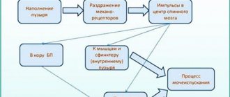
General mechanisms (pathogenesis) of disorders of excretory function of the kidneys
Urinary disorders are the result of disorders of the following processes:
• Filtration (formation of primary urine in renal corpuscles),
• Reabsorption (transport of ions, fluid, proteins, amino acids, glucose and other substances from the lumen of the renal tubules into the lumen of the capillaries of the secondary network),
• Secretion (transport of ions, fluid and a number of other substances into the lumen of the tubules).
Causes and mechanisms of filtration failure
The filtration process is carried out in the initial part of the nephron - the renal glomeruli, where primary urine is formed. The driving force behind the filtration process is effective filtration pressure , which is created as a result of the difference between the hydrostatic pressure in the capillaries of the glomerulus (~ 50-60 mm Hg), facilitating the passage of blood plasma elements through the filter membrane (through the capillary wall into the lumen of the capsule Shumlyansky-Bowman), and oppositely acting forces created by the oncotic pressure of the blood plasma (~ 25 mm Hg) and the pressure in the Shumlyansky-Bowman capsule (~ 15 mm Hg):
Rf = Rgk – (Ron + Rm),
Рф – effective filtration pressure,
Pgk – hydrostatic blood pressure in the capillaries of the glomerulus,
Ron – oncotic pressure of blood plasma,
Рм – pressure of primary urine in the Shumlyansky-Bowman capsule.
Hydrostatic pressure in the capillaries of the glomerulus is a value that depends mainly on the muscle tone of the walls of the afferent and efferent arterioles.
The resistance of the glomerular afferent arteriole varies with systemic blood pressure. The oncotic pressure of blood plasma depends on the protein content in it.
The pressure in the cavity of the Shumlyansky-Bowman capsule is determined by the patency of the renal tubules and urinary tract, and also depends on intrarenal pressure.
On average, filtration pressure is about 20 mmHg. The filtering membrane consists of three layers: capillary endothelium, basement membrane and epithelial cells (podocytes) of Bowman's capsule, covering the outer surface of the basement membrane with their legs.
The glomerular filtration rate is determined by determining the clearance of substances that are only filtered in the kidneys, but are not reabsorbed or secreted in the renal tubules (for example, the polysaccharide inulin).
Clearance (from English clear
- purify) corresponds to the volume of blood plasma that is completely cleared of this substance in 1 minute.
To determine clearance, it is necessary to establish the concentration of the substance used, for example inulin, in urine (M) and blood plasma (K) and the amount of urine (D) released in 1 minute:
Clearance = M/K x D [ml/min].
In clinical settings, they often resort to determining the glomerular filtration rate by the clearance of endogenous creatinine (a product of the breakdown of muscle proteins). Normally, the glomerular filtration rate for creatinine is 125-130 ml/min for men and 110-115 ml/min for women.
Mechanisms of impaired glomerular filtration. Impaired glomerular filtration is accompanied by either a decrease or an increase in filtrate volume.
Reasons for decreased glomerular filtrate volume.
• Decrease in effective filtration pressure in hypotensive conditions (arterial hypotension, collapse, etc.), ischemia of the kidney (kidneys), hypovolemic conditions. When systolic blood pressure drops below 50 mm Hg. filtration stops completely.
• An increase in the oncotic pressure of blood plasma as a result of an increase in the concentration of proteins, which can occur with increased protein synthesis (for example, with multiple myeloma), the introduction of protein drugs or blood thickening.
• Reduced filtration surface area (observed with necrosis of the kidney or part thereof, myeloma, chronic glomerulonephritis and other conditions).
• Reduced permeability of the filtration barrier due to thickening or compaction of the basement membrane or its other changes (chronic glomerulonephritis, diabetes mellitus, amyloidosis and other diseases).
• Increased pressure in the cavity of Bowman's capsule due to increased intrarenal pressure due to interstitial edema or obstruction of the tubules and urinary tract.
Nephritic syndrome
Nephritic syndrome is a symptom complex caused by diffuse damage to the renal tissue of inflammatory origin with the involvement of all parts of the nephrons, interstitial tissue and blood vessels in the pathological process.
One of the most common forms of pathology in this category is glomerulonephritis.
Acute glomerulonephritis is a disease, usually of infectious-allergic or immunoautoaggressive origin.
Chronic glomerulonephritis in 10–20% of patients is the outcome of acute glomerulonephritis, and in 80–90% it is a primary chronic disease.
Etiology . The causes of acute glomerulonephritis can be infectious and non-infectious factors.
Infectious agents: streptococci (usually β-hemolytic streptococcus of group A), viruses (for example, hepatitis, measles, rubella), toxoplasma.
Non-infectious factors: endogenous (for example, tumor Ag; Ag formed as a result of massive tissue damage - for example, in burn disease, long-term tissue crush syndrome, etc.); exogenous (for example, some antibiotics, non-narcotic analgesics, vaccines, blood serum, alcohol, organic solvents).
Nephrotic syndrome
Nephrotic syndrome is a symptom complex characterized by massive proteinuria (more than 3 g of protein/day), hypo- and dysproteinemia, hyperlipidemia, hypercholesterolemia, widespread edema and edema of the serous cavities.
Causes.
Primary nephrotic syndrome is caused by kidney pathology: glomerulonephritis (acute and chronic), glomerulosclerosis, lipoid nephrosis, membranous glomerulopathy, drug-induced kidney damage.
Secondary nephrotic syndrome develops with extrarenal pathology: chronic infection (tuberculosis, syphilis, viral hepatitis, etc.), diseases of the blood system (leukemia, lymphogranulomatosis, etc.), tumors, diseases of immune autoaggression (rheumatoid arthritis, scleroderma, vasculitis, etc.) , diabetes mellitus, intoxication (gold, mercury, bismuth, NSAIDs, insect and snake venoms), nephropathy in pregnant women, renal vascular thrombosis, etc.
The pathogenesis is caused by an increase in the permeability of the basement membrane of the glomerular capillaries for protein. This may be due to the damaging effects of immune complexes, lysosomal enzymes, and reactive oxygen species (released by neutrophils and monocytes).
With any mechanism of damage, there is an increased intake of blood plasma proteins (mainly albumin) into the glomerular filtrate, and pronounced proteinuria (more than 3 g of protein/day, in some cases - up to 50 g of protein/day). The consequence of this is hypoproteinemia (less than 60 g of protein/l), mainly due to a decrease in albumin content.
Hypoproteinemia → causes a decrease in colloid-osmotic pressure of the blood → increased release of fluid from the vessels into the intercellular space → development of hypovolemia → increased activity of the RAAS → increased aldosterone production → decreased excretion of Na+ ions in the urine, ↑ Na+ concentration in the blood → ↑ osmotic pressure of the blood → stimulation of secretion ADH → water retention in the body → development of edema .
Stimulation of ADH secretion under the influence of hypernatremia is accompanied by an increase in blood plasma volume, its dilution occurs, hypoproteinemia and hypoonkia increase. As a result, excess water is not retained in the bloodstream, but moves into the tissue, which contributes to a further increase in edema, i.e. a “vicious circle” arises.
Hypoproteinemia is combined with dysproteinemia, since along with albumin, the content of γ-globulins in the blood often decreases, which can also enter the urine.
Rice. 1. Etiological factors of nephrotic syndrome.
Hyperlipidemia develops due to increased levels of low and very low density lipoproteins (VLDL) with normal or reduced levels of high density lipoproteins (HDL). of cholesterol and triacylglycerols in the blood plasma increases
The development of hyperlipidemia is caused by two mechanisms: increased production of lipoproteins in the liver and impaired VLDL catabolism. It is assumed that reduced catabolism of lipoproteins may be due to the loss of certain substances in the urine (for example, lipoprotein lipase ).
In nephrotic syndrome, many transport proteins are lost in the urine. In this regard, the levels of a number of microelements (Fe, Cu and Zn), vitamin D metabolites, thyroid and steroid hormones are reduced in the blood plasma.
Thromboembolic complications pose a danger in nephrotic syndrome . Hypercoagulation is caused by an increase in the content of procoagulants - fibrinogen and plasma factors V (proaccelerin) and VIII (antihemophilic globulin A), a decrease in the content of antithrombin III, a weakening of the activity of the fibrinolytic system (decreased plasminogen content, increased activity of a2-antiplasmin).
Infectious complications caused by a decrease in the level of immunoglobulins A and G due to their loss in the urine.
Kidney failure
Renal failure is a syndrome that develops as a result of a significant decrease or cessation of excretory function, as well as disruption of other processes in the kidneys.
Depending on the speed of occurrence and further development, acute and chronic renal failure are distinguished.
Uremia
Uremia (urinary bleeding, from the Greek uron
- urine and
haima
- blood) is a syndrome consisting of self-poisoning (autointoxication) of the body with metabolic products that are normally excreted by the kidneys.
Cause: renal failure.
Pathogenesis.
• Hyperazotemia (accumulation of urea, uric acid, creatinine in the blood).
• Accumulation of toxins in the blood - products of protein decay (phenol, indole, skatole, cresol), purines (guanidine, etc.).
• Intoxication of the body with an excess of ammonium compounds (ammonia and its derivatives), formed during the transformation of urea in the intestine under the influence of ureases.
• Damage to cell membranes and enzymes by the above agents, which is accompanied by disruption of the energy supply to cells of organs and tissues.
• Cerebrovascular accident.
• Overhydration of the brain.
• Brain hypoxia.
• Ionic imbalance (hyperkalemia, hypermagnesemia, hypo- or hypernatremia, hypocalcemia).
• Acidosis (the result of inhibition of acid and ammoniogenesis, hemodynamic disorders and gas exchange in the lungs).
• Disruption of electrophysiological processes in the cells of organs and tissues, including the brain and heart, which underlies loss of consciousness during coma, aggravation of disorders of the cardiovascular, respiratory and other physiological systems.
Manifestations. In the initial stages of uremia, there are virtually no symptoms. As the severity of kidney dysfunction increases, the following appear: lethargy, bitterness in the mouth (urea accumulates in saliva), the smell of ammonia from the mouth (when urea is broken down by oral bacteria, ammonia is released), nausea, vomiting, diarrhea, itchy skin, irritability, muscle atrophy, toxic uremic pericarditis, anemia, clonic convulsions, Kussmaul respiration (caused by acidosis with a severe decrease in alkaline reserve), coma.
Kidney stone disease
Kidney stones (nephrolithiasis) is a chronic disease characterized by the formation of dense stones (stones) in the kidney tissue, calyces and pelvis.
Nephrolithiasis is one of the most common manifestations of urolithiasis, which means the formation of urinary stones not only in the kidneys and pelvis, but also in other parts of the urinary tract.
Most urinary stones contain calcium salts (oxalates, sulfates, carbonates, phosphates); stones are also formed from uric acid (urate) and cystine.
In addition to crystalline compounds, urinary stones include organic impurities - proteins, glycosaminoglycans, desquamated epithelium, tissue detritus, etc.
Etiology.
• Exogenous causes: “hard” drinking water, dietary habits (presence in food of large amounts of oxalic and uric acids, nucleoproteins, deficiency of vitamins A and B6, hypervitaminosis D); long-term intake of vitamin C (formation of oxalates).
• Endogenous causes: infections (urinary tract, gastrointestinal tract, reproductive system, etc.), metabolic disorders (gout, myeloma, etc.), endocrinopathies (mainly hyperparathyroidism).
Conditions for the development of nephrolithiasis.
• Reducing the content in urine of solubilizers (agents that maintain salts in a dissolved state: urea, creatinine, xanthine, citrates, etc.), inhibitors of salt crystallization (inorganic pyrophosphate), complexing agents (Mg2+, citrates).
• Increased content in the urine of factors that trigger the process of crystallization of salts in the urine (mucoproteins, pyruvic acid salts, collagen, elastin, sulfonamides).
• Change in urine pH (at a pH below 5, urates - uric acid salts - are precipitated, at a pH above 7 - phosphates).
• Increased content of stone-forming salts (mainly calcium) in the urine.
• Disorders of urine outflow.
Pathogenesis. The mechanism of formation of urinary stones is not completely clear. There are two theories - crystallization and “matrix ” theory.
Crystallization theory - the formation of a kidney stone is the result of the precipitation of crystals of certain substances (salts, uric acid, cystine) from a saturated solution (this is facilitated by the appropriate pH and the lack of crystallization inhibitors). The crystallization process is accompanied by the formation of dense conglomerates that can include various organic substances and structures.
The “matrix” theory - the process of stone formation depends on the appearance of crystallization centers, the role of which can be played by proteins (for example, fibrin), blood clots, cellular detritus, etc.
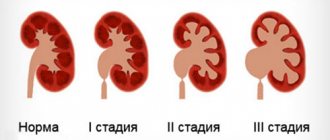
Consequences. The most significant: hydronephrosis with atrophy of the kidney (kidneys), pyelonephritis, nephrosclerosis, kidney abscesses, renal colic.
Introduction
The kidneys occupy one of the central places in the regulation of the constancy of the internal environment of the body. Basic functions of the kidneys.
1. The excretory (excretory) function is carried out by the renal nephrons - the removal of metabolic products and toxic substances.
2. Homeostatic – maintaining a constant internal environment of the body (water-electrolyte balance, CBS, systemic blood pressure, bcc).
3. Incretory – synthesis and release of biologically active substances into the blood (renin, erythropoietin, active form of vitamin D, prostaglandins, bradykinin, etc.).
4. Metabolic - the kidneys take an active part in the metabolism of proteins, fats and carbohydrates.
General etiology and pathogenesis of renal dysfunction
Causes of kidney diseases
I. By origin: primary (hereditary and congenital) and secondary (acquired) factors.
Mechanisms.
There are always two components found in stones: organic and mineral. In this regard, there are two points of view on the mechanism of stone formation. They are formulated in the form of crystallization and colloid theories. According to the crystallization theory, the formation of stone begins with the process of crystallization of salts. At the same time, organic components (fibrin, collagen, cellular detritus) will also be included in the composition of the stone. The authors of the colloidal theory believe that first an organic matrix is formed on which salts crystallize.
Consequences: hydronephrosis with atrophy of the kidney (kidneys), pyelonephritis, nephrosclerosis, kidney abscesses, renal colic.