Cholinergic synapse[edit | edit code]
Source:
Visual Pharmacology
.
Author
: X. Lulman.
Per. with him. Ed.
: M.: Mir, 2008
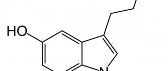
Acetylcholine - Vyacheslav Dubynin (Doctor of Biological Sciences, M.V. Lomonosov Moscow State University)
Acetylcholine
(ACh) - a transmitter in postganglionic synapses - accumulates in high concentrations in the vesicles of the axoplasm of the nerve ending. ACh is formed from choline and activated acetic acid (acetyl coenzyme A) under the action of the enzyme acetylcholine transferase. Highly polar choline is actively taken up by the axoplasm. There is a special transport system on the membrane of the cholinergic axon and nerve endings. The mechanism of mediator release has not been fully disclosed. Vesicles are anchored in the cytoskeleton by the synapsin protein in such a way that their concentration near the presynaptic membrane is high, but there is no contact with the membrane. When excitation occurs, the Ca2+ concentration in the axoplasm increases, protein kinases are activated, and phosphorylation of synapsin occurs, leading to the detachment of vesicles and their binding to the presynaptic membrane. The contents of the vesicles are then released into the synaptic cleft. Acetylcholine instantly passes through the synaptic cleft (the ACh molecule is about 0.5 nm long, and the width of the cleft is 30-40 nm). On the postsynaptic membrane, i.e., the membrane of the target organ, ACh interacts with receptors. These receptors are also excited by the alkaloid muscarine and are therefore called muscarinic acetylcholine receptors (M-cholinergic receptors). Nicotine mimics the effect of acetylcholine on receptors at ganglion synapses and the end plate. Nicotine excites cholinergic receptors at ganglion synapses and the end plate of the motor neuron, which is why this type of receptor is called nicotinic acetylcholine receptors (N-cholinergic receptors).
In the synaptic cleft, acetylcholine is quickly inactivated by a specific acetylcholinesterase located in the cleft, as well as by a less specific serum cholinesterase (butyrylcholinesterase) found in the blood serum and interstitial fluid.
Based on their structure, method of signal transmission and affinity for various ligands, M-cholinergic receptors are divided into several types. Let's consider M1, M2 and M3 receptors. M1 receptors are found on nerve cells, such as ganglia, and their activation promotes the transition of excitation from the first to the second neuron. M2 receptors are located in the heart: the opening of potassium channels leads to a slower diastolic depolarization and a decrease in heart rate. M3 receptors play a role in maintaining the tone of smooth muscles, for example, in the intestines and bronchi. Excitation of these receptors leads to activation of phospholipase C, membrane depolarization and increased muscle tone. M3 receptors are also located in gland cells, which are activated by phospholipase C. In the brain there are different types of M-cholinergic receptors that play a role in many functions: transmission of excitation, memory, learning, pain sensitivity, control of brain stem activity. Activation of M3 receptors in the vascular endothelium can lead to the release of nitric oxide NO and thus dilate blood vessels.
Acetylcholine deficiency
The cholinergic system is the part of the brain that produces acetylcholine. According to one version, damage to this zone is associated with the development of senile dementia of the Alzheimer's type. Cholinergic effects cause the start of neurodegenerative processes, manifested by disorders of short-term and long-term memory, disorders of speech and cognitive functions, loss of the ability to navigate the environment, and the inability to self-care.
Lack of acetylcholine also plays a role in the development of idiopathic parkinsonism. Acetylcholine interacts with the neurotransmitter dopamine to promote smooth movement. When there is an imbalance between acetylcholine and dopamine, a person's movements become imprecise, causing:
- limiting the pace and range of movements;
- involuntary fast rhythmic oscillatory movements in different parts of the body;
- postural instability – inability to maintain body position;
- lack of muscle responses to stimuli.
In the peripheral nervous system, the neurotransmitter causes contraction of the cardiac and skeletal muscles. The destruction of acetylcholine receptors by factors of humoral and cellular immunity provokes the development of myasthenia gravis, an autoimmune disease manifested by episodes of muscle weakness and fatigue. The disease occurs due to an autoimmune attack on postsynaptic acetylcholine receptors, resulting in disruption of neuromuscular signaling. In the treatment of myasthenia gravis, drugs are used that inhibit (slow down) the activity of cholinesterase (an enzyme that breaks down acetylcholine). The use of these drugs enhances the effects of acetylcholine on the endocrine glands, heart, nerve ganglia, smooth and skeletal muscles.
The role of acetylcholine in the formation of tobacco addiction has been proven. When nicotine reaches the brain structures, acetylcholine receptors are activated. Over time, the brain gets used to the fact that nicotine, similar to acetylcholine, is now in excess. Accordingly, since there is a lot of this substance, it is necessary to adapt the process of synthesis of mediators to the new reality. To correct this, brain neurons reduce the production of acetylcholine. When their own reserves of acetylcholine are no longer enough, the smoker will reach for a cigarette to cheer up, concentrate, and calm down. That is, in a smoker, the functional duties of acetylcholine are transferred to nicotine.
Acetylcholine[edit | edit code]
Source:
Clinical Pharmacology by Goodman and Gilman Volume 1
.
Editor
: Professor A.G.
Gilman Ed.
: Practice, 2006.
Acetylcholine
(lat. Acetylcholinum) - a mediator of the nervous system, a biogenic amine related to substances formed in the body.
Acetylcholine plays an important role as a mediator of the central nervous system. It is involved in the transmission of impulses in different parts of the brain, with small concentrations facilitating and large concentrations inhibiting synaptic transmission. Changes in acetylcholine metabolism can lead to impaired brain function.
Acetylcholine is a mediator of nerve impulse transmission to the muscle. With a lack of acetylcholine, the strength of muscle contractions decreases.
The endings of the nerve fibers for which it serves as a mediator are called cholinergic, and the receptors that interact with it are called cholinergic receptors. Cholinergic receptors of postganglionic cholinergic nerves (heart, smooth muscles, glands) are designated as m-cholinergic receptors (muscarine-sensitive), and those located in the area of ganglionic synapses and in somatic neuromuscular synapses are designated as n-cholinergic receptors (nicotine-sensitive). This division is associated with the characteristics of the reactions that occur during the interaction of acetylcholine with these biochemical systems: muscarinic-like in the first case and nicotine-like in the second; m- and n-cholinergic receptors are also located in different parts of the central nervous system.
Storage and release of acetylcholine[edit | edit code]
When microelectrode recording of electrical potentials of the postsynaptic membrane of the neuromuscular synapse, Fatt and Katz (1952) identified spontaneous small (0.1-3 mV) depolarizing potentials that occurred randomly approximately once per second. The authors called these potentials miniature endplate potentials. Their amplitude was significantly below the threshold for the development of an action potential. They increased under the influence of the AChE inhibitor neostigmine and were blocked by tubocurarine (a competitive blocker of N-cholinergic receptors); therefore, they were due to the release of acetylcholine. In this regard, it was suggested that acetylcholine is released from presynaptic endings in fractional constant portions - quanta. The morphological substrate of quanta, synaptic vesicles, was soon discovered (De Robertis and Bennett, 1955). When an action potential arrives at the axon terminal of a motor neuron, 100 or more quanta (vesicles) of acetylcholine are released (Katz and Miledi, 1965). The patterns of acetylcholine storage and release studied at the neuromuscular junction are also applicable to other cholinergic synapses with fast transmission.
It is estimated that each vesicle contains from 1000 to 50,000 molecules of acetylcholine, and the presynaptic terminal of a motor neuron contains 300,000 or more vesicles. In addition, it is possible that a fairly significant amount of acetylcholine is diffusely dissolved in the axoplasm. Recording of currents of single channels of the postsynaptic membrane of the neuromuscular synapse with constant application of acetylcholine showed that one molecule of this mediator causes a potential of the order of 3 x 10”7 V. It follows from this that even the minimum (according to calculations) amount of acetylcholine in one vesicle is 1000 molecules — is sufficient to induce a miniature endplate potential (Katz and Miledi, 1972).
Exocytosis of acetylcholine and other mediators from presynaptic terminals is suppressed by botulinum toxin and tetanus toxin - poisons of Clostridium botulinum and Clostridium tetani, respectively. These anaerobic spore-forming organisms produce some of the most potent toxins known (Shapiro et al., 1998). Clostridium toxins, consisting of heavy and light chains linked by disulfide bridges, bind to an as yet unknown receptor at the cholinergic terminal and are then transported through endocytosis into the cytosol. The light chain is a zinc-containing endopeptidase that, upon activation, hydrolyzes core components of the SNARE complex involved in exocytosis. Different types of botulinum toxin destroy different proteins of the presynaptic membrane (syntaxin-1 and SNAP-25) and synaptic vesicles (synaptobrevin). Botulinum toxin A as a drug is discussed in Chapter. 9 and 66.
Tetanus toxin is a poison of central action: it is retrogradely transported along the axons of motor neurons into the bodies of these neurons in the spinal cord, then passes into the inhibitory neurons associated with motor neurons and blocks the exocytosis of the transmitter from the latter. This is what leads to the cramps characteristic of tetanus. The venom of the black widow spider, α-latrotoxin, binds to transmembrane proteins of presynaptic terminals called neurexins, causing massive exocytosis of synaptic vesicles (Schiavo et al., 2000).
Excess acetylcholine
In most cases, an imbalance in the level of a neurotransmitter means its deficiency, not its excess. But sometimes an excess of acetylcholine occurs, especially if you take active measures to increase its levels.
An increase in acetylcholine levels may be caused by exposure to organophosphate pesticides, insecticides, or nerve agents.
These chemicals lead to increased levels of acetylcholine in the nervous system and cause symptoms such as:
- pulmonary obstruction syndrome;
- sweating;
- weakness;
- headache;
- fainting;
- diarrhea and nausea;
- change in mental status;
- muscle cramps;
- spasms;
- paralysis;
- stopping breathing.
Exposure to chemicals that affect acetylcholine balance may occur through the skin, respiratory or digestive system. The most likely route for these compounds to enter the body is through the ingestion of pesticides through vegetables and fruits, as well as through contact with household products used to kill insects.
An imbalance in acetylcholine levels aggravates Parkinson's disease. Normally, the body maintains a balance of acetylcholine and dopamine, another neurotransmitter that controls muscle contractions. Parkinson's disease is a neurodegenerative disease that causes involuntary movements, tremors, and disturbances in mood and thinking.
The exact cause of the disease is not known. But it has been shown that in patients with this syndrome, dopamine levels decrease, and because of this, acetylcholine levels increase. In this case, the muscles become too “excited”, and symptoms such as jerking movements and tremors appear.
Some drugs to treat Parkinson's disease block the action of acetylcholine. Dopamine levels return to normal and symptoms disappear. These drugs are called anticholinergics. Other symptoms of Parkinson's disease, such as memory complications, are also possibly related to acetylcholine levels.
Read also[edit | edit code]
- Anatomy and physiology of the nervous system
- Parasympathetic nervous system
- Sympathetic nervous system
- Synaptic transmission
- Cholinergic receptors and synapses
- Cholinomimetics
- Anticholinergics
- Nicotine
- M-cholinergic receptor stimulants
- M-cholinergic receptor blockers
- Acetylcholinesterase inhibitors (AChE)
- Poisoning with acetylcholinesterase blockers
- Nerve transmission at neuromuscular synapses and autonomic ganglia N-cholinergic receptors
- Muscle relaxants
- Agents acting on the autonomic ganglia
- Ganglion stimulators
- Ganglioblockers