Scalenus syndrome (also called scalenus syndrome) is a group of symptoms that include pain, numbness and weakness in the neck, shoulder or arm. The cause of symptoms is compression or damage to nerves or blood vessels in the costoclavicular space. The costoclavicular space is located between the collarbone and the upper rib, on both sides of the body. Most of the vessels (arteries and veins) and nerves supplying the arm pass through this space. Narrowing of this space can cause compression of nerves and blood vessels, which interferes with the normal functioning of the upper limb. The cause of the narrowing can be various conditions, such as injury, obesity, congenital anomalies, and poor posture. But sometimes it is not possible to find out the specific reason for the narrowing.
Causes of the disease
To function properly, blood vessels and nerves need a certain amount of space. Compression of the vessels in the costoclavicular space can lead to injury or, in rare cases, loss of a limb. In order for nerves and blood vessels to function properly, they need adequate space. When compression occurs, their function is accordingly impaired. Compression of the blood vessels at the outlet of the chest can impair blood flow to and from the arm. It can also promote the formation of a blood clot, which can further slow or completely block blood flow through the damaged vessel. If a clot ruptures, it can travel down into the arm, blocking small blood vessels in the arm. Sometimes, the clot migrates to the lungs, a life-threatening condition called pulmonary embolism. Nerves also need space to be able to stretch when the arm moves. If the nerve as it exits the chest wall is compressed or cannot move freely, the patient will not be able to move the arm as normal. Pain and sensory disturbances in the hand often accompany this condition.
Risk factors for scalene muscle syndrome (SMS):
- Gender – Women are more likely to have SLM syndrome than men.
- Age – The syndrome most often develops between the ages of 20-50.
- Diseases - SLM - syndrome is often associated with another disease, such as damage to the rotator cuff, osteochondrosis of the cervical spine, injury to the brachial plexus, diabetes mellitus, hypothyroidism.
SCALENE MUSCLE SYNDROME
Scalenus syndrome
(syn.:
Naffziger syndrome, costoclavicular compression syndrome, cervical rib syndrome, scalenus syndrome
) is a pain syndrome caused by a narrowing of the gap between the anterior and middle scalene muscles and compression of the neurovascular bundle of the upper limb passing through it. It develops gradually, most often at the age of 30-40 years. The occurrence of the syndrome is associated with bone anomalies, pathological changes in the scalene muscles themselves, as well as with the neuroreflex effect on muscles due to osteochondrosis and diseases of the internal organs. Damage to the anterior scalene muscle may be caused by tension in the muscles of the shoulder girdle during certain types of work activity. Tension and hypertrophy of the anterior scalene muscle, and sometimes its fibrous degeneration, causes a decrease in the interscalene and costoclavicular spaces. This leads to irritation of the roots of the brachial plexus (C8-D1) and the appearance of tortuosity of the subclavian artery and vein. As a result, pain syndrome, vascular and neurovegetative disorders develop. On examination, attention is drawn to swelling in the supraclavicular region on the side of the scalene muscle syndrome, and an increase in the vascular pattern of the veins on the chest and in the shoulder area. Characterized by a short neck, possible presence of rudimentary cervical ribs, and curvature of the cervical spine.
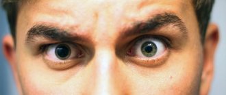
Scalene muscle syndrome most often manifests itself as a complex of symptoms affecting the nervous, arterial and venous systems and very rarely with one of them. The most constant symptom is pain in the arm, mainly in the ulnar zone of the forearm and hand. They can be spontaneous, but more often occur during work. The pain is sharp, shooting, sometimes burning with a large area of distribution, usually intensifies during sleep. They are often combined with acroparesthesia in the ulnar, less often in the radial zone of the hand and forearm. Movement disorders are characterized by increased fatigue or decreased muscle strength in the distal parts of the hand, especially in the IV-V fingers, less often in the proximal parts. In the muscles of the ulnar zone of the forearm and hand, especially the eminence of the little finger, hypotonia and malnutrition are noted. Autonomic-vascular disorders are detected in the form of Raynaud's syndrome - sensations of coldness, paleness, blueness, and sometimes redness of the skin, mainly in the distal parts of the arm. Bernard-Horner syndrome is sometimes observed (see Bernard-Horner syndrome). Many patients complain of pain in the heart area, which occurs or intensifies with deep inhalation or exhalation, turning the body or moving the left hand. Circulatory failure may occur in the vertebral and basilar arteries, usually in the form of repeated cochleovestibular crises. Headaches, mainly of the occipital localization, are often observed. The pulsation of the subclavian and radial arteries is weakened or absent. Auscultation reveals a systolic murmur in the supraclavicular region. A decrease in blood pressure in the affected limb is typical, sometimes to zero.
The Lange test helps to recognize the syndrome of the anterior scalene muscle - the disappearance of pulsation in the radial artery when abducting and raising the arm upward while simultaneously turning the head in the opposite direction. Palpation reveals a hypertrophied, tense anterior scalene muscle, as well as tenderness of the supraclavicular and subclavian points. Temporary regression of symptoms after infiltration of the anterior scalene muscle with novocaine solution is of important diagnostic significance. Vascular compression in Scalene muscle syndrome can cause thrombosis of the subclavian artery itself or its distal branches, the formation of post- and stenotic aneurysms with the possibility of embolism, and circulatory disturbances in the subclavian vein (see Paget-Schretter syndrome). The state of blood circulation in the subclavian artery system is studied using thermography (see), aortography (see), volumetric sphygmography or vasoreography (see Rheography) with simultaneous Lange test, and in the subclavian vein system - using phlebography (see).
There are conservative and surgical treatment methods. In the initial period of the disease, novocaine infiltration is most effective at the attachment site of the anterior scalene muscle, relieving its spasm. Analgesics, sympatholytics, drugs that improve peripheral blood flow and reduce muscle tension, physiotherapeutic procedures (diadynamic currents or iontophoresis with novocaine, trimecaine on the neck and shoulder girdle, massage of the collar zone of the affected limb) are also used. Indications for surgical treatment may be the severity of vascular and neurological disorders, especially pain, and vegetative-vascular disorders, resistance to conservative treatment and progression of the course, the presence of bone anomalies and complications, as well as electrophysiological and radiological data. research. Surgical treatment is aimed at restoring and improving blood flow through the arterial and venous vessels, as well as eliminating the factor of mechanical compression of the brachial plexus and autonomic fibers in the space between the scalenes. Scalenotomy or scalenectomy is used, often in combination with periarterial sympathectomy, partial or complete resection of the cervical ribs or hypertrophied transverse processes of the VII cervical vertebra, according to indications - intersection of the fibers of the middle scalene muscle, reconstructive operations on the subclavian artery and vein in cases of violation of their patency, etc.
Forecast
in cases of timely diagnosis and treatment, favorable.
Bibliography:
Anichkov M. N. and Lev I. D. Clinical and anatomical atlas of aortic pathology, L., 1967;
Bogolepov N. K., Burd G. S. and Seleznev A. N. Changes in the nervous system in scalenus syndrome, Zhurn, neuropath, and psychiat., t. 74, no. 6, p. 843, 1974; Bosnev V. Shoulder-arm syndrome, trans. from Bulgarian, Plovdiv, 1978, bibliogr.; Kipervas I. P. Neurovascular syndromes of the shoulder girdle and arms, M., 1975, bibliogr.; Pokrovsky A.V. et al. The effectiveness of the operation of crossing the anterior scalene muscle in scalenus syndrome (scalenus syndrome), Zhurn, neuropath, and psychiat., v. 76, no. 8, p. 1172, 1976; Popelyansky Ya. Yu. Cervical osteochondrosis, compression and reflex syndromes, p. 136, M., 1966; Yumashev G. S. and Furman M. E. Osteochondrosis of the spine, M., 1973, bibliogr., Brain's diseases of the nervous system, ed. by JN Walton, Oxford, 1977; Naffziger H. S. a. Grant WT Neuritis of the brachial plexus mechanical in origin, the scalenus syndrome, Surg. Gynec. Obstet., u. 67, p. 722, 1938.
A.H. Seleznev.
Causes
SLM occurs as a result of compression of the nerves or vessels at the exit of the chest. Main reasons:
- Trauma – A traumatic episode may cause damage to the bone or soft tissue at the thoracic outlet. Most people with SLM have a history of one or another episode of an accident, injury at work or at home.
- Congenital anomalies, such as an extra rib or a tight ligament connecting the spinal column to a rib, can reduce the costoclavicular space.
- Poor posture - sagging shoulders or excessive forward tilt of the head can put compression on the area where nerves and blood vessels exit the chest.
- Frequent, repetitive movements can wear down the tissues and lead to SLM. An example would be movements associated with raising the arm (the collarbone lowers and the costoclavicular gap decreases), such as swimming, baseball, tennis, weightlifting.
- Other causes: Weight gain (pregnancy or obesity), overdeveloped neck muscles (from weightlifting or martial arts), or holding your arms in one position for a long time (working on a computer) can put additional pressure on nerves and blood vessels. Diseases that impair nerve function, such as hypothyroidism and diabetes, may be predisposing factors for neurogenic SLM.
Scalenus syndrome as a cause of numbness and cold hands
The scalene muscles are three powerful muscles of the neck (anterior, middle, posterior). When at least one of them is shortened, compression of the nerves occurs, which, in turn, causes pain or numbness in the arm, problems with the shoulder joint, and glenohumeral periarthrosis. About the reasons for the shortening of these muscles and effective methods of treatment.
About the scalene muscles of the neck
The scalene muscles begin posteriorly from the transverse processes of the cervical vertebrae, stretch vertically along the neck and are attached to the two uppermost ribs, the first and second, deep behind the collarbone. This group of muscles is shaped like steps, hence the name.
Why is shortening of the scalene muscles dangerous?
The main feature is that
between them pass the subclavian artery and the nerves of the cervical plexus, which are directly connected to the arms
.
If the muscles are healthy, then nothing threatens the arteries and nerves - healthy muscles are elastic, soft, covered with a dense slippery membrane - fascia. This ensures they slide smoothly relative to each other and protects them from any pinching when moving the head and neck. However, with pathology - shortening - everything changes: the anterior scalene muscle spasms, this causes pain, and a muscle stretched like a string can pinch the nerves and artery
.
Therefore, the first danger of shortening the scalene muscles is radicular pain, akin to that which occurs when the nerves are compressed (squeezed) by hernia and osteophytes of the cervical spine
. We turned our head unsuccessfully, tension in the shortened muscle arose, pressure on the nerve increased, and pain arose. But that is not all.
Blood vessels in the costoclavicular space (at the exit from the chest) may also be pinched. This may impair blood flow to the arms, hence
numbness in the hand, sensory impairment, vertebral artery syndrome.
Often, glenohumeral periarthrosis begins with shortening of the scalene muscles. Gradually, neck instability develops and the head moves forward.
What follows is a typical scenario. To compensate for balance and center of gravity, the muscles in the back of the neck are forced to tighten and become overloaded. Due to overload, painful trigger points are formed and myofascial syndrome develops. If left untreated, this leads to compensatory curvature of the spine in the thoracic and lumbar regions as it is forced to seek balance. Curvature of the spine, in turn, disrupts the distribution of axial loads on the intervertebral discs, displacement of the vertebrae and, thus, opens the way to the formation of protrusions and disc herniations in the cervicothoracic region.
Timely consultation with a doctor, early detection and proper treatment can avoid all these serious complications of shortening of the scalene muscle. Therefore, it is useful to regularly undergo examination by an experienced kinesiologist, who, using a special functional muscle test, determines the locations of spasmed, weakened and shortened muscles, and then, based on the identified problems, draws up an individual treatment program using the method of applied kinesiology.
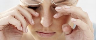
How signs should alert you. Characteristic signs of scalene muscle syndrome:
- Pain in the neck and shoulder, especially when bending to the side and turning.
The pain may radiate from the shoulder down the arm and into the little and ring fingers.
Sometimes it spreads to the chest or back of the head. It often intensifies at night, as well as when turning the head, moving the arm to the side and taking a deep breath (the scalene muscles contract when taking a deep breath and pull the upper ribs and chest upward).
- Numbness or tingling in the fingers, hands, entire arm, muscle weakness.
- Shifting the head forward.
— Restriction of movement of the chest, since the first and second ribs, to which the scalene muscle is attached, are fixed.
When the blood vessels are compressed, swelling of the hand, cold hand syndrome, pale or blue skin, and a weak pulse in the hand are typical.
How does shortening of the scalene muscles develop?
Long-term overload leads to muscle shortening, which often occurs against the background of weakening of the antagonist muscles - the long extensors of the neck.
. The main function of the long neck extensors is to support the head and its weight when bending forward.
So, in modern man, the long extensors of the neck are chronically overstrained and weakened.
When the long extensors of the neck weaken, other muscles, mainly the scalenes (as the main antagonist), take on increased load and shorten them, and the same
scalene syndrome is formed.
Next important question
: why do our neck muscles weaken and spasm, and why has this problem become relevant for most people, especially in the last few years?
The number one reason is a constantly bent neck forward.
.
The muscles in the back of the neck tense every time we tilt our head forward - to look at a smartphone, read a magazine, when we cook in the kitchen or sit for a long time at the computer with our head stretched forward. In modern humans, the long extensors that support the head experience chronic overload.
Reason number two is weakness of the lower back muscles, a sedentary lifestyle.
Modern man spends most of his life sitting. How are we sitting? Leaning on the back of a chair or in a banana pose, slouching. The back is rounded, the lower back is relaxed, bends back, the head is stretched forward. In this position, there are simply no physiological bends
, the lumbar and cervical spine are in a constant kyphotic state. And we spend several hours a day in this position.
As a result, the back muscles (back extensors, quadratus, lumbar muscles) weaken, and a person cannot support his lower back in a sitting position. In such conditions, the long neck extensors are also not included in the work, the short neck extensors work instead, the head is supported by short muscles, and not by long ones, the load is distributed unevenly across all discs, and is shifted more to the 5th, 6th, 7th intervertebral discs.
If this situation is repeated day after day, the long neck muscles weaken and shorten, gradually causing vertebral instability.
Fixing your hands in one position for a long time (working on a computer) can put additional pressure on nerves and blood vessels
.
The syndrome occurs and occurs against the background of the muscle due to sports overtraining, especially in those sports where there are frequent repetitive movements associated with raising the arm (the collarbone drops and the costoclavicular space decreases), for example, swimming, baseball, tennis, weightlifting.
How to restore health to your neck muscles?
Scalene syndrome often resembles a cervical hernia with nerve compression. Therefore, at the first stage of treatment, the most important thing is to establish the source of the problems.
This cannot be done using MRI. Shortening of the scalene muscles can only be detected by a special functional muscle test, which is carried out by an experienced specialist in applied kinesiology.
Each person may have their own reasons for the development of spinal pathologies and pain. The task of applied kinesiology is to identify them, and then, based on examination data, to create an individual program of exercises aimed at restoring the full functionality of shortened, weakened, idle muscles, and eliminating muscle imbalances
.
Doing this with regular strength exercises is dangerous and ineffective. To work with weakened muscles and a sore spine, special rehabilitation equipment is needed - biomechanical strength decompression kinesi simulators. They are widely used in rehabilitation after injuries and operations on the spine and joints, as they allow the restoration of weakened muscles with minimal stress on the musculoskeletal system.
Therapeutic exercises strengthen weakened neck muscles, the upper back in the area of the cervical and upper thoracic vertebrae, from which the long extensors of the neck and shoulder girdle begin. At the same time, spasms are relieved, blood flow and nutrition of the spine improves, pain, dizziness and other unpleasant symptoms disappear, and blood pressure stabilizes. And all this without the use of drugs or expensive hardware technologies.
Stretching and strengthening the shoulder and pectoral muscles helps relieve pressure on the nerves and blood vessels in the costoclavicular space.
Specialist consultation is required.
Doctor’s consultation + diagnostics using applied kinesiology – 1000 rubles.
Call, write! Tel.: (843) 570-55-25 , WhatsApp: 79655968085 or VKontakte group .
Follow us on Instagram (photos and videos from classes):
Read us on Yandex Zen:
Don't miss out on the fun! Subscribe to our news:
Subscribe to news from the Kazan Kinesitherapy Center
Center promotions, therapeutic exercises and useful tips from our specialists on how to independently maintain the health of your joints and spine without medications
Similar articles:
Osteochondrosis of the thoracic spine. How to identify and treat?
Back pain in the area of the shoulder blades. Causes and effective treatments
Shrugs with dumbbells. Back exercises at home
Symptoms
Symptoms of SLM vary depending on whether the blood vessels or nerves are compressed.
Symptoms of nerve compression
- Pain or tenderness in the neck, shoulder, arm
- Numbness or tingling in the neck, shoulder, arm
- Muscle weakness or areas of muscle failure in the arm
- Difficulty performing fine motor tasks; fast fatiguability
Arterial Compression Symptoms
- Swelling of the hand
- Pain or deep tenderness in the neck, shoulder, or arm
- Numbness, tingling, or heaviness in the arm
- Changes in skin color - the hand or fingers turn pale
- Changes in skin temperature – the arm or fingers are cooler than the rest of the skin
- Small black spots on fingers
- Weak or absent pulse in the arm
- Pulsating tumor in the collarbone area
Venous Compression Symptoms
- Swelling of the hand
- Pain or deep tenderness in the neck, shoulder, or arm
- Numbness, tingling, or heaviness in the arm
- Changes in skin color - the hand or fingers become blue
- Seal in the area of the subclavian vein
- Visually noticeable venous network on the chest.
Complications
For a long time, some patients reflexively react to pain with muscle fixation. They have to hold a rigid certain position in order to reduce pain. But, unfortunately, this reflex muscle defense aggravates the course of the disease and, ultimately, leads to increased pain. Muscle defence, in turn, then requires separate treatment. It is important to begin treatment for SLM in its early stages. If left untreated, SLM can cause significant nerve or vascular damage, including limb loss.
Clinically Relevant Anatomy
Anatomy of the knee joint
The knee joint consists of two main joints - the patellofemoral and tibiofemoral. The patella is located in the intercondylar fossa. Its articular surface (back side) is covered with cartilage, which slides along the cartilage of the front of the femur (in the intercondylar fossa). In this case, the movement of the joint is accompanied by minimal resistance due to the synovial fluid, which washes the articular surfaces and is produced by the synovium and the inner part of the joint capsule. It is also known that some joint capsules generate synovial fluid. The capsule of the knee joint is attached to the edges of the cartilaginous surface of the patella, so only its articular surface and the intercondylar fossa of the femur are in contact with synovial fluid.
Collateral ligaments fuse with the capsule and contribute to joint stability. On the anterior surface of the patella, between the patellar tendon (attached to the kneecap) and the skin, there is an additional bursa (prepatellar bursa), which usually does not come into contact with the knee joint capsule and allows the patellar tendon to glide better. At the level of the tibial tuberosity there is a similar bursa (infrapatellar bursa). When the knee joint is affected, these bags can become hyperproductive. This may be due to increased pain in the front of the knee.
Friends, on July 17, a webinar by Georgy Temichev “The Knee Joint” will take place. Find out more...
Each ligament provides support and protection to the knee joint, and these ligaments also indirectly strengthen each other. The two ligaments that are most associated with the patellofemoral joint are the two collateral ligaments (lateral and medial) because they fuse with the knee capsule. The supracondylar-patellar and menisco-patellar ligaments form the medial and lateral retinacular-patellar part of the ligament complex, which provides medial and lateral attachment of the quadriceps tendon at the level of the patella.
Diagnostics
Diagnosing SLM can be difficult. Symptoms vary in nature and severity, depending on the individual. In addition, the symptoms are similar to those that occur with other diseases (for example, damage to the rotator cuff, diseases of the cervical spine leading to compression of the roots, etc.). Medical history and physical examination help to clarify the onset of the disease, the nature of symptoms, and their dependence on body position. In addition, there are external signs of this disease (swelling of the arm, discoloration, loss of sensitivity, limited range of motion in the shoulder). During a physical examination, the doctor may carry out procedures (manipulations) to identify symptoms (pulse pressure in the arms in various positions of the arm, both sick and healthy).
Instrumental research methods
- Electromyography (EMG) – EMGs help test how nerves and muscles are functioning. Small needle electrodes are inserted into the muscle where the problem is. Electrodes measure the electrical activity of the muscle innervated by a particular nerve. The pathological response of a muscle provides information about the condition of the nerve going to that muscle. In addition, EMG allows you to determine the speed of impulse transmission along a nerve fiber. This is done using electrodes placed on the skin. The speed of impulse conduction along each nerve fiber has a certain average value, and a deviation indicates damage to the nerve fiber.
- X-ray allows you to diagnose bone changes in the chest and ribs (the presence of an additional cervical rib).
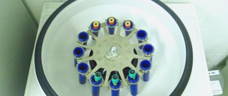
- Laboratory examinations - general blood tests, blood for hormones, blood for sugar help in diagnosing SLM.
- Magnetic resonance imaging (MRI) - allows you to visualize the soft tissues of the body MRI to find out the cause of compression of a nerve or vessel.
- Computed tomography (CT) allows you to more clearly visualize changes in bone tissue.
- Ultrasound examination – Using an ultrasonic wave, it is possible to visualize soft tissues and blood vessels, the presence of blood clots and stenoses.
- Angiography – X-ray examination using contrast is used to diagnose vascular lesions. Angiography of arteries and veins is used to diagnose blocks and other problems in the blood vessels.
Treatment
Drug treatment
- NSAIDs are non-steroidal anti-inflammatory drugs. These medications help reduce pain and inflammation (swelling and redness). For example: aspirin, ibuprofen (Advil), naproxen (Aleve), and celecoxib (Celebrex) movalis
- Muscle relaxants – Often used to treat muscle spasms, these medications can relieve pain by relaxing the muscles. Cyclobenzaprine (Flexeril), carisoprodol (Soma), diazepam (Valium), methocarbamol (Robaxin), and tizanidine (Zanaflex).
- Neuropathic drugs - The principle of their action is based on changes in the neurotransmitter transmission of pain impulses to the spinal cord and brain. Medicines that may help reduce pain by affecting neurotransmitters include fluoxetine (Prozac), sertraline (Zoloft), paroxetine (Paxil), citalopram (Celexa), venlafaxine (Effexor), amitriptyline (Elavil), imipramine (Tofranil), desipramine (Norpramine) ), doxepin (Sinequan), and amoxapine (Ascendin).
- Opioids - Narcotic analgesics are used only for very severe pain after conventional analgesics have been exhausted. A combination of opiates with NSAIDs is possible (to enhance the analgesic effect).
Exercise therapy
Physical therapy is one of the most important components of SLM treatment. Selecting certain exercises helps improve posture and proper distribution of muscle loads. Exercises help increase range of motion in the limb. Stretching and strengthening the shoulder and pectoral muscles can help increase and relieve pressure on the nerves and blood vessels in the costoclavicular space. • There is a wide range of motion, both passive and active.
Physiotherapy
Various physiotherapeutic techniques can relieve swelling, inflammation, restore blood circulation and reduce compression of nerves.
Manual therapy
The use of certain manual therapy techniques allows you to mobilize the spine and ribs and increase the range of motion in the shoulder joint.
Blockades
Sometimes used for differential diagnosis and treatment. But, taking into account the anatomical features of this area, injections should be carried out by a doctor with experience in performing such manipulations.
Acupuncture (acupuncture)
Acupuncturists believe that a healthy body contains channels through which energy flows. When these channels close, the energy is blocked, which leads to various diseases. Needles are inserted into certain points (biologically active). In certain cases, acupuncture can reduce pain and restore conduction along nerve fibers.
Massage can help relieve stress and relax tense muscles. Massage helps increase blood flow to body tissues and helps muscles get rid of metabolic waste products.