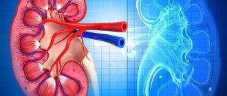
Main indications for examination of the urinary system
- Assessment of individual renal function.
- Visualization of a “non-functioning” kidney during intravenous urography.
- Demonstration of ectopic renal tissue.
- Detection of congenital anomalies.
- Assessment of renal artery patency.
- Diagnosis of renovascular hypertension.
- Kidney injury.
- Acute and chronic renal failure.
- Urinary tract infection.
- Assessment of renal obstruction.
- Preoperative assessment of renal function.
- Assessment of bladder function.
- Detection of vesicoureteral reflux.
- As an alternative method to intravenous urography in patients with an allergic reaction to iodine.
- Preparation for kidney transplantation.
Why do you need a comprehensive ultrasound of the urinary organs?
If you are interested in what is included in an ultrasound of the urinary system, then the procedure involves assessing the condition and function of the kidneys, ureters, and bladder. Their study using ultrasound is prescribed if the patient has complaints about certain symptoms:
- pain in the lower abdomen;
- urinary incontinence;
- pain when urinating;
- pain in the kidney area;
- frequent urination;
- urinary retention;
- frequent urge to urinate;
- swelling of the lower extremities and area around the eyes;
- blood or pus in the urine.
The reason for performing an ultrasound may be high blood pressure, the need to monitor the condition after surgery on the urinary organs and monitor the course of chronic diseases. A study may be prescribed if unsatisfactory and alarming results from urine and blood tests have been obtained. If necessary, the procedure is combined with an ultrasound of the abdominal cavity, if it is necessary to make an accurate diagnosis.
Ultrasound helps identify the following diseases:
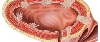
- urolithiasis (ultrasound is one of the preferred methods for diagnosing urolithiasis);
- developmental anomalies of the urinary organs;
- tumor and cystic formations;
- inflammatory processes (pyelonephritis, glomerulonephritis);
- vascular disorders;
- inflammation of the urinary tract (urethritis) or bladder (cystitis).
Techniques
Angionephroscintigraphy
allows:
- obtain qualitative and quantitative information about the state of renal blood flow;
- assess the functional state of various parts of the kidney;
- separately evaluate the transit function of the radiopharmaceutical in the parenchyma, renal pelvis, and ureters, which is important in the diagnosis of obstructive nephropathy;
- obtain an image of the organs of the urinary system, the blood pool, present the kinetics of radiopharmaceuticals in them graphically, qualitatively and quantitatively evaluate the data obtained.
Examination of patients with urolithiasis
Laboratory methods for diagnosing urolithiasis include:
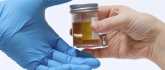
General urine analysis
Urinalysis is a routine method of evaluating patients with urolithiasis. Acidity, density, the presence of cylinders and protein, leukocytes and erythrocytes, and salts are assessed. Leukocytes in the urine indicate an inflammatory process in the urinary tract. The presence of red blood cells in a urine test is common in urolithiasis, however, it can also be an early sign of tumors.
Blood chemistry
Gives an idea of kidney function and allows you to assess the likelihood of developing urolithiasis. For the diagnosis of ICD, the content in the blood is important:
- electrolytes and creatinine - an imbalance indicates a dysfunction of excretion in the kidneys;
- phosphorus - excess phosphorus in the blood is excreted by the kidneys, which promotes stone formation;
- calcium – excess is observed in hyperparathyroidism, leading to increased excretion by the kidneys – hypercalciuria and stone formation; • uric acid (an increase indicates a disorder of purine metabolism - gout or hyperuricosuria);
- parathyroid hormone, which is closely related to the level of calcium in the blood.
Biochemical study of 24-hour urine for metabolic disorders
This important study provides information about the chemical composition of urine. This information is absolutely necessary for selecting effective specific prevention of urolithiasis relapse. The study can also identify patients who are at high risk of developing stones in the future.
A biochemical study of daily urine (collected during the day) is carried out - the level of pH (acidity), calcium, oxalates, uric acid salts, sodium, phosphates, citrates, magnesium, creatinine, and the overall volume of urine is determined.
Study of the level of calcium, oxalates, urates (uric acid salts) in urine:
An increase in the level of one of the three components (calcium, oxalate salts, uric acid) in daily urine indicates a predisposition to the formation of stones in the genitourinary system.
Hypercalciuria
Hypercalciuria can be divided into absorptive, resorptive, and hypercalciuria resulting from increased renal calcium loss, based on blood and urine test results associated with a calcium-restricted diet.
Resorptive hypercalciuria is observed in primary hyperparathyroidism, which, if possible, requires parathyroidectomy (removal of the parathyroid glands).
Absorptive hypercalciuria occurs as a result of the following conditions: calcium-restricted diet, thiazide diuretics, oral calcium-binding substances, and phosphate supplements.
Hypercalciuria resulting from increased renal calcium loss is much less common than absorptive hypercalciuria and is usually associated with secondary hyperparathyroidism and controlled with thiazide diuretics.
Another method for diagnosing hypercalciuria, when hyperparathyroidism has been excluded, is a blood test against the background of a hypocalcium diet (with this diet it is recommended to consume calcium 600-800 mg/day), moderately limiting the consumption of foods containing oxalates, and taking thiazide diuretics.
Although it should be remembered that limiting the intake of calcium-depleted foods increases the risk of stone formation as a result of a secondary increase in oxalate absorption (increased urine oxalate levels). A reduced intake of calcium from food leads to a decrease in oxalate-binding substances in the digestive tract, increasing free oxalates, thereby increasing the ability to absorb oxalates. The end product of this process is the formation of stones in the genitourinary system.
Hyperoxaluria
Hyperoxaluria (excessive urinary excretion of oxalic acid salts) may be primary (a rare genetic disease), secondary (acquired as a result of insufficient calcium intake), intestinal origin (as a result of malabsorption, associated with chronic diarrhea or short bowel syndrome), or idiopathic. Limiting oxalate intake and taking vitamin B6 are the main recommendations for patients with idiopathic hyperoxaluria. Intestinal hyperoxaluria is the most treatable type of oxaluria, and a diet enriched with calcium leads to positive results.
Calcium citrate
Calcium citrate is a recommended supplement because citrate helps to further reduce stone formation. Calcium therapy serves an oxalate-binding role, reducing the absorption of oxalates from the digestive tract. Calcium is an essential element in food, especially if foods contain high levels of oxalate salts (oxalates). However, you should not add vitamin D, as this enhances calcium absorption, reducing the amount of calcium in the gastrointestinal tract that can bind to oxalates. The optimal 24-hour urine oxalate level is 20 mg/day or less.
Hyperuricosuria
Hyperuricosuria (excessive urinary excretion of uric acid salts) predisposes to the formation of calcium-containing stones, since monosodium urate may lead to malabsorption of macromolecular inhibitors or may serve as a nidus for heterogeneous growth of calcium oxalate crystals. Gout is a metabolic disease that accelerates stone formation, characterized by high levels of uric acid in the blood serum. Treatment for gout includes potassium citrate, allopurinol (prevents the formation of uric acid, reduces its concentration in body fluids), and a combination of these. In general, patients with urate stones and hyperuricemia (high levels of uric acid in the blood) are treated with allopurinol; Patients with calcium urate stones are treated with citrates. The optimal 24-hour urine uric acid level is 600 mg/day or less.
Examination of sodium and phosphorus levels in urine
- Excess sodium excretion may contribute to the development of hypercalciuria. Elevated urine sodium levels are almost always associated with poor diet. Decreasing sodium (salt) intake may decrease urinary calcium excretion, thereby decreasing calcium saturation.
- Elevated phosphate levels are a marker of a subtype of absorptive hypercalciuria known as renal phosphate loss (absorptive hypercalciuria type III). Renal phosphate loss is identified by elevated urine phosphate levels, low blood phosphate levels, elevated blood levels of 1,25-vitamin D3 (calcitriol), and hypercalciuria. This type of hypercalciuria is rare and does not respond to standard treatments.
- The above laboratory parameters are examined only when the degree of absorption hypercalciuria type III is high. Any patient with hypercalciuria who has low blood phosphate levels and high urine phosphate levels may have this condition. A blood test for 1.25-vitamin D3 confirms or excludes this pathology. When blood phosphate levels are low, supplemental phosphate intake is used, then the activation of vitamin D, initially caused by hypophosphatemia, is reduced. All this corrects hypercalciuria, which is ultimately vitamin D dependent.
Study of citrate and magnesium levels in urine
- Magnesium and especially citrate are important chemical inhibitors of stone formation. Hypocitraturia is one of the most common metabolic defects that predisposes to genitourinary stone formation, and some authors recommend citrate as primary or adjunctive therapy in almost all patients diagnosed with calcium-containing genitourinary stones. The normal level of urinary citrate, according to many laboratories, is considered to be 320 mg/day, but the optimal level of urinary citrate in healthy individuals is closer to the average level (640 mg/day). Monitoring the pH (relative density) of urine is necessary to titrate and optimize citrate intake. A pH level of 6.5 is generally considered optimal. When urine pH is more than 7.0, there is a possibility of calcium phosphate sediment formation.
- Liquid or powder citrate preparations are recommended when absorption is impaired or for chronic diarrhea. Lemon juice concentrates are a good source of citrates. In addition, large amounts of lemon juice increase the need for fluid.
- The properties of magnesium as a stone formation inhibitor are less pronounced than those of citrate.
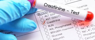
Testing the level of creatinine in urine
Urine creatinine is monitored over 24 hours. Most people excrete 1-1.5 g of creatinine daily.
Volume of urine excreted
Patients with urolithiasis should aim to pass up to 2 liters of urine per day to reduce the risk of stone formation. Patients with cystine stones or in resistant cases need to excrete up to 3 liters of urine for adequate prevention.
Determination of relative density (pH) of urine
Some types of stones, such as those composed of uric acid or cystine, are pH-sensitive. This means that such urinary stones form only in conditions of increased acidity of urine. Calcium phosphate and struvite genitourinary stones form when the urine pH is alkaline. Urine pH-metry is a diagnostically important method for diagnosing urolithiasis; in addition, it is possible to diagnose the latent course of urolithiasis in some patients. Instrumental methods for diagnosing urolithiasis
Plain radiography of the abdominal cavity
Plain radiography is an easy-to-perform test that allows you to identify X-ray positive stones, which include stones with a high calcium content, within a few minutes.
Urate, indinavir-induced and cystine stones of the urinary system are not detected by X-ray (called X-ray negative), so additional diagnostic methods may be required.
Kidney ultrasound
Renal ultrasound is a routine method in the diagnosis of urolithiasis. Allows to identify X-ray negative stones (urate, cystine stones) in the kidneys. It should be remembered that detecting ureteral stones using ultrasound is extremely difficult and is possible with large stones at the initial and distal ends of the ureter.
Intravenous urography
Intravenous urography, well known as intravenous pyelography, remains the standard method of examining patients with urolithiasis in many clinics. It also has the advantage of obtaining information about the anatomy of the genitourinary tract and the functioning of the kidneys.
However, it has certain pros and cons, as well as the need for additional preparation:
- Sometimes, with complete obstruction, the study can take up to 4 hours and be uninformative. - For good visualization, bowel preparation is necessary (maximum clearance of gases)
- A contrast agent is administered, which may cause an allergic reaction in some people and may also be nephrotoxic.
- Quite significant radiation exposure.
- Spiral CT of the kidneys without contrast enhancement is currently the best method for diagnosing acute renal colic.
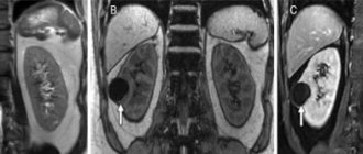
Spiral CT scan of the kidneys without contrast enhancement
Spiral CT of the kidneys without contrast enhancement is the most sensitive method for diagnosing urolithiasis. It detects well not only X-ray positive, but also X-ray negative stones.
- Speed of execution. In many institutions, renal CT is the method of choice in cases of suspected acute renal colic.
- Allows you to diagnose pathologies other than urolithiasis (aortic aneurysm, abdominal tumors);
- Safety, since no contrast agent is used, which may be poorly tolerated by patients. Moreover, the contrast agent, filling the entire collector system of the kidney, makes it difficult to identify stones in the urinary system. Therefore, CT of the kidneys is not used as a screening method for detecting urolithiasis.
- Stones of the urinary system that are not visualized on plain urography and are clearly identified by CT scan of the kidneys may indicate the urate nature of the stone. This makes it possible to carry out a differential diagnosis between calcium and urate stones, and thereby prescribe the necessary therapy. Hounsfield units on kidney CT also help diagnose urate stones (the density of urate stones is different from other types of stones).
- Disadvantages of non-contrast CT - no data on renal function
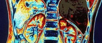
Contrast-enhanced spiral CT scan of the kidneys
A contrast-enhanced CT scan of the kidneys is performed only after a non-contrast CT scan of the kidneys. Allows you to assess the functional state of the kidneys, identify anomalies and structural features of the kidneys, ureters, and clarify the blood supply. Allows you to plan surgical treatment.
Preparing for the study
- Ensuring patient comfort, correct positioning, immobility and emptying of the bladder before the examination.
- Ensuring hydration level, which is achieved by drinking 200 ml of water 30 minutes before the test.
- separately evaluate the transit function of the radiopharmaceutical in the parenchyma, renal pelvis, and ureters, which is important in the diagnosis of obstructive nephropathy.
- Stop taking diuretics one day before angionephroscintigraphy. Before the study (2-3 days before), it is not advisable to conduct X-ray contrast studies or take medications that can block the secretion process in the tubules.
Treatment of infectious processes of the urinary system
If a urinary tract infection is detected, the patient is prescribed treatment, which can be carried out both in outpatient and inpatient settings. The form of patient management depends on the severity of his condition, the characteristics of the pathology, the presence of concomitant diseases and complications.
It is important to understand that treatment of infectious pathology must be carried out by a physician, urologist or nephrologist. Patients should not try to cope with the disease on their own, as the lack of proper therapy can provoke progression of the disease and cause serious complications.
Treatment of infections of the excretory system begins with the appointment of an appropriate regimen. The patient is advised to limit physical activity; in severe cases, strict bed rest is prescribed. To stimulate diuresis, the patient should drink up to 2.5 liters of fluid daily.
An important component of treatment is a special diet. Marinades and smoked dishes, which adversely affect the condition of the kidneys, are completely excluded from the diet. The patient is advised to consume more foods containing vitamin C, which helps acidify urine.
Drug treatment of infections involves the use of antibacterial drugs or drugs from the sulfonamide group. The choice of drug depends on the sensitivity of the pathogen to certain groups of drugs.
Additionally, symptomatic therapy is provided to eliminate the clinical picture of the disease and improve the patient’s condition. For this purpose, analgesics, antipyretics, and antispasmodics are used. If necessary, herbal and physical therapy is carried out. In addition to general treatment, local administration of anti-inflammatory drugs through the urethra into the bladder is practiced.
Bring with you
- Passport.
- Referral for research.
- Discharge summaries, results of ultrasound, scintigraphy, CT and MRI along with discs (preferably for the last three months), as well as other data that allows you to study the history of your disease in more detail.
- The results of our research along with illustrations (if you visit us again).
- If you are undergoing a study at the expense of the compulsory health insurance fund (CHI), you must additionally provide a referral in form 057/u-04, a medical policy and SNILS (more details in the “Compulsory Medical Insurance” section).
Attention!
Dear patients, we remind you that immediately after the injection of radiopharmaceuticals you become a source of ionizing radiation, which means that contact with you is associated with receiving radiation exposure. Taking this into account, while waiting for a scan, you must use the toilet located in the patient waiting room; it is extremely undesirable to go outside the patient waiting room or walk along the corridor. For the same reason, there should be no accompanying persons in the room where you are waiting for the scan. Warn them about this in advance and offer to wait for you in the laboratory lobby or outside the laboratory.
Do not take pregnant women or small children as accompanying persons!
From the patient room, at the time calculated by the doctor, the patient is invited to the tomograph for scanning.
Attention!
During scanning, objects containing metal (chains, belts, jewelry, etc.) should not be on the patient’s body. We kindly ask you to remove all these items from yourself before the start of the study, so as not to waste time when placing yourself in the tomograph. During the scan, you must be in a state of complete rest, breathing should be even, calm, and you cannot move. It is important to remember that any movement can affect the quality of the image.
After the scan, the patient awaits a conclusion and is sent home.