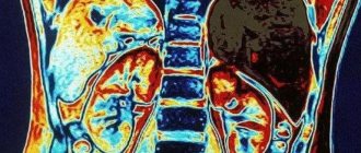
The kidneys are the most important organs of the urinary system. It is for this reason that any, even the most minor, deviations in their functioning can cause disruptions throughout the body, which will not have the best effect on the well-being and quality of life of the patient as a whole.
Nowadays, there are various diagnostic techniques, thanks to which specialists are able to identify the pathological process at the very beginning of its occurrence and prescribe the correct treatment, which will be the key to a quick recovery.
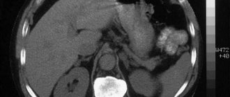
One of the most popular methods that is widely used nowadays is CT scan of the kidneys with contrast. And, if in the recent past this type of examination was not carried out in every clinic, and only in large cities, now you can undergo a CT scan in many medical centers.
general information
Even though CT scans of the kidneys with contrast are performed quite often, many people do not know what this diagnostic method is. In this regard, patients have many concerns and questions related to the upcoming scan.
Some patients deliberately refuse to undergo scanning, citing the fact that CT scans pose a potential health hazard due to the fact that X-rays are used to perform the scan.
However, all worries about the safety of CT are completely in vain, since scanning with a contrast agent does not pose any threat to life and health. In addition, if necessary, diagnostics can be carried out several times in a row within one week, because the radiation to which the body is exposed during the study is very insignificant.
This research method is carried out in almost the same way as conventional computed tomography without the use of a contrast amplifier. The difference comes down to the fact that to perform a CT scan of the kidneys with contrast, a special substance is injected intravenously into the patient. It is used to ensure that the resulting images of organs are of the highest quality. Only clear images can be the key to making a correct diagnosis and eliminating any errors.
Contrast is administered immediately before the start of the study. After the patient is positioned on a special retractable tomograph table, he will be injected with a contrast amplifier, which very quickly spreads through the bloodstream. All manipulations are performed under the strict guidance of a radiologist, so this event should not cause any concern. If the patient begins to feel worse, the administration of the drug will be suspended and the patient will be provided with the necessary medical care.
Methods for detecting abnormalities in kidney function
Nephrologist (in some cases – urologist)
) to confirm or clarify the diagnosis, in addition to general blood and urine tests, has in its arsenal a sufficient number of methods and means.
Kidney examination
is always comprehensive, because many of the observed symptoms may indicate other diseases, incl. oncological
Therefore, to make an accurate diagnosis, as a rule, both laboratory and instrumental methods are used:
Laboratory diagnostics. Includes blood tests (general and biochemical) and urine tests (general, according to Nechiporenko, according to Zimnitsky, Amburger’s test, etc.). If there are deviations in the general tests, the necessary additional tests are prescribed, each of which allows us to draw conclusions about the existing pathology.
- Ultrasound diagnostics (ultrasound).
- X-ray diagnostics (infusion urography, retrograde pyelography, computed tomography, etc.).
- Magnetic resonance imaging (MRI).
- Endoscopic diagnostics (cystoscopy, chromocystoscopy).
- Radioisotope diagnostics (radioisotope renography, scintigraphy).
Our clinic staff will carry out the necessary diagnostics, if necessary, and prescribe appropriate treatment.
Indications for diagnostics
Kidney CT with contrast is performed only for certain indications. The study is usually prescribed after undergoing ultrasound diagnostics, when the identified diagnosis is in doubt and either its confirmation or refutation is required. Very often, a CT scan is prescribed after surgery in order to assess the quality of the surgical intervention.
The main indications for undergoing a CT scan of the kidneys with contrast may include:
- prolapse of the urinary organs;
- presence of cysts;
- inflammatory diseases;
- pathology of the organ structure;
- violation of metabolic processes;
- injuries received;
- evaluation of ongoing treatment;
- preparation for planned surgery;
- pyeloectasia;
- kidney infarction;
- the presence of neoplasms and metastases.
If the patient has indications for a study using contrast, the attending physician will tell you in more detail what kind of diagnosis it is and for what purpose it will be carried out.
Types of technical inspection
An important step in the diagnosis of kidney disease
is to determine the degree of their functionality and the structural state of internal tissues using the following methods:
- radiography of the organ after preliminary administration of an indicator substance, which, when removed from the body, stains the kidney tissue, allowing one to assess their functional abilities;
- Ultrasound of the kidneys, includes ultrasonography and Dopplerography, determining the structural state of the organ tissues and its vascular network, size and foreign inclusions, characterizing the functional state of the kidneys;
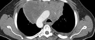
- tomography, CT and MRI, are based on the use of X-ray examinations with varying intensities of radiation exposure to the body, allowing a thorough examination of the structural features of the organ.
Contraindications for undergoing computed tomography with contrast
Despite the relative safety, CT scan of the kidneys with contrast has some contraindications, which include:
- Children under 2 years of age. X-ray radiation has a negative impact on the development of the child’s body, so even the slightest radiation is a strict contraindication to this type of diagnosis.
- Diabetes.
- Contrast intolerance. Generally, the contrast enhancer is well tolerated by patients. However, in some cases, contrast can still cause allergies, so people with intolerance to this type of scanning will not be allowed.
- Diseases of the urinary organs. Any disturbances in the functioning of the kidneys may cause the contrast agent to be unable to be excreted from the body, resulting in a toxic effect. Therefore, any diseases of the urinary organs are a strict contraindication to conducting contrast studies.
- Pregnancy. Even if a woman is not sure about her pregnancy, the specialist will definitely refer the patient to take a pregnancy test. This measure is necessary because X-ray radiation has a negative effect on the developing fetus. If it turns out that a woman is pregnant, she will be scheduled to undergo magnetic resonance imaging without the use of a contrast amplifier.
- Endocrine pathologies.
- Obesity. If the patient weighs more than 120 kg, a conventional CT scanner will not be suitable for examination. For scanning, you will have to give preference to an open type device.
- Presence of mental illness. Such patients will not be able to stay in a confined space for a long time. Alternatively, a contrast-enhanced renal CT scan can be performed while the patient is in a state of drug-induced sleep.
This type of research has many contraindications. For this reason, it is very important to inform the radiologist before starting the scan about any potential contraindications, because the specialist must take into account even the most minor nuances.
Directions for diagnosing kidney diseases
The examination is based on a laboratory basis for determining the correspondence of the concentrations of blood and urine components, including the following methods:
- biochemical analysis;
- blood test according to the general list;
- urine analysis for general components;
- microscopic examination of urine.
The most effective blood indicators for pathology of the urinary system are the following elements:
- total protein, which determines the total amount of protein structures in the blood, which decrease with increasing excretion through the kidneys;
- creatinine, an indicator of the intensity of energy metabolism in tissues, which is excreted through the kidneys, when its range fluctuates, allows you to diagnose the disease;
- urea is formed as a result of protein metabolism and is utilized through the kidneys, therefore its concentration in the blood characterizes their functional state;
- albumin, a protein component of the blood that is involved in the formation of a blood clot and is excreted in the urine, fluctuations in the content of which indicate a pathological functional abnormality of the kidneys.
Urinalysis indicators include such detectable elements as protein, glucose, nitrites, density and acidity level of the medium. Microscopy determines the content of red blood cells, leukocytes, epithelium, casts and bacteria that are part of the settled liquid sediment.
How to properly prepare for a kidney CT scan with contrast
A significant advantage of kidney CT with contrast is that no serious preparatory measures will have to be taken before the diagnosis. Experts provide a set of recommendations that should be followed to obtain the clearest images.
So, 2-3 days before the scan, you should remove from your diet any foods that can cause intestinal dysfunction. 8 hours before the start of the study, the patient should not take any food, and the restriction on the consumption of clean water does not apply.
Immediately before scanning, you will need to remove any metal jewelry from your body, as well as items of clothing with metal inserts. You will have to leave any gadgets outside the office, as they can interfere with the operation of the tomograph, which will not have the best effect on the quality of the resulting images.
How to prepare for a kidney ultrasound procedure?
Ultrasound diagnosis of the kidneys is carried out provided that the bladder is full and the intestines are freed from fecal matter and gases. This way, ultrasonic waves can easily penetrate the liquid medium and provide clear visualization of kidney tissue and other organ elements.
In order to prepare for a kidney ultrasound, you must follow the following recommendations:
- Limit the consumption of foods that cause increased gas formation for 3 days before the ultrasound.
- Perform a cleansing enema or use laxatives the day before the procedure (if you have intestinal problems).
- Drink approximately 1 liter of regular drinking water and refrain from going to the restroom for 1-2 hours before the examination.
You can clarify the need for certain preparatory stages directly from your doctor.
Progress of CT examination
Before starting a kidney CT scan with contrast, the radiologist conducts a preparatory conversation with the patient, asks about his well-being, and talks about how to contact the medical staff during the study.
The specialist also studies the patient’s medical record, which contains notes about all previous diseases. Before scanning, the patient undergoes an allergy test for contrast. If after it is performed a rash, redness appears on the skin, and the patient experiences severe itching, then a CT scan of the kidneys with contrast will be strictly contraindicated.
If no signs of intolerance to contrast were detected, then the patient is escorted to a separate room where the tomograph is located, after which the drug is administered intravenously.
After all the preparatory manipulations, the table will be pushed inside the tomograph and the device will begin its work. During the CT scan of the kidneys with contrast, the radiologist will be in the next room, but you can always contact him through a special microphone.
Sometimes during the course of the study, patients begin to experience a significant deterioration in well-being, which in most cases is associated with psycho-emotional experiences. In such a situation, you must inform your doctor about any deterioration. If there are compelling reasons for this, the scanning progress will be stopped.
During operation of the device, the scanning part will perform rotational movements in order to take a large number of pictures. During CT scanning, specific noises may be made, this is quite normal, there is no need to be afraid of it.
The duration of a CT scan with contrast is about 30 minutes. After the study comes to an end, the patient can immediately leave the office and wait to receive a report from the radiologist.
Advantages and disadvantages of kidney CT with contrast
Before going for a CT scan using contrast, every patient should have an understanding of the main advantages and disadvantages of the procedure.
The advantages of this type of research include:
- absolute painlessness of diagnosis;
- non-invasive;
- prompt receipt of scan results;
- making an accurate diagnosis.
Regarding the issue of CT disadvantages, they also exist:
- it is possible that an allergy to contrast may occur;
- undesirability of conducting research on children;
- high cost of diagnostics.
CT with contrast in exceptional cases causes any complications. In isolated cases, patients show signs of an allergic reaction, but this is rather an exception to the rule.