- What does a CT scan of the adrenal glands show?
- CT with contrast
- Indications
- Contraindications
- Preparing for the examination
- How is the procedure performed?
- Decoding the results
- How often can you do it
CT scan of the adrenal glands is a modern, informative, non-invasive examination method that will make it possible with a high degree of probability to identify benign and malignant neoplasms of the adrenal glands at the earliest stages of the development of the disease.
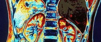
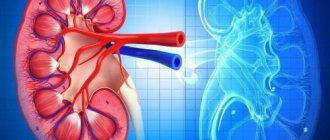
The adrenal glands are located in the abdominal cavity in close proximity to the upper pole of the kidney. Their examination is often difficult due to their small size. Computed tomography of the adrenal glands allows you to obtain layer-by-layer images of any organ or tissue, even if the anatomical formation is located deep in the body. During one examination, you can obtain about 100 images. The doctor can set the thickness of the sections arbitrarily, depending on the type of pathology for which the examination is being carried out. Thanks to this method, it is possible to detect adrenal tumors in the early stages of the disease in 95-97% of cases.
What does a CT scan of the adrenal glands show?
In the photographs obtained during the examination, you can see:
- adrenal glands, kidneys, lymph nodes and other soft tissues of the area being examined;
- benign tumors of the adrenal glands - adenomas;
- lipomas, cysts, hematomas;
- malignant neoplasms of the adrenal glands;
- foci of necrosis of tumor tissue with the formation of an area of calcification;
- involvement of nearby lymph nodes in the pathological process.
Using the images, you can distinguish a benign neoplasm from a malignant one, determine the prevalence of the pathological process, and the involvement of nearby tissues in the pathological process.
What's better?
Many people are interested in which method is better for problems with the adrenal glands. Both studies provide quite a lot of information about problems with the adrenal glands . They make it possible to identify the disease and identify the cause of the malfunction. MRI is a safer method, since the human body is not exposed to radiation. With CT scanning, radiation can change the structure of cells. Naturally, each method has a number of features.
MRI has the following features over CT:
- MRI is safer and does not contain x-rays.
- Visualizes soft tissue well.
- Allows you to determine whether a tumor is benign or malignant.
- It can be carried out in the 2nd and 3rd trimester of pregnancy.
- Possible for children.
CT, in turn, has a harmful effect on the body and is prohibited for pregnant women, as well as children under 12 years of age.
Despite a number of advantages, MRI also has disadvantages:
- It is necessary to stay in the tomograph for a long time.
- Loud noise when the tomograph is operating.
- Cannot be used by people with metal implants.
MRI is usually performed after receiving the results of ultrasound and CT. CT scans are also much cheaper. Typically, doctors try to reduce the patient's expenses as much as possible.
With a CT scan, a specialist can see not only the organ, but also the X-ray density, which changes in some diseases. Magnetic resonance imaging does not show bone tissue. Typically, doctors prefer to prescribe a CT scan for problems with the adrenal glands. MRI is used if the patient is prohibited from exposure to X-rays.
CT scan of the adrenal glands with contrast
The examination is always performed using an iodine-based contrast agent, since without contrast enhancement, neither the adrenal glands themselves nor tumors in them will be visible on the images.
There are two main methods for administering a contrast agent during CT scanning of the adrenal glands.:
- a single intravenous injection immediately before the procedure or during the examination after the first series of images is taken (without contrast);
- bolus administration: the drug is injected into the blood throughout the entire examination using a special mechanical dispenser - an injector.
After administration of an iodinated contrast agent, the patient may experience nausea, flushes of warmth or heat in the body, and a metallic or sour taste in the mouth. These reactions do not pose a potential danger to the patient's health, so the examination can be continued.
Alarming symptoms indicating the development of adverse drug reactions are:
- dizziness;
- swelling of the face;
- sore throat;
- unusual (pathological) excitement;
- itchy skin, rash;
- bronchospasm;
- drop in blood pressure.
If any of the symptoms of intolerance to an iodine-containing drug appear, you must inform medical staff to receive timely medical attention.
The risk of developing intolerance to contrast agents is high in those patients who have previously had allergic reactions to drugs containing iodine, as well as to seafood.
How is an MRI of the adrenal glands done?
Preparing for the study does not take much time. Typically, experts recommend adhering to the following rules:
- a day before the procedure, avoid foods that are too fatty and spicy, fast food, alcohol, soda, and foods that cause increased gas formation;
- 6-8 hours before the diagnosis, you need to completely refuse food in order to scan on an empty stomach;
In some cases, when the study needs to be done immediately, the scan is done without prior preparation of the patient.
Clothes worn by a person inside the tomograph should not restrict movement, tighten the body, or contain metal parts. Also, the patient should not be wearing metal jewelry, watches, and his pockets should not contain any electronic devices or items made of metal or alloys.
Order of conduct
The scanning procedure itself is carried out in several stages:
- Preliminary consultation. The doctor examines the patient and finds out whether an MRI scan can be performed.
- The administration of a contrast agent is carried out immediately before diagnosis. It is done in cases where it is necessary, for example, when vascular and tumor pathologies are suspected.
- The scanning procedure itself. The person lies down on a movable couch, on which he is fixed with special belts. During the 15-20 minutes that the procedure lasts, the patient must remain completely still in order to obtain clear images.
- After the diagnosis is completed, the couch moves out of the tomograph tube, and the person is released from the belts. Now you need to wait a little to make sure that the scan was successful and all the data was recorded.
- Processing the results. The specialist processes the received data, on the basis of which he prints out or records images on an electronic medium, and, if necessary, builds a three-dimensional image of the problem area, which allows for a clearer visualization of the pathology.
After these manipulations, the person can lead a normal lifestyle, undergo further examination and treatment. The data obtained from MRI scanning in most cases makes it possible to make an accurate diagnosis even in the early stages of the disease, so you can begin treatment of the disease in a timely manner and avoid many negative consequences.
This article contains general information only, is not scientific material and should not be construed as a substitute for medical advice.
Indications for the procedure
You can get a referral from your doctor for a computed tomography scan of the adrenal glands with contrast in the following cases::
- benign and malignant neoplasms of the adrenal glands;
- decreased or increased blood pressure;
- differential diagnosis of adrenal hyperplasia and adenoma;
- deepening of the voice in women, excess hair growth on the face and body;
- an increase in the size of the mammary glands in men;
- a sharp increase in body weight for no apparent reason;
- decreased muscle strength, muscle weakness;
- damage to the abdominal lymph nodes (lyphogranulomatosis, metastases).
Indications for CT scanning of the adrenal glands
CT allows you to quickly examine the adrenal glands for the presence of a pathological process. More often, this type of diagnosis is prescribed as a clarifying method if the presence of tumors is suspected, but other reasons for the malfunction of this gland are also possible. A doctor will suspect adrenal pathology if a person experiences the following symptoms:
- sudden fluctuations in body weight for no apparent reason;
- increased sweating;
- excessive growth of body hair;
- the appearance of bronze spots or depigmentation of areas of the skin;
- in women, the voice becomes rough, and in men, the mammary glands grow;
- muscle weakness;
- cardiopalmus;
- blood pressure surges;
- infertility (can also be a consequence of a malfunction of the pituitary-adrenal system).
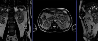
Enlargement of both adrenal glands according to CT data (indicated by arrows)
As a result of a CT scan of the adrenal glands, the following diseases can be diagnosed:
- benign and malignant tumors;
- adrenal cysts;
- Itsenko-Cushing syndrome;
- Addison's disease;
- primary hyper- and hypoaldosteronism;
- hemorrhage in the adrenal cortex (Waterhouse-Friderichsen syndrome);
- congenital anomalies of organ development.
Contraindications for examination
All contraindications for performing a CT scan of the adrenal glands are divided into absolute and relative.
Absolute contraindications include:
- pregnancy at any stage;
- patient weight over 120 kg.
Relative contraindications:
- age up to 12 years;
- allergy to drugs containing iodine;
- feeding the baby with breast milk;
- chronic renal failure;
- multiple myeloma.
The administration of a contrast agent is contraindicated in the following cases::
- pregnancy and lactation;
- presence of diabetes mellitus;
- insufficiency of kidney and liver function;
- intolerance to iodine-based drugs.
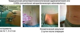
CT
CT (computed tomography) is a method based on X-rays . It can be used to examine the adrenal glands and identify various types of pathologies, as well as neoplasms. Early diagnosis of adrenal diseases is quite important. They produce hormones . If the adrenal glands are removed without constant medical intervention and the use of hormonal drugs, a person faces death.
Indications for using this method are:
- Diagnosis of neoplasms.
- If tumors are present, determine the location and stage of development.
- Diagnosis of cysts.
- Determining the location of injury after an accident.
- Presence of metastases.
- Endocrine diseases.
CT scanning is contraindicated during pregnancy and breastfeeding. It is also not recommended for use in children under 12 years of age. In this case, the study is carried out only on an urgent basis. When performing a procedure with a contrast agent, the procedure is prohibited for patients with an allergy to iodine, hypothyroidism and bronchial asthma. CT scanning of the adrenal glands is not recommended more than once a year.
No special preparation is required for the study . Basically, the procedure is performed on an empty stomach. A few days before the CT scan, you should not consume gas-forming foods or alcohol.
During the procedure, doctors receive a three-dimensional image of the organ being examined. The photographs are taken in the form of sections. From here, the specialist can examine each piece of the organ and form an overall picture.
Preparing for a CT scan of the adrenal glands
If you plan to undergo an adrenal gland examination, you will need to abstain from eating for 6 hours before the procedure. This is due to the fact that the contrast agent can only be administered on an empty stomach. This reduces the likelihood of nausea and vomiting in response to the introduction of an iodine-containing drug into the blood.
Preparation for a CT scan of the kidneys and adrenal glands does not require the patient to follow a special diet or cleanse the intestines using an enema and drugs such as Fortrans.
Contraindications and risks
Computed tomography of the adrenal glands cannot be prescribed if the patient has the following contraindications:
- child's age up to 3 years;
- pregnancy;
The use of contrast expands this list to include:
- allergic reaction to iodine and drug components;
- late stages of diabetes mellitus;
- severe form of renal failure.
To prevent side effects in other patients, clear recommendations for preparing for CT should be followed.
How is a CT scan of the adrenal glands done?
The procedure itself takes no more than 10 minutes, since the adrenal tissue does not retain the contrast for long. The bulk of the time is spent preparing the patient for the procedure.
Preparation includes:
- changing clothes: metal fasteners, buttons, clothing trim elements can leave shadows on the pictures and significantly complicate diagnosis;
- injection of a contrast agent in a jet or placement of a catheter for bolus administration of a contrast agent into a vein in the patient’s arm;
- placing the patient in the tomograph, fixing the patient in a stationary position using belts and pillows.
Before turning on the tomograph, the medical staff leaves the room, but continues to monitor the patient through the window from the adjacent office. The patient can contact medical staff at any time using voice communication or a special panic button.
During the entire examination, it is necessary to remain absolutely still , since even the smallest movements can affect the quality of the resulting images and reduce the information content of the CT scan.
After the examination is completed, the catheter is removed from the patient’s arm and the patient is asked to change into his own clothes.
What to do before the procedure itself
Before diagnosis, the patient is placed on a special moving table and then sent to a tomography machine. During the examination, in order to avoid obtaining unclear images, it is forbidden to move. Sometimes (for mental disorders or if necessary) the patient is secured with belts. At the doctor's command, the person must not breathe for several seconds. During a CT scan, a specialist examines the condition of the kidneys and adrenal glands on the monitor and assesses the extent of their damage.
Afterwards, the result can be transferred to disk and decrypted on your doctor’s computer. In any case, the doctor evaluates the image and, taking into account the information received, confirms or refutes the diagnosis.
Attention!
This article is posted for informational purposes only and under no circumstances constitutes scientific material or medical advice and should not serve as a substitute for an in-person consultation with a professional physician.
For diagnostics, diagnosis and treatment, contact qualified doctors! Number of reads: 4436 Date of publication: 08/21/2018
Urologists - search service and appointment with urologists in Moscow
How often can a CT scan of the adrenal glands be done?
CT scanners use X-rays to produce images. The radiation dose that a patient receives during one examination of the adrenal glands does not pose a threat to human health. However, radiation doses received over a short period of time add up and can lead to the development of radiation sickness or increase the risk of developing cancer. Accordingly, such an examination can be repeated no earlier than after 6 months, and only if there are serious indications.
The optimal interval between examinations is 12 months.
How is a CT scan done with and without contrast?
Before the examination, the patient must remove all metal objects; there should also be no metal elements on clothing in the scanning area.
The essence of tomography is to pass X-rays through the body and process the resulting data using special equipment. The patient is placed on a couch that is connected to a scanning device.
When contrast enhancement is used, the patient is given intravenous Omnipaque or Ultravist before scanning. Otherwise, all stages of the procedure are similar to native CT (without contrast). During the diagnostic period, it is important to remain motionless.
When the beam tube of the tomograph moves, a layer-by-layer examination of tissue occurs in the form of images - tomograms. They are processed by a computer program and produces the final image. The result can be printed or recorded on media.
The entire procedure usually takes about half an hour. Upon completion, the patient needs to drink as much plain water as possible, avoid eating salty and spicy foods, so as not to create additional stress on the kidneys and remove the contrast agent from the body as soon as possible