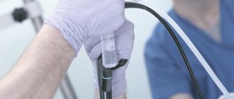
Cystoscopy is an examination of the inner surface of the bladder using an endoscopic system.
Cystoscopy is one of the main methods used to diagnose neoplasms and other diseases of the bladder. The study is carried out using a cystoscope, which is inserted into the urethra and then advanced into the bladder.
In medical practice, 2 types of cystoscopes are used: rigid and movable.
The rigid cystoscope has practically been replaced by flexible equipment, but it is used for retrograde catheterization of the ureters and transurethral resection of the prostate gland.
Using a movable cystoscope, you can observe the internal structure of the bladder on the monitor screen. A movable cystoscope consists of a thin, hollow metal tube 30 cm long, equipped with a light source and an optical system.
The equipment allows the doctor, using the magnification function, to examine the condition of the mucous membrane of the urethra and bladder. The cystoscope is equipped with a large number of different optical fibers and lenses that transmit a complete image of these organs from the inside
In addition, the cystoscope has special channels that allow delivery directly into the bladder of the instruments necessary for the manipulation (biopsy forceps, loops for removing polyps, needles for aspiration, etc.).
1
Ureterorenofibroscope Pentax FUR-9RBS
2 Pentax FUR-9RBS ureterorenofibroscope
3 Cystoscopy in MedicCity
History of cystoscopy of the bladder
Even in ancient times, scientists dreamed of being able to see the internal structure of organs without cutting into the patient. The Italian physician Bozzini discovered in 1805 how the functioning of the urinary tract could be observed using a light source.
German urologist Maximillian Nitze in 1877-79. proposed to combine light and an optical device in one device. The device, which was a tube with a light bulb attached to the inner end, was passed into the bladder. The bladder was then filled with fluid.
This made it possible to examine the bladder much more conveniently and accurately.
Types of cystoscopes
The cystoscopes used for this study are of two types: flexible and rigid.
Currently, experts prefer a flexible option. However, retrograde catheterization of the ureters is still performed using a rigid cystoscope.
A movable (flexible) cystoscope is a thin hollow metal tube 30 cm in length. This tube is equipped with an optical system and a light source. This device transmits an image of the internal structure of the bladder on the monitor screen.
Cystoscopy can be performed not only to examine the bladder, but also for therapeutic purposes. This procedure is used to treat urolithiasis. A special lithotripter crushes stones, which are removed through the ureter through an installed catheter.
Chromocystoscopy is also performed. Using a solution of a dye injected into the bladder, the functioning of the kidneys is assessed separately and the degree of patency of the ureters.
With the help of cystoscopy, it is possible to identify neoplasms that are not visible under normal lighting. During the study, special fluorescent drugs are introduced, which accumulate in the cancer tissue in a significant volume. This allows us to determine the nature of tumors in the bladder.
Preparation for cystoscopy
Preparation for cystoscopy involves proper hygiene of the genital organs, which reduces the number of complications. Therefore, immediately before the bladder examination procedure, the doctor treats the patient’s genitals with an antiseptic.
Carrying out cystoscopy
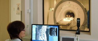
Survey cystoscopy of the bladder is one of the most common examinations. This type of cystoscopy is used in cases where the information obtained through ultrasound of the bladder or CT scan of the bladder is not enough to make a diagnosis. Cystoscopy of the bladder in women is much easier than in men, since they have a shorter and wider urethra.
Cystoscopy of the bladder in men, due to the fact that their urethra is much longer, is performed under general anesthesia. In this case, local anesthesia is used for women.
Cystoscopy of the bladder is most often performed in the supine position on a urological chair or a special table with footrests. If the legs or pelvic bones are affected, cystoscopy is performed in the lateral position.
Before examining the bladder, a warm solution of novocaine or lidocaine gel or another anesthetic is injected into the urethra for pain relief. Sometimes it is allowed to take analgesics.
For better passage, the end of the cystoscope is treated with glycerin or petroleum jelly and carefully inserted through the urethra. As soon as the device reaches the bladder, residual urine is removed from the organ and its volume is measured. Then the capacity of the bladder is determined by filling its volume with furatsilin solution. Usually this is about 200 ml of solution. If there is blood or pus in the bladder, the bladder is washed before cystoscopy.
During cystoscopy, the wall and apex of the bladder are observed, then the left and right side walls, the back wall and the bottom. Cystoscopy is performed to determine the presence of ulcers, stones and foreign bodies in the bladder, and to assess the nature of the existing discharge.
Cystoscopy of the bladder in women is sometimes performed together with chromocystoscopy. For this type of study, a 0.4% solution of indigo carmine is injected intravenously and the time taken for it to be excreted in the urine is checked.
If necessary, a bladder biopsy is performed during cystoscopy.
During surgical cystoscopy of the bladder in women, urethromy is performed, bladder tumors are removed, and a catheter is installed in the ureter.
If a man has a urethral stricture, bougienage (dilation of the urethra) or urethromy is performed.
1 Cystoscopy in MedicCity
2 Cystoscopy in MedicCity
3 Consultation with a urologist
How to prepare for the examination
Cystoscopy does not require special preparation. If it is performed under anesthesia, then you need to abstain from food 10-12 hours before the appointed time. 3-4 hours before the procedure, you should avoid consuming any liquids. Since you cannot drive after anesthesia, you should arrive at the clinic in another way. If the examination of the bladder walls is carried out without anesthesia, it is enough to simply appear on an empty stomach.
Attention! Before the procedure, external toilet of the genital organs should be performed.
Bladder diseases for which cystoscopy is indicated
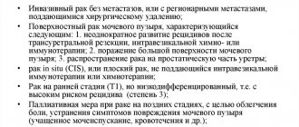
Cystoscopy is indicated for the following diseases:
- cystitis;
- tuberculosis;
- urinary disorders;
- hematuria (using cystoscopy you can see in which area of the bladder the bleeding is located);
- suspicion of urolithiasis;
- fistulas in the vagina and bladder;
- presence of atypical cells in urine samples;
- BPH;
- malignant and benign formations of the bladder and urethra;
- bladder injuries (with the help of research, you can find out the area and nature of ruptures in the mucous membrane of the bladder);
- suspicion of the presence of a foreign body in the bladder (bladder cystoscopy) helps to accurately determine the size and location of the foreign body and determine a treatment regimen.
Contraindications for cystoscopy
Contraindications are divided into general and local.
Local contraindications
Cystoscopy is not performed in the presence of inflammatory processes and blood in the urine. In this case, cystoscopy of the bladder can lead to the development of even greater inflammation.
General contraindications:
- renal failure;
- myocardial infarction;
- pregnancy.
Complications after cystoscopy
If cystoscopy is not performed professionally, the following complications may occur:
- infections in the bladder cavity;
- the appearance of cystitis or bladder erosion;
- traumatic complications (urethral ruptures, which occur during cystoscopy in men);
- Bladder perforation (puncture of the bladder wall, accompanied by leakage of urine).
In order to avoid unpleasant bladder diseases, cystoscopy should be performed only in a well-established medical clinic!
Contraindications for cystoscopy
Like any procedure involving intervention in the human body, cystoscopy has a number of contraindications.
It is prohibited in the following cases:
- renal failure and myocardial infarction;
- violation of the patency of the urethra;
- inflammatory processes of the bladder that are in the acute stage;
- menstruation and pregnancy;
- the use of contrast agents is prohibited for patients in a state of post-traumatic shock and with impaired renal excretory function.
After cystoscopy
After cystoscopy, the following unpleasant sensations may be observed (which disappear over time):
- pain and burning in the bladder (to reduce these symptoms, it is recommended to drink more fluids);
- frequent urination;
- urinary retention;
- lower back pain;
- chills, fever;
- bloody discharge in the urine (should disappear after a couple of days).
If the discharge does not disappear within 2 days after cystoscopy, then you should urgently seek advice from a urologist.
1 Consultation with a urologist after cystoscopy
2 Consultation with a urologist after cystoscopy
3 Consultation with a urologist after cystoscopy
What tests should a woman undergo if she has cystitis?
| № | Analysis name | What does it mean for cystitis? | Deadline | Price |
| 1 | General urine analysis with sediment microscopy | Increased number of white blood cells Presence of red blood cells or blood Presence of mucus, columnar epithelium Salts, bacteria | 1 day | 500 rub. |
| 2 | Urine culture for microflora | Increased number of bacteria or fungi: Escherichia coli, enterococcus, streptococcus, Proteus, Klebsiella | 5–7 days | 1600–1900 rub. |
| 3 | Urinalysis according to Nechiporenko | Exceeding the number of leukocytes more than 2000, sometimes more than 1000 erythrocytes | 1 day | 700 rub. |
| 4 | PCR urine test or Femoflor | Detects DNA of mycoplasmas, ureaplasmas, chlamydia, gonococcus, trichomonas in venereal cystitis in urine. Or a large number of opportunistic intestinal microflora, fungi, herpes viruses. | 1 day | 300 rub. for an infection. |
| 5 | Vaginal smear and test for sexually transmitted infections | In some cases, inflammation in the genital tract and STIs are detected | 1 day | 900 rub. |
Tests for cystitis in women, if the disease is chronic, must be supplemented by a study of the immune status, ultrasound diagnostics of the kidneys, ureter and bladder.
In some cases, cystoscopy is prescribed - examination of the mucous membrane of the bladder and urethra using a special cystourethroscope device, which is inserted directly into the bladder cavity.
In the multidisciplinary clinic “MedicCity”, paramount importance is attached to the treatment of urological diseases such as urethritis, cystitis, impotence, prostatitis, varicocele, etc. Professional doctors, high-quality diagnostics and modern treatment methods, an individual and delicate approach to each patient - that’s what we do We are rightfully proud.
If you read reviews about cystoscopy on the Internet, they are full of all sorts of stories about the unpleasantness of this type of examination. Thanks to reviews of cystoscopy, you can find medical institutions where procedures such as cystoscopy are performed at a high professional level. The cost of this service varies. However, the main selection criterion should still be the professionalism of urologists and good recommendations from the medical institution itself.
In our clinic, when performing cystoscopy, we use an anesthetic drug - Cathejell with Lidocaine. To ensure patient comfort, this type of examination is performed under sedation (sleep therapy).
If you are interested in the price of cystoscopy, you can see it here.
Difficulty urinating
Causes of difficulty urinating in women
Difficulty urinating is a condition that occurs in a number of urological and neurological diseases.
Cystitis
Acute cystitis is the most common urological pathology among women. Due to the anatomical features of the female urethra (proximity of the vagina and rectum, short length and large width, facilitating the penetration of infection into the bladder), virulence of the pathogen, violation of personal hygiene rules, decreased local immune response, concomitant diseases and other reasons, acute cystitis often becomes chronic relapsing form[3].
With chronic cystitis, bladder overactivity may develop with a decrease in its capacity and an increase in pressure inside, as well as increased activity of the muscle that compresses the bladder. All these pathological changes can lead to frequent urge, spasms and pain when urinating[2].
Surgical and diagnostic interventions
Pain, pain, weak or intermittent stream, imperative urge to urinate can be a consequence of surgery on the pelvic organs and kidneys, or the consequences of cystoscopy [4].
Pregnancy
The growing uterus puts pressure on the organs of the urinary system, making it difficult to release urine. This condition cannot be considered a pathology, however, it can also bring a lot of inconvenience. Difficulty in the outflow of urine during pregnancy may be a predisposing factor for the development of urolithiasis, the formation of stones in the bladder, and the development of pyelonephritis.
Kidney and bladder stones
The growth of stones in the renal pelvis and bladder stones can become a mechanical obstruction to the outflow of urine. Usually the urine becomes cloudy, and when the stones move, it becomes mixed with blood. And urination disorders are accompanied by severe pain in the lumbar region and fever.
Postmenopausal changes
Urinary disorders in women during menopause are associated with estrogen deficiency, which leads to atrophic changes in the mucous membranes, weakening of the musculo-ligamentous apparatus of the pelvis, which causes organ prolapse, and impaired blood supply to the choroid plexuses of the urethra. This results in urinary incontinence, leakage, increased nocturnal urine output, involuntary bladder muscle contractions, and an overactive bladder[5].
Overactive Bladder
This is a fairly common cause of urinary disorders among women and men[6]. With this pathology, involuntary activity of the muscular lining of the bladder is observed when it is filled. This causes frequent imperative urge to urinate, urine is released in small portions, with pain. The stream may be intermittent and weak.
An overactive bladder may have neurogenic causes (Parkinson's disease, multiple sclerosis, Alzheimer's disease, organic brain damage) and be considered as an independent disease associated with impaired innervation of the bladder. To make this diagnosis, it is necessary to exclude a number of other diseases[3].
Hyporeflex bladder
When the sensitivity of the bladder decreases and overstretches, a condition called a hyporeflex bladder can occur. In this condition, the patient is forced to urinate in a thin, sluggish stream, pressing on his stomach, thus helping to empty the bladder. Chronic urinary retention occurs, which is dangerous for the development of infection in the urinary tract, the spread of this infection to the kidneys in an ascending way, the formation of stones in the bladder, the development of renal failure up to the complete loss of kidney function. This is already a life-threatening condition.
Urethral stricture
Narrowing of the urethra in women can occur as a consequence of injuries to the pelvic organs, inflammatory diseases, the appearance of adhesions between the urethra and vagina, etc. Depending on the degree of stricture, urine may leak in a weak stream or intermittently.
Injuries and tumors of the spine and brain
Disturbances in the outflow of urine due to injuries and neoplasms of the central nervous system are associated with the innervation of the bladder, urethra and pelvic muscles, which leads to discoordination of the act of urination. This condition is called neurogenic bladder and most often presents with difficulty urinating (in 64%)[7].
More rare causes are:
- blood clots or mucus in the urinary tract,
- entry of foreign objects into the urinary tract.
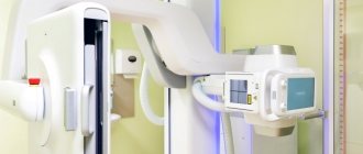
Ultrasound of the bladder: preparation
If you are having a bladder ultrasound, preparation for the study should be done carefully. Otherwise, the results may be distorted or incomplete. However, preparation before an ultrasound of the bladder does not involve any complex or burdensome actions: it is enough to follow a few simple recommendations. Preparing a patient for an ultrasound of the bladder includes several stages.
Ultrasound of the bladder: step-by-step preparation for the study
- During the day, refrain from eating foods that contribute to the formation of gases in the intestines. These include raw vegetables, fruits, legumes, baked goods, and sweets.
- Before going to bed you need to drink an adsorbent. Consult your doctor about dosage.
- You must come to the procedure with a full bladder. To do this, an hour before the session you need to drink 0.5-1 liter of non-carbonated liquid. Or accumulate urine throughout the day by gradually drinking liquid in small portions. It is recommended not to urinate for 3 hours, but if the urge is too strong, you can empty your bladder a little and then drink a small amount of liquid.
Bring a diaper or towel, as well as a napkin, to the examination.
The more thoroughly you prepare before an ultrasound of the bladder, the better the results will be. Before preparing, consult your doctor: perhaps he will give some individual recommendations based on your state of health or the presence of any diseases.