Price list Doctors clinic
Ultrasound of the bladder with determination of residual urine is a diagnostic method that allows not only to assess the condition and structure of the organ, but also to determine the usefulness of its emptying. The study is carried out in two stages: first, the doctor performs a conventional ultrasound examination, determines the condition and volume of the organ. The patient should then urinate, and then measure the volume of urine remaining in the bladder after voiding. We can speak of normal function of the urinary system if the volume of urine does not exceed 30 ml.
At the Family Doctor clinic, ultrasounds are performed by highly qualified diagnosticians. We use modern ultrasonic equipment from leading global manufacturers. High-quality images, as well as the professionalism of specialists, allow us to quickly and accurately assess the functions of the urinary system and select effective treatment regimens.
Indications for the study
Ultrasound of the bladder with residual urine may be prescribed in the following cases:
- frequent urge to urinate;
- the presence of impurities, sediment, flakes in the urine;
- change in urine color;
- pain in the lower abdomen and/or groin, pain;
- difficulty urinating;
- weak jet pressure;
- male infertility of unknown origin, sexual dysfunction.
The reason for the study may be unsatisfactory results of laboratory tests, suspicion of prostate adenoma or prostatitis in men.
An indication for ultrasound with determination of residual urine may also be the need to monitor the treatment. So, the procedure is repeated after taking measures to treat prostate adenoma.
Bladder examination algorithm: how to prepare for an ultrasound for women and men?
Ultrasound of the bladder in women and men: how to prepare correctly? This question worries all patients who are faced with the need to undergo an ultrasound examination of the genitourinary system for the first time. To answer this question, it is necessary to take into account all the features of the procedure.
In a man, the urethra passes through the prostate. With its diseases (adenoma, prostatitis), the urethra is compressed by the tissues of the enlarged gland, which causes incomplete emptying of the bladder. After a standard ultrasound procedure, the man is asked to urinate, and the residual amount of urine is measured by inserting an ultrasound probe into the rectal cavity.
Therefore, a man needs to do 2 enemas: one the night before, the other in the morning before the procedure, so that the lower part of the large intestine is free and does not contain fecal matter.
DETAILS: What disability group if there is no kidney
In women, if there are no contraindications, the most accurate source of information about the condition of the bladder is the transvaginal ultrasound method. For urgent indications, it is carried out even during menstruation and in the first trimester of pregnancy.
Preparation
Ultrasound is a painless and safe examination method, so no careful preparation is required. However, it is important to come to the diagnosis with a full bladder. This is necessary both for better visualization of the organ and for the second stage - assessing the volume of urine after emptying. Therefore, there are two ways to prepare:
- 1-2 hours before the test, you need to drink about 1-1.5 liters of liquid - clean water without gas, tea, unsweetened compote - and not urinate.
- You should stop urinating 3-4 hours before, but there is no need to increase the volume of fluid you drink.
- If the patient cannot hold urine, then you should come to the clinic in advance and continue taking fluids at the ultrasound room.
The day before the ultrasound, it is important to adhere to a gentle diet - avoid dishes, foods and drinks that cause increased gas formation, including black bread, fresh cabbage, legumes, soda, alcohol, strong tea and coffee.
Why is ultrasound performed only on a full bladder?
An ultrasound scan is done of a full bladder - when it is maximally full, the inside is seen better, since in an empty state it is wrinkled and difficult to see by a specialist.
Before an ultrasound of the bladder, it is necessary to fill it with liquid, then the walls will straighten and stretch without unnecessary folds. When the organ is full, it can bring the intestinal loops closer to the abdominal cavity, to its front part.
Filling of the organ should occur in natural conditions and at a pace, but in some cases the patient may not know about the need for an ultrasound, or if the procedure is emergency and there is no time to prepare according to the rules. In this case, the bladder needs to be filled quickly and efficiently.
To do this, just drink a liter of clean water an hour before the intended procedure. Instead of water, it is allowed to use tea or fruit drink, juice or herbal infusions. Experts do not recommend drinking milk or highly carbonated drinks to ensure the reliability of the examination results.
The bladder of all people is different in volume, and if after an hour the patient does not have the urge to deurinate, then it is worth drinking another half liter of liquid. It is important to prevent overflow; if the urge to urinate appears long before the procedure, you can visit the toilet, but then be sure to replenish the lost volume of water.
As a rule, early urination indicates excellent functioning of the kidneys, which produce urine. Experts advise not to empty your bladder for several hours before the ultrasound, so as not to drink excess water and not put a strain on your kidneys. This is only possible if the patient has a good knowledge of his excretory system.
Contraindications to the procedure
Ultrasound with determination of residual urine is carried out in one of three ways:
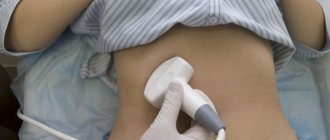
- transabdominal - through the anterior abdominal wall;
- transrectally - through the rectum;
- transvaginally - through the vagina.
The first method has no contraindications, with the exception of acute inflammatory diseases of the skin of the abdomen. Transrectal examination is not performed for anal fissures, exacerbation of hemorrhoids, proctitis, paraproctitis. The transvaginal method is not suitable for inflammatory diseases of the genital tract, and is also not used in women who have not begun to be sexually active.
Overactive bladder in women: treatment (medications)
To reduce hyperactivity syndrome, medications are used to help relax the walls of the organ. This:
- tolterodine (“Detrol”);
- oxybutynin in the form of a skin (transdermal) patch (“Oxytrol”);
- oxybutynin in;
- trospium;
- solifenacin;
- darifenacin;
- fesoterodine.
The main method for diagnosing female genital organs is ultrasound examination. Ultrasound, thanks to a special sensor that emits ultrasound, allows you to find out the state of women's health. Through computer data processing, the doctor can see a high-quality image of the internal organs on the screen.
During the initial examination or for preventive purposes, the procedure can be performed at almost any time of the cycle, excluding the period of menstruation.
If a woman has previously been diagnosed with any diseases of the genitourinary system or has symptoms indicating pathology, ultrasound is prescribed on certain days or performed several times in order to be able to quickly monitor the patient’s condition.
If there are signs of pregnancy, the procedure is carried out 14 days after ovulation, however, a delay in menstruation can also indicate the appearance of a cyst in the ovaries or uterus.
The most favorable time for ultrasound examination is considered to be the period after menstruation (the first 3-6 days).
As a rule, during one procedure, the doctor examines not only the uterus, but also the ovaries, fallopian tubes, and cervix.
If the diagnosis is aimed at assessing the condition of the ovaries or corpus luteum, it is repeated several times during one menstrual cycle (usually on days 21-24 or 14-16). Preparing for an ultrasound of the pelvic organs for women involves certain actions. It directly affects how accurately the diagnosis will be made.
Depending on the method of the procedure, preparation for a gynecological ultrasound of the pelvis for women may differ.
For example, before a transabdominal examination, it is recommended to drink plenty of fluids to ensure that the bladder is as full as possible.
Other methods of ultrasound diagnostics require diet, preliminary bowel cleansing and other preparatory measures. How to prepare for a pelvic ultrasound for women, depending on the purpose of the diagnosis?
This ultrasound examination technique is carried out using a special sensor (vaginal). Transvaginal diagnosis is carried out not only in gynecology, but even in urology (if the doctor suspects the presence of genitourinary diseases).
Preparing for a pelvic ultrasound for women does not require filling the bladder with fluid or taking any medications to cleanse the intestines. To carry out the diagnosis, you will need a disposable diaper/towel to lie on.
DETAILS: Neurogenic bladder in women and men: treatment, symptoms
If a doctor examines a pregnant woman, her bladder needs to be reasonably full. To do this, you should carry out appropriate preparation by drinking approximately 500 ml of liquid before the test (1-1.5 hours before).
What else you should know before the examination:
- It is impossible for there to be gases in the peritoneal organs during the study, so for 2-3 days it is worth reducing the consumption of foods that cause them (baked goods, vegetables/fruits, sugar, dairy products).
- It is not recommended to perform cleansing enemas before the procedure.
- You can eat in the evening before the diagnosis, but no later than 6 hours.
- To eliminate gases, it is allowed to take activated carbon or Enzistal.
Decoding the results
The results of the procedure will be issued in writing on average within 15 minutes after the study. The doctor will draw up a conclusion that reflects the condition, structure of the organ, characteristics and size, as well as the volume of residual urine. In general, such a study allows you to obtain the same information as a standard ultrasound - detect inflammatory diseases (signs of acute or chronic cystitis), suspect urolithiasis, see tumors and neoplasms. Additionally, the doctor will determine how much residual urine remains in the bladder after emptying.
If during the examination a residue of more than 30 ml is detected, the specialist will give recommendations on further actions. If prostate adenoma is suspected, additional diagnostic methods, consultation with a urologist, and laboratory tests may be required.
Indications
Frequent urination
The bladder is examined in the case of:
- pathological processes in various parts of the urinary system;
- determination of the source of erythrocyturia;
- complaints characteristic of prostatic hyperplasia, cystitis, kidney stones and urolithiasis;
- establishing the cause of acute urinary retention and other dysuric symptoms;
- suspicion of the presence of benign or malignant neoplasms;
- suspicion of diverticulosis;
- traumatic lesions of the bladder;
- control of therapy of various types of therapy.
Preparing for an abdominal ultrasound
Preparation for an abdominal ultrasound should begin approximately one day before the examination. Abdominal ultrasound is best performed in the morning, on an empty stomach or after 6-8 hours of eating!
Preliminarily exclude the following products from the diet: legumes in any form, carbonated drinks, milk, sweets and baked goods, black bread, fermented milk products (including cottage cheese), raw vegetables and fruits, caffeine-containing spirits, alcohol, fish and meat - fatty varieties (products that increase gas formation in the intestines).
For patients with problems with the gastrointestinal tract (constipation), it is advisable to take enzyme preparations and enterosorbents (for example, festal, mezim-forte, activated carbon or espumizan, 1 tablet 3 times a day) during this period of time, which will help reduce the manifestations of flatulence.
If you are taking medications, notify the ultrasound doctor about this.
Bladder volume and filling
The volume of urea is 350-750 ml in men, 250-550 ml in women.
The volume decreases if scars form on the walls, which in turn arise due to inflammation. Developmental pathologies of neighboring organs (uterine fibroids, prostate adenoma) also reduce the volume of the bladder.
After operations on the abdominal organs (removal of the appendix, intestinal surgery) it leads to adhesions that reduce the ability of the bladder walls to stretch.
In pregnant women, the organ narrows and elongates. Its volume decreases, so expectant mothers feel the need to go to the toilet more often.
An increase in the volume of the bladder occurs when it overflows with urine, and this occurs when the urethra is blocked by a stone or tumor, as well as the growth of the prostate gland.
Structure of the bladder
The ultrasound machine “sees” the bladder right through. The organ is a round-shaped muscular sac consisting of several layers:
- The inner mucous layer is folded, but as it fills, the folds smooth out, except for the fold near the ureter (to prevent urine from flowing back out). The mucous membrane is very sensitive to infections that enter the body through the urethra.
- The submucosal layer is located between the mucous membrane and the muscular layer. It is replete with nerve endings and blood vessels, so any foreign body (infection, stones, sand) causes a burning sensation, frequent urge to go to the toilet, and nagging pain.
- The muscle layer consists of smooth muscles lying in three layers, connected into one large compressor muscle, responsible for pushing urine out. Around the ureter, the muscles form circular sphincters - peculiar valves responsible for the release of urine into the ureter.
- The serous layer covers the entire surface of the bladder.
Preparing for an ultrasound of the gallbladder
Preparation should begin approximately one day before the test. Ultrasound of the gallbladder is best performed in the morning, on an empty stomach or after 6-8 hours of eating!
Preliminarily exclude the following products from the diet: legumes in any form, carbonated drinks, milk, sweets and baked goods, black bread, fermented milk products (including cottage cheese), raw vegetables and fruits, caffeine-containing spirits, alcohol, fish and meat - fatty varieties (products that increase gas formation in the intestines).
The peculiarity is that for the study you need to take products that stimulate the contraction of the gallbladder. I usually use full-fat sour cream (20%) and dark chocolate for these purposes. In such cases, an ultrasound of the gallbladder is done twice - on an empty stomach and after consuming choleretic products, which usually takes longer.
