Glaucoma is a group of eye diseases that cause damage to the optic nerve.
Degenerative eye pathology is one of the leading causes of chronic blindness.
Open angle glaucoma (OAG) is the most common form. Also known as chronic OAG. Refers to a group of eye diseases that lead to damage to the optic nerve head with progressive loss of retinal ganglion cells and their axons. It is the most dangerous due to its asymptomatic course and loss of visual acuity.
Slit examination reveals typical changes in the optic nerve. Damage to the optic nerve results in decreased peripheral vision and eventually lead to blindness.
OAG is controlled with medications; in severe cases, surgical therapy is prescribed.
Causes of open angle glaucoma
The open-angle form of the disease has many causes, with 60 to 70% of cases having no identifiable cause and called primary open-angle glaucoma (POAG).
⅔ of patients with this pathology have increased IOP - >21 mm Hg. Water drainage is insufficient, while production by the ciliary body is normal. Secondary OAG occurs due to developmental abnormalities, scarring, trauma or infection, blockage of channels with iris pigment (pigment dispersion syndrome) or abnormal protein deposits (pseudoexfoliation syndrome).
We recommend reading: Closed-angle and open-angle glaucoma
POH develops due to compression of the optic nerve by tumors or hematomas, dysfunction of the outflow of intraocular fluid and degenerative processes in the eye.
Primary prevention of glaucoma
To minimize the effect of negative factors, it is necessary to control the following conditions:
- Ophthalmological diseases. Injuries to the eye area and cataracts can provoke a negative reaction to increased IOP due to disturbances in the circulation of intraocular fluid. To prevent glaucoma, it is recommended to promptly treat concomitant eye diseases.
- Diet. A monotonous, unbalanced diet can have a significant negative impact on the nutrition and hemodynamics of the eyes. A deficiency of protein foods, vitamins and minerals is extremely undesirable. To fully replenish nutrients, the diet must include lean meat, fish, plant foods, milk and dairy products.
- Physical activity. Moderate loads help activate all processes, including accelerating blood flow and gas exchange. Regular performance of a set of physical exercises minimizes the risk of developing hypoxia of the visual system. At the same time, it is worth considering that heavy lifting and excessive stress can provoke instability of intraocular pressure.
- Prevention. If you have a family history, as well as existing ophthalmological diseases, you must regularly undergo a medical examination with an ophthalmologist. Glaucoma does not manifest itself in any way in the initial stages, and changes in the fundus can develop very slowly. Only an experienced specialist has the opportunity to identify the disease in the early stages, when treatment is most effective and the prognosis is favorable.
Risk group
Factors that quickly lead to the development of POP:
- Age—incidence increases with age, most often after age 65 (and rarely before age 40).
- Family history. It is believed that there is incomplete penetrance and variable expressivity of the genes involved.
- Race – The disease is 3–4 times more common in Afro-Caribbean people.
- Ocular hypertension is a major risk factor for the disease, with 9% of patients developing glaucoma within five years if left untreated.
Also at risk are patients with the following pathologies: myopia (myopia), central retinal vein occlusion, retinal detachment and retinitis pigmentosa, diabetes and systemic hypertension.
Surgical treatment of glaucoma
Surgical treatment of open-angle glaucoma involves sinustrabeculectomy. This is a non-penetrating filtering operation that is carried out in several stages. Usually the interval between operations is 4-6 weeks.
During the operation, the doctor creates a new path for the outflow of intraocular moisture from both chambers of the eyeball. A filtration cushion is created under the sclera and mucosa, which stabilizes the pressure. A modification of the technique is the implantation of a mini-shunt, that is, a miniature drainage, under the scleral flap. Thanks to its design, a stable outflow of liquid is ensured.
After surgery, the eye is covered with a bandage for several days. Drop therapy is mandatory. Superficial sutures are removed after 7-10 days. During the rehabilitation period, it is recommended to limit the consumption of salt and pickled foods, as well as alcohol. Do not rub your eyes for 10 days. It is important to protect the operated area from water and dust. It is recommended to sleep on the side opposite to the eye in which the intervention was performed. To avoid injury, it is better to limit physical activity.
Advantages of sinustrabeculectomy:
- the patient returns home almost immediately after the operation;
- The procedure for operating one eye takes 20 minutes;
- painlessness;
- quick rehabilitation (1-3 weeks);
- lack of bed rest;
- minimal restrictions during the recovery period.
A positive effect after sinustrabeculectomy is observed in 60-80%. In other patients, there is a need to repeat the operation. After treatment, you need to undergo examinations twice a year in order to promptly identify complications and other disorders in the functioning of the visual system.
Classification of open-angle glaucoma
There are 4 stages of the disease:
- on the first, excavation of the second pair of cranial nerves is observed;
- on the second - the depression reaches the edge, vision from the periphery narrows by 10 degrees;
- third - excavation almost everywhere, a large percentage of visual perception is lost;
- on the fourth - blindness, but light perception is preserved.
Regardless of the stage of development, the open-angle form of the disease is divided into compensated, subcompensated and uncompensated. Based on the speed of the disturbance, stable and unstable forms of OAG are distinguished.
Symptoms of open angle glaucoma
The main problem with the pathology is damage to the optic nerve. The pathophysiology is not fully understood, but there is a progressive loss of retinal ganglion cells and axons. In the early stages it affects only the peripheral field of vision, but as it progresses it affects central vision, leading to loss of acuity, which can lead to severe impairment and complete loss of visual perception.
For most OAG, optic neuropathy is associated with elevated IOP. This has given rise to the hypothesis of retinal ganglion apoptosis, the rate of which is influenced by hydrostatic pressure on the optic nerve head. Optic neuropathy results in characteristic changes in the optic disc and loss of visual field.
In the open-angle form, the outflow through the trabecular meshwork worsens (the role of which is to absorb aqueous humor). This is a chronic degenerative obstruction that is painless.
IOP is not always elevated.
Early primary symptoms of open-angle glaucoma are rare. Typically, the patient becomes aware of visual field loss when optic nerve atrophy occurs. Signs of OAG:
- The head of the optic nerve (disc) is a slightly vertically elongated circle with a central depression called the cup. The sensorineural rim is the tissue between the rim of the cup and the rim of the disc and consists of the axons of retinal ganglion cells. Characteristic changes in the optic nerve include an enlarged cup, thinning or tearing of the neurosensory rim, and hemorrhage.
- Changes in the visual field caused by damage to the optic nerve are manifested by arcuate and paracentral scotomas. Problems with twilight vision and rainbow glare appear.
- Discomfort, pain in the eyes due to increased IOP and redness periodically occur in the eyes.
Factors for development
Knowing your predisposition to increased intraocular pressure helps you take control of the disease in time and minimize possible consequences in the form of damage to the optic nerve and loss of vision. Here are some of the factors that increase your chance of getting glaucoma:
- Hereditary predisposition (if one of your blood relatives has this disease, then you have a greater risk of increased intraocular pressure than those whose family does not have this problem). This is often explained by the specific shape of the angle of the anterior chamber of the eye, which can be inherited, like other constitutional features.
- Eye injuries. After damage to the eyeball (for example, when it is contused as a result of an impact), conditions can be created for a deterioration in fluid circulation and an increase in pressure.
- Advanced stages of cataract. If the lens, which has become cloudy to the point of almost complete loss of vision, is not removed, it will continue to swell and gradually collapse. Particles of the lens enter the angle of the anterior chamber and impair the normal outflow of fluid.
- Inflammatory diseases of the choroid (iridocyclitis) can also cause disturbances in the outflow of intraocular fluid.
- Endocrine problems (certain types of disorders in the production of thyroid hormones, pituitary hormones, long-term diabetes mellitus with the presence of vascular changes in the retina).
- Previous circulatory disorders in the eye. Prolonged oxygen starvation associated with them can provoke the formation of new defective vessels that disrupt the filtration of moisture in the corner of the anterior chamber of the eye.
- New growths in the eye (which are large enough to partially block the outflow of aqueous humor).
If you have a predisposition to glaucoma, then you will need to visit an ophthalmologist and monitor intraocular pressure regularly and with the frequency prescribed by the doctor after the examination.
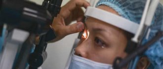
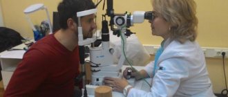
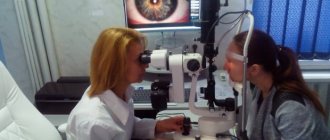
Diagnosis of open angle glaucoma
The ophthalmologist will carefully examine the eye for the presence of glaucoma, concomitant pathologies and conduct a differential diagnosis. Methods for examining a patient to make a diagnosis:
- Gonioscopy is a method used to measure the angle between the cornea and the iris.
- Measuring the thickness of the cornea.
- Tonometry is an objective measurement of IOP based on the assessment of corneal resistance to indentation. A range of 10–21 mmHg is considered normal.
- Examination of the optic disc is a direct marker of disease progression. The examination is carried out using a slit lamp. The pupil is dilated with drops.
- Measuring the visual field to clarify how far the pathology has progressed. The test is called perimetry.
End stage glaucoma
Glaucoma is an eye disease characterized by high eye pressure and damage to the optic nerve. The final stage of the disease is called terminal glaucoma.
It is expressed by severe pain in the eyes, which can radiate to the face and head, lacrimation and nausea. At the terminal stage of the disease, irreversible processes already occur in the eye, and complete blindness occurs.
Therefore, at the first symptoms of the disease, you need to contact a specialist and begin treatment.
What it is?
End-stage glaucoma is the last stage of the disease. It is characterized by an irreversible loss of objective vision. In this case, visual acuity decreases and only light perception is preserved. Blindness gradually sets in.
In terminal glaucoma, excavation of the optic disc, kinking of retinal vessels and retinal edema are detected. Small hemorrhages appear on the nerve disc due to the formation of blood clots in the vessels. If absolute glaucoma causes severe pain in the eyes, it is called terminal painful glaucoma.
This pain does not go away with medication, and it can only be relieved by surgery.
Causes of terminal glaucoma
There are the following reasons:
- genetic factor;
- heart diseases;
- high pressure surges;
- pathologies of the endocrine and nervous system;
- high pressure inside the eyes;
- glaucoma therapy was not started in a timely manner;
- late diagnosis of the disease;
- inadequately selected therapy.
Symptoms of terminal glaucoma
Symptoms of glaucoma.
The following symptoms are identified:
- severe pain (absolute painful glaucoma);
- irradiation of pain to the face and head;
- pain in the eyes;
- lacrimation;
- redness of the eyeballs;
- the appearance of photophobia;
- severe nausea;
- urge to vomit;
- swelling of the facial skin around the eyes;
- redness of the skin that surrounds the eyes;
- there is no reaction to pupil light;
- loss of vision.
Diagnosis of terminal glaucoma
When the first symptoms of the disease appear, you should definitely consult an ophthalmologist. He will collect all the patient’s complaints and find out how the disease began.
He will also conduct an objective examination, measure eye pressure and examine the fundus.
After this, he will prescribe additional tests, conduct differential diagnostics with other eye diseases and make a preliminary diagnosis.
Additional research methods:
- tonometry;
- perimetry;
- gonioscopy;
- optical coherence tomography;
- Heidelberg laser retinotomography;
- laser polarimetry;
- Ultrasound of the eyes.
Treatment methods for terminal glaucoma
At this stage of the disease, laser therapy and surgery are used for treatment.
If the patient has the first symptoms of the disease, you should not try to cure him on your own, but urgently need to contact a specialist.
The doctor will collect complaints, conduct an eye examination and prescribe special treatment methods. To cure terminal glaucoma, patients are prescribed drug therapy, surgical treatment methods and folk remedies.
Each patient is prescribed a specially selected dietary diet.
Drug treatment
Patients with absolute glaucoma are prescribed the drugs listed in the table:
| Group | Name |
| Cholinomimetics | "Pilocarpine", "Fotil" |
| Sympathomimetics | "Tyramine", "Ephedrine", "Clonidine" |
| Prostaglandins | "Xalatamax", "Glauprost", "Xalatan" |
| Adrenergic blockers | "Arutimol", "Ocumed" |
| Carbonic anhydrase inhibitors | "Azopt", "Trusopt" |
| Antihypertensive drops | "Mexidol" |
| Combination drugs | "Cosopt", "Xalacom", "Azarga" |
Surgical methods of treating absolute glaucoma
Laser treatment is widely used in the treatment of glaucoma.
Laser methods are widely used.
These include traction laser operations, transscleral laser cyclocoagulation, laser iridotomy, peripheral iridoplasty and papilloplasty.
All these operations are based on the use of a laser beam, with which it is possible to perform surgery without cutting the walls of the eye. This operation is not painful and is performed quickly.
Features of operations:
- Traction laser operations are performed in the area of the trabecular network of the corners of the anterior chambers of the eyes. They are based on the effect of a laser coagulant in the trabecular area, which leads to improved outflow of intraocular fluid.
- Transscleral laser cyclocoagulation is based on thermal destruction of part of the ciliary body. As a result, the production of aqueous humor and the pressure inside the eyes are reduced.
- Laser iridotomy is used as an additional surgical intervention after intraocular surgery. It is not used if there is swelling or clouding of the cornea, or if the patient has a shallow anterior chamber of the eye.
- Peripheral iridoplasty and papilloplasty are based on the fact that light laser coagulants are applied to the periphery of the iris. As a result, the angle of the anterior chamber expands.
Other surgical methods
The following methods are also used:
- Diathermocoagulation and cryopexy of the ciliary body. With the help of these operations, the production of aqueous humor is reduced, eye pain is reduced and intraocular pressure is reduced.
- Optociliary neurectomy. It allows you to relieve pain in a short time, reduce intraocular pressure and preserve the eyeball.
- Removal of the eye if organ-preserving operations are not possible.
Complications with absolute glaucoma and after operations
The first symptoms of the disease occur when the cornea swells and its nerve endings become irritated. As a result of such pathological changes, the tissues of the cornea become very sensitive to various infectious pathologies. Therefore, very often patients experience complications of terminal glaucoma.
These include inflammatory processes of the cornea, inflammatory damage to the iris, and perforation of the cornea. Complications often occur after surgical interventions.
Often there is severe eye bleeding, the inability to reduce intraocular pressure, increased pain in the eyes and gaping of the postoperative wound.
Source: //EtoDavlenie.ru/glaznoe/terminalnaya-glaukoma.html
Treatment of open-angle glaucoma
Treatment does not have to begin immediately if a slightly elevated IOP is detected. Given the possibility of fluctuations in intraocular pressure, the patient should undergo tonometry 2-3 times. If IOP is elevated at any time of the day, treatment should begin immediately.
Drugs that lower IOP are prescribed by your doctor. The ophthalmologist indicates the dosage and treatment regimen. Take medications strictly according to the prescribed regimen.
Typically, beta blockers have been the preferred option. In 2000, doctors began prescribing prostaglandin analogs because they were just as effective but had fewer side effects. A prostaglandin analogue should be used to treat patients with early or moderate OAG.
Control of IOP may require combining these medications or combining them with others (eg, sympathomimetics, carbonic anhydrase inhibitors, or miotics). As a rule, drugs are administered one at a time.
For urgent reduction of IOP and before surgery, Mannitol 20% (up to 500 ml) is administered by slow intravenous infusion until IOP is reduced satisfactorily. Acetazolamide by intravenous injection can also be used to urgently lower IOP.
Laser and surgical treatment are considered if medications do not help. The following operational methods are used:
- argon laser trabeculoplasty;
- selective or cyclodiodic trabeculoplasty;
- iridotomy;
- trabeculectomy;
- canaloplasty;
- trabeculotomy.
Treatment
Unfortunately, today there are no methods that can cure or cure glaucoma, but it must and can be treated and controlled. Depending on the type, stage and severity of the disease, the ophthalmologist will prescribe glaucoma drops, recommend laser treatment, or suggest surgery.
As a rule, treatment begins with the prescription of eye drops of various types, which have one property - they reduce intraocular pressure and, thereby, prevent damage to the optic nerve. But these drops need to be dripped constantly, according to a schedule, because... their period of action in the eye is limited.
Some drops may cause a feeling of discomfort and burning in the eye, but this is not a reason to discontinue the drops on your own without first consulting with an ophthalmologist about the possibility of replacing them with others.
Regardless of treatment method, early diagnosis of glaucoma is the best way to avoid blindness. Therefore, preventive examinations by an ophthalmologist are simply necessary for the early detection of glaucoma, especially if you have risk factors for developing this disease.
The asymptomatic and painless course of the disease determines the frivolous attitude of some patients to the prescribed treatment, who drip drops irregularly, or even stop altogether, forget to purchase a new bottle of drops in advance, and do not appear at the appointed time for a follow-up examination with an ophthalmologist.
This attitude to treatment is the reason for the progression of glaucoma and, as a consequence, irreversible vision loss. Patient adherence to treatment is one of the main factors for success in the fight against glaucoma.
Don't be afraid of doctor's orders. If the doctor says that you will need to use eye drops 1-3 times a day for a long time or even for life, do not refuse it, citing a busy work schedule, fear of side effects, or the high cost of the medicine. Compliance with the drop instillation regimen is the most important tool in preventing complications of the disease.
If the doctor prescribes an operation (laser or surgical), then in your case, instilling drops does not provide sufficient prevention of complications. This depends on the severity and aggressiveness of the glaucoma.
Do not be afraid to resort to surgery, because for some types of increased intraocular pressure, this is the only way to avoid complications of the disease and stop vision loss.
An ophthalmologist can prescribe not only treatment that reduces pressure in the eye, but also drugs that protect optic nerve cells from damage, as well as vitamins, if necessary. Prevention of consequences should include the following measures:
- Regular visits to a specialist;
- Compliance with doctor's orders.
Prevention
No modern treatment methods will help get rid of the disease forever. But preventive measures will help delay the onset of complete blindness.
Recommendations for the patient:
- eat foods rich in vitamin A;
- read in good lighting conditions;
- If you lose twilight vision, consult a doctor and wear glasses;
- protect your eyes from the harmful effects of ultraviolet radiation;
- visit an ophthalmologist at least 2 times a year after 45 years;
- choose the right glasses or contact lenses to correct myopia or astigmatism.
Characteristic features of absolute glaucoma and its treatment
Absolute glaucoma is a terminal condition of glaucoma, its final stage, in which vision is completely lost and blindness occurs. This degree of the disease is characterized by the detection of irreversible phenomena in all parts of the eyeball, absolute atrophy of the optic nerve.
The level of intraocular pressure (IOP) and the condition of the anterior ciliary vessels make it possible to determine the degree of compensation for glaucoma. In the initial stage of glaucoma, the IOP level does not exceed 28 mm. Hg Art.
(compensatory stage of glaucoma). The stage of decompensation indicates an IOP above 28 mm. Hg Art. (the “cobra” symptom, in which there is dilation of the superficial vessels of the eyeball).
In this case, swelling of the cornea and other eye tissues may also occur.
Absolute glaucoma is a severe and unfavorable outcome of all clinical manifestations of glaucoma, which result in complete and final blindness. With constantly elevated IOP, the normal functioning and metabolism in the tissues of the organ of vision is disrupted. This leads to atrophic changes and gradual loss of function. Severe pain may occur.
Dystrophic changes in the eye are expressed in damage to the cornea in the form of keratitis or ulcers of the cornea of the eye.
The process may be complicated by the addition of an infection, which may result in a complication in the form of a perforation of the cornea.
During the occurrence of perforation, hemorrhagic manifestations are observed when the posterior arteries rupture and the membranes of the eye (or part of them) are pushed out of the eyeball under blood pressure.
Treatment of absolute glaucoma
The treatment method for absolute glaucoma is only surgical, when it is necessary to eliminate unbearable pain caused by degenerative changes in nerve endings. Surgery is usually prescribed after unsuccessful attempts at conservative treatment. Most often, distraction therapy and increased use of miotic drugs, as a rule, do not produce results.
Reputable ophthalmologists believe that the use of conventional operations used to treat glaucoma does not give the expected effect.
Thus, complications are observed in the form of wound gaping, bleeding, a sharp increase in IOP, and increased pain.
That is why, based on many years of practice and world experience, it was decided that the optimal option for surgical treatment of absolute glaucoma is optociliary neurectomy.
This operation is technically simple in the practice of ophthalmic surgery, while at the same time representing a very effective means of eliminating pain. In most cases, the eye can be preserved as a cosmetic organ.
Of course, there are contraindications for this operation:
- The patient's serious condition.
- Dystrophic changes in the cornea.
- Malignant neoplasms.
Also, you should be especially careful in case of trophic disorders of the cornea, since in this case optociliary neurectomy may have an adverse effect.
At the same time, with dolorous absolute glaucoma, the operation of ciliarotomy and diathermocoagulation of the ciliary nerves without transection of the optic nerve can be very beneficial for the effective elimination of pain and influence on trophic processes in the cornea of the eye. This operation is much less dangerous, although it is more technically complex. According to the observations of doctors, the postoperative course is favorable in terms of eliminating pain and improving the general condition of the patient.
In some cases, for medical reasons, enucleation of the eyeball is performed, i.e., its removal. Subsequently, intraocular prosthetics can be performed to eliminate the cosmetic defect.