Retina for myopia
In this article
- Retina for myopia
- Symptoms of pathological changes in the retina
- Pathology of the retina in myopia
- Major retinal diseases
- What is retinal detachment
- How to prevent retinal detachment due to myopia: laser coagulation
- How is retinal detachment diagnosed?
- Other symptoms of retinal detachment due to myopia
Myopia is not only changes in the fundus of the eye, it is also the thinning of the retinal pigment epithelium and the accompanying degenerative changes. Among the first changes are redistribution of pigment and blanching of the optic nerve head. Atrophy then occurs either around the disc or on one side of it. Further changes appear on the temporal side of the disc in the shape of a crescent. The size of these changes corresponds to the area of thinning of the retina. In this case, the progression of myopia is accompanied by further stretching of the fundus, a decrease in blood supply to the choroid and an increase in chorioretinal degeneration. All these changes that occur in the retina during the development of myopia have been studied for a long time. One thing remains unclear: whether elongation of the eye somehow affects the condition of the retina or not. Research on this issue is still ongoing.
Movement of light in the eye
Structure of the human eye
The human eye is a remarkable achievement of evolution and an excellent optical instrument. The sensitivity threshold of the eye is close to the theoretical limit due to the quantum properties of light, in particular the diffraction of light. The range of intensities perceived by the eye is , the focus can move quickly from a very short distance to infinity. The eye is a lens system that forms an inverted real image on a light-sensitive surface. The eyeball is approximately spherical in shape with a diameter of about 2.3 cm. Its outer shell is an almost fibrous, opaque layer called the sclera. Light enters the eye through the cornea, which is the transparent membrane on the outer surface of the eyeball. In the center of the cornea there is a colored ring called the iris (iris) with the pupil in the middle. They act like a diaphragm, regulating the amount of light entering the eye. The lens is a lens consisting of a fibrous, transparent material. Its shape and, therefore, focal length can be changed with the help of the ciliary muscles of the eyeball. The space between the cornea and the lens is filled with aqueous fluid and is called the anterior chamber. Behind the lens is a clear, jelly-like substance called the vitreous. The inner surface of the eyeball is covered with the retina, which contains numerous nerve cells - visual receptors: rods and cones, which respond to visual stimuli, generating biopotentials. The most sensitive area of the retina is the macula, which contains the largest number of visual receptors. The central part of the retina contains only densely packed cones. The eye rotates to examine the object being studied.
Rice. 1. Human eye
Refraction in the eye
The eye is the optical equivalent of a conventional photographic camera. It has a lens system, an aperture system (pupil) and a retina on which the image is captured.
The lens system of the eye is formed from four refractive media: the cornea, the aqueous chamber, the lens, and the glass body. Their refractive indices do not differ significantly. They are 1.38 for the cornea, 1.33 for the aqueous chamber, 1.40 for the lens and 1.34 for the vitreous (Fig. 2).
Rice. 2. The eye as a system of refractive media (numbers are refractive indices)
Light is refracted in these four refractive surfaces: 1) between the air and the anterior surface of the cornea; 2) between the posterior surface of the cornea and the water chamber; 3) between the water chamber and the anterior surface of the lens; 4) between the posterior surface of the lens and the vitreous body. The strongest refraction occurs on the anterior surface of the cornea. The cornea has a small radius of curvature, and the refractive index of the cornea differs most from the refractive index of air. The refractive power of the lens is less than that of the cornea. It accounts for about one-third of the total refractive power of the eye's lens systems. The reason for this difference is that the fluids surrounding the lens have refractive indices that are not significantly different from the refractive index of the lens. If the lens is removed from the eye, surrounded by air, it has a refractive index almost six times greater than in the eye. The lens performs a very important function. Its curvature can be changed, which provides fine focusing on objects located at different distances from the eye.
Reduced eye
A reduced eye is a simplified model of a real eye. It schematically represents the optical system of a normal human eye. The reduced eye is represented by a single lens (one refractive medium). In a reduced eye, all the refractive surfaces of the real eye are summed algebraically to form a single refractive surface. The reduced eye allows for simple calculations. The total refractive power of the media is almost 59 diopters when the lens is accommodated for vision of distant objects. The central point of the reduced eye lies 17 millimeters in front of the retina. A ray from any point on the object enters the reduced eye and passes through the central point without refraction. Just as a glass lens forms an image on a piece of paper, the lens system of the eye forms an image on the retina. This is a reduced, real, inverted image of the object. The brain forms the perception of an object in an upright position and in real size.
Accommodation
To see an object clearly, it is necessary that after the rays are refracted, an image is formed on the retina. Changing the refractive power of the eye to focus near and distant objects is called accommodation. The most distant point to which the eye focuses is called the far point of vision - infinity. In this case, parallel rays entering the eye are focused onto the retina. An object is visible in detail when it is placed as close to the eye as possible. The minimum clear vision distance is about 7 cm with normal vision. In this case, the accommodation apparatus is in the most tense state. A point located at a distance of 25 cm is called the point of best vision, since in this case all the details of the object in question are visible without maximum strain on the accommodation apparatus, as a result of which the eye may not get tired for a long time. If the eye is focused on an object at a near point, it must adjust its focal length and increase its refractive power. This process occurs through changes in the shape of the lens. When an object is brought closer to the eye, the shape of the lens changes from a moderately convex lens shape to a convex lens shape. The lens is formed by a fibrous jelly-like substance. It is surrounded by a strong flexible capsule and has special ligaments running from the edge of the lens to the outer surface of the eyeball. These ligaments are constantly tense. The shape of the lens is changed by the ciliary muscle. The contraction of this muscle reduces the tension of the lens capsule, it becomes more convex and, due to the natural elasticity of the capsule, takes on a spherical shape. Conversely, when the ciliary muscle is completely relaxed, the refractive power of the lens is weakest. On the other hand, when the ciliary muscle is in its maximum contracted state, the refractive power of the lens becomes greatest. This process is controlled by the central nervous system.
Rice. 3. Accommodation in a normal eye
Presbyopia
The refractive power of the lens can increase from 20 diopters to 34 diopters in children. The average accommodation is 14 diopters. As a result, the total refractive power of the eye is almost 59 diopters when the eye is accommodated for distance vision, and 73 diopters at maximum accommodation. As a person ages, the lens becomes thicker and less elastic. Consequently, the ability of a lens to change its shape decreases with age. The power of accommodation decreases from 14 diopters in a child to less than 2 diopters between the ages of 45 and 50 years and becomes 0 at the age of 70 years. Therefore, the lens almost does not accommodate. This disturbance of accommodation is called senile farsightedness. The eyes are always focused at a constant distance. They cannot accommodate both near and far vision. Therefore, to see clearly at various distances, an old person must wear bifocals with the upper segment focused for distance vision and the lower segment focused for near vision.
Refraction errors
Emmetropia . It is believed that the eye will be normal (emmetropic) if parallel light rays from distant objects are focused into the retina when the ciliary muscle is completely relaxed. Such an eye clearly sees distant objects when the ciliary muscle is relaxed, that is, without accommodation. When focusing objects at close distances, the ciliary muscle contracts in the eye, providing a suitable degree of accommodation.
Rice. 4. Refraction of parallel light rays in the human eye.
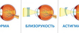
Hypermetropia (hyperopia). Hyperopia is also known as farsightedness. It is caused either by the small size of the eyeball or by the weak refractive power of the eye's lens system. Under such conditions, parallel light rays are not refracted sufficiently by the lens system of the eye for the focus (and therefore the image) to be on the retina. To overcome this anomaly, the ciliary muscle must contract, increasing the optical power of the eye. Consequently, a farsighted person is able to focus distant objects on the retina using the mechanism of accommodation. There is not enough accommodation power to see closer objects. With a small reserve of accommodation, a farsighted person is often unable to accommodate the eye sufficiently to focus not only close, but even distant objects. To correct farsightedness, it is necessary to increase the refractive power of the eye. To do this, convex lenses are used, which add refractive power to the power of the eye's optical system. Myopia . In myopia (or nearsightedness), parallel light rays from distant objects are focused in front of the retina, despite the fact that the ciliary muscle is completely relaxed. This happens due to the eyeball being too long, as well as due to the refractive power of the optical system of the eye being too high. There is no mechanism by which the eye can reduce the refractive power of its lens less than is possible with complete relaxation of the ciliary muscle. The process of accommodation leads to deterioration of vision. Consequently, a person with myopia cannot focus distant objects on the retina. The image can only focus if the object is close enough to the eye. Therefore, a person with myopia has limited range of clear vision. It is known that rays passing through a concave lens are refracted. If the refractive power of the eye is too great, as in myopia, it can sometimes be neutralized by a concave lens. Using laser technology, it is also possible to correct excessive corneal convexity. Astigmatism . In an astigmatic eye, the refractive surface of the cornea is not spherical, but ellipsoidal. This occurs due to too much curvature of the cornea in one of its planes. As a result, light rays passing through the cornea in one plane are not refracted as much as rays passing through it in another plane. They do not gather in a common focus. Astigmatism cannot be compensated by the eye using accommodation, but it can be corrected using a cylindrical lens that will correct an error in one of the planes.
Correction of optical anomalies with contact lenses
Recently, plastic contact lenses have been used to correct various vision anomalies. They are placed against the front surface of the cornea and are secured by a thin layer of tears that fills the space between the contact lens and the cornea. Hard contact lenses are made of hard plastic. Their dimensions are 1mm in thickness and 1cm in diameter. There are also soft contact lenses. Contact lenses replace the cornea as the outer surface of the eye and almost completely cancel out the portion of the eye's refractive power that normally occurs on the front surface of the cornea. When using contact lenses, the anterior surface of the cornea does not play a significant role in the refraction of the eye. The front surface of the contact lens begins to play the main role. This is especially important in individuals with abnormally formed corneas. Another feature of contact lenses is that, by rotating with the eye, they provide a wider area of clear vision than regular glasses. They are also more convenient to use for artists, athletes, etc.
Visual acuity
The human eye's ability to see fine details clearly is limited. The normal eye can distinguish different point light sources located at a distance of 25 arc seconds. That is, when light rays from two separate points enter the eye at an angle of more than 25 seconds between them, they are visible as two points. Beams with smaller angular separation cannot be distinguished. This means that a person with normal visual acuity can distinguish two points of light at a distance of 10 meters if they are 2 millimeters apart.
Rice. 7. Maximum visual acuity for two point light sources.
The presence of this limit is provided for by the structure of the retina. The average diameter of the receptors in the retina is almost 1.5 micrometers. A person can normally distinguish two separate dots if the distance between them in the retina is 2 micrometers. Thus, in order to distinguish between two small objects, they must excite two different cones. At least there will be 1 unexcited cone between them.
Sources:
https://www.all-fizika.com/article/index.php?id_article=1982
What zones does the retina divide into?
The retina of the eye is divided into two main areas:
- The central zone (other names: macula, macula) is the main part of the retina, on which the quality of human vision depends by 90%. Its function is to provide subject and color perception of the surrounding world. A larger number of cones are concentrated here, which are responsible for the perception of color diversity. This is why any pathological changes cause major vision problems.
- Peripheral zone. Rods are located here - receptors that provide peripheral, spatial and twilight vision. If pathology occurs in this area, then the field of vision narrows significantly, and some areas disappear.
Pathology of the retina in myopia
There are many types of diseases, as well as the causes of their occurrence. One of them is myopia.
Retinal detachment can occur in anyone, and patients with high myopia are at particular risk. With high myopia, a large eyeball provokes stretching of the retina and disruption of its nutrition. The latter causes a deterioration in metabolic processes in tissues and blood circulation, which causes hypoxia and retinal dystrophy.
There is peripheral and central chorioretinal dystrophy. In the most serious case, a “dry” or “wet” lesion of the macular region of the retina occurs. This significantly impairs vision and even leads to blindness. If the adverse changes in the retina are severe, the patient is diagnosed with myopic disease.
Myopic disease occurs quite rarely. As a rule, high myopia remains a harmless pathological change in refraction.
Astigmatism and myopia
The main determinants of refraction are the focusing ability of the cornea and lens and the length of the eyeball.
Myopia is usually denoted by a minus sign. Mild is 0 to -1.5 D, moderate is -1.5 to -6.0 D, and severe is -6.0 D or more. Clear vision is only possible at close range.
At birth, most babies are diagnosed with hypermetropia, but as they grow, by the age of 5-8 years, emmetropia also appears. This is a physiological process in which the refractive state of the eye changes in magnitude and decreases in size.
Myopia is considered a common perceptual defect in childhood. It is characterized by blurriness of objects seen side by side and usually results from abnormal lengthening of the visual axis. The prevalence of the disease has nearly doubled worldwide over the past two decades, and clinical onset times are decreasing at an alarming rate. It is well known that the younger the patient acquires the disease, the faster it progresses. In addition, there are a number of epidemiological studies that show that the disease is more common in urban areas, among highly trained professionals, educated patients, computer users, university students, and is associated with high intelligence.
The exact etiology is still unknown, and there are no generally accepted methods of prevention or treatment.
The risk is especially great when a decrease in acuity is diagnosed by more than minus six diopters.
| The higher the degree of myopia, the higher the risk of pathological complications such as macular degeneration, retinal detachment, cataracts and glaucoma. |
Many people may be familiar with the phrase: “Don’t sit in front of a computer or TV for a long time, otherwise you will damage your eyesight!” This is probably due to the fact that the most common and long-standing hypothesis about the etiology of the development of the disease was that excessive proximity to books or television leads to overstrain of structures. Accommodative fatigue occurs when the ciliary muscle weakens due to overuse. This causes the clear lens to lose its focusing ability, causing hyperopic defocus when looking at pictures and text.
It was also found that children with two myopic parents had the most negative spherical equivalent refraction and the longest axial length.
Major retinal diseases
Macular degeneration (central retinal degeneration) is one of the most serious eye diseases, leading to complete loss of central vision. With this disease, a gray transparent spot first appears in front of the eye, which darkens over time and loses transparency, as a result of which the person cannot see the objects he is looking at. One of the reasons for the development of macular degeneration is high myopia.
Peripheral dystrophy is diagnosed during an examination of the fundus with special lenses. According to statistics, this disease in the vast majority of cases (40%) develops in people with myopia. Its main danger lies in the absence of symptoms at the development stage, so it is usually diagnosed by chance. However, if the patient does not regularly visit the ophthalmologist’s office, and pathological processes begin to develop in the periphery of the retina, then due to retinal detachment, visual acuity will occur unexpectedly for the patient. Often people come to the ophthalmologist only when the retinal detachment has reached the central zone and there is an accompanying feeling of a veil before the eyes. This is considered an advanced stage, and in this case only surgery will help, which cannot guarantee complete restoration of visual functions.
Dystrophic changes in the retina can be detected by an ophthalmologist through a thorough examination of the fundus. As a rule, if there is a suspicion of problems with the retina, the specialist suggests conducting an in-depth examination of the peripheral area. The laser surgeon is responsible for the latter. There are two options for the development of events: either the specialist will recommend dynamic observation, or will carry out restrictive laser coagulation, which allows you to limit the affected areas of the retina. This procedure allows you to avoid complex and dangerous operations and permanent loss of vision.
The main reasons for improper focusing of light in myopia
The development of myopia often begins due to the fact that the refractive power of the optical system does not correspond to the length of the axis of the eye. With such an ophthalmological pathology, the focusing of bright flashes of stripes falling on the organ of vision occurs not on the surface of the retina that perceives light, as happens in people with normal vision, but in front of it.
The shape of the surface of the cornea can hardly be called ideally spherical, and its thickness is not uniform. As a result, visual images can become defocus not only in the center of the inner retina of the eyeball, which perceives glare, but also at its periphery.
Speaking about the main reasons for focusing light rays in front of the retina, we should highlight:
- An overly elongated eyeball. Because the distance from the top of the cornea to the membrane is too large, axial myopia occurs. This prevents glare from hitting the central part of the shell, causing it to focus in front of it.
- Irregular shape of the cornea. In this situation, strong refraction of light rays occurs, leading to their focusing not where it should be to ensure normal visibility. The rays reach the surface of the retina already defocused, as a result of which the sharpness and clarity of the image deteriorates.
I would like to note that ophthalmological studies are ongoing today, which will allow us to determine the optimal peripheral myopic defocus. Thanks to this, in the future it will be possible to develop the most accurate, and most importantly, individual scheme for the correction and treatment of myopia for each patient.
You may be interested in: Top 6 devices for treating myopia
What is retinal detachment
One of the most serious eye diseases is retinal detachment. Usually, retinal detachment takes a person by surprise, as no special symptoms arise.
If it is not recognized in time, you may be left without vision. The disease develops rapidly. However, it is practically not curable without surgical intervention.
There are many methods of surgical removal of retinal detachment. They differ in the mechanism of action on the affected area, some of them combine well with each other.
Drops, ointments and tablets will not help with this disease. In this way, the patient only delays time, which is why the detachment spreads throughout the entire eye. The sooner you contact an ophthalmologist, the greater the results of treatment and the better the quality of vision after surgery. The goal of surgical procedures is to block the retinal tear.
Retinal detachment is the process of separation of the rods and cones (neuroepithelium) from the pigment epithelium. During the process, the nutrition of the outer layers of the retina is disrupted, as a result of which the person rapidly loses vision. According to its type, retinal detachment can be dystrophic, traumatic and secondary. The latter type is not an independent clinical form, it is just a consequence of certain diseases.
Dystrophic retinal detachment (primary) involves the occurrence of a tear or breaks in the retina. It usually occurs close to the center of the eye, where thinning and degeneration most often occur. There is no immunity from retinal detachment, but there are preventive measures. These include laser coagulation of areas of the retina that may cause detachment. Unfortunately, not all patients consider these procedures necessary. Especially during a period when no alarming symptoms arise.
What is myopia in simple words?
Myopia is the same as myopia: a visual impairment in which a person sees well up close, but blurry at a distance.
IMPORTANT! According to statistics, such a pathology is present in every third inhabitant of the planet, regardless of gender and age.
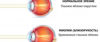
If with normal vision the image is projected on the retina, then with myopia it is projected in front of it.
Causes
From a physiological point of view, myopia develops due to changes in the shape of the eyes, disruption of the lens ligaments or its displacement, due to the congenital anatomical features of the organ.
Factors that provoke the development of myopia are:
- hereditary predisposition;
- inflammatory and infectious eye diseases;
- constant increased strain on the eyes (working at a computer, reading in the dark);
- poor eye hygiene (improper use of contact lenses, use of low-quality cosmetics);
- violation of intraocular circulation;
- incorrect selection of vision correction products (glasses or contact lenses);
- acute deficiency of vitamins and minerals.
Symptoms
Myopia is characterized by a gradual decrease in vision, so a person does not immediately notice that he has begun to see worse, but a deterioration in visual acuity is the main sign of myopia.
To see something in the distance, a person has to strain his eyes, squint, which can cause headaches, increased fatigue, dryness and discomfort of the eyes.
How to prevent retinal detachment due to myopia: laser coagulation
Timely preventive measures significantly reduce the risk of retinal detachment due to myopia or any other disease.
As mentioned above, the main preventive measure is laser coagulation. This is a fairly simple and not painful procedure, but if the eyes are very sensitive, pain may still occur. In this case, factors such as:
- volume of coagulation and zones of its implementation;
- model of laser equipment;
- features of the anatomy of the palpebral fissure;
- correct choice of contact lens;
- surgeon experience.
If the patient knows that the pain threshold is low or that he is uncomfortable during the procedure, it is better to warn the doctor about this in advance. The pain can be reduced with the help of special medications and then the procedure will take place without any difficulties.
How is retinal detachment diagnosed?
Diagnosis of retinal detachment cannot be made without a complete ophthalmological examination of the eyes. It includes checking vision and intraocular pressure, examining the fundus using contact and non-contact methods.
Complementing the diagnostics:
- perimetry - determination of visual fields using special equipment;
- Ultrasound scanning: helps to identify retinal detachment through blind areas of the eye, allows you to determine the height of the shell, relief, contents under it, thickness of the shells and other significant parameters;
- electrophysiology - “cardiogram of the eye”, which allows you to determine the degree of damage to the retina;
- optical coherence tomography – its result is a two- or three-dimensional image;
- X-ray computed tomography allows you to visualize the structures of the eye down to the smallest detail;
- MRI allows you to assess the degree of damage to the eye and create a 3D image of it.
How to understand that retinal detachment has occurred
If you compare the human eye to a camera, the main sign of retinal detachment is a “scratch” on the edge of the photographic image. The picture is still visible, but its quality is seriously compromised. But this is not the main problem: fluid leaks through the retinal tear, which flows under the retina and separates it from the choroid. If we continue the analogy with photographs, it looks as if near the scratch the emulsion swells and separates from the substrate. In this case, a person develops a gray veil at the edge of his field of vision. The speed at which this veil spreads depends on the location of the rupture. Sometimes a few hours are enough for the field of vision to be completely covered by the “curtain”; in other cases, the spread of the veil to the central area of the visual field occurs over months.
Other symptoms of retinal detachment due to myopia
Another symptom of retinal detachment is “morning improvement” - a sign in which a person, after a long period of inactivity, becomes able to see well: the veil fades or decreases. By lunchtime the patient's condition worsens, and by evening everything reaches its peak.
If the retinal tear is in the upper parts of the eye, then due to the descending fluid, retinal detachment occurs very quickly in myopia. If the gap is at the bottom, then the detachment slowly rises and occurs less rapidly, but the scars will be more pronounced, since there will be much more time for their occurrence.
To prevent retinal detachment due to myopia and other diseases of the visual organs, you need to regularly visit an ophthalmologist and carefully follow all his recommendations.
Farsightedness and myopia: features of the diseases
The human eye is designed in such a way that during visual function, light rays pass through the cornea and lens, are refracted and focused precisely on the retina to further form a visual image. Depending on whether the patient is farsighted or nearsighted, the focus shifts and the image does not fall on the retina.
With farsightedness, light rays are refracted in such a way that they fall behind the surface of the retina. As a result, the patient sees nearby objects blurred.
With myopia, light rays are refracted and do not reach the retina, so the patient has difficulty seeing into the distance.
If vision correction is required, myopia and farsightedness are most often eliminated using modern laser techniques, which allow the pathology to be quickly and accurately eliminated. For severe hypermetropia and myopia, phakic or intraocular lenses are used. The correction method is selected individually, so each patient must undergo a comprehensive examination.