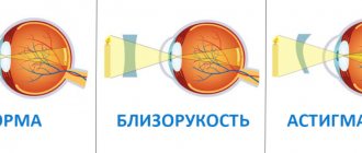
Myopic astigmatism is a combination of astigmatism and myopia. Astigmatism is a vision defect in which the refractive power of the eyes is impaired due to the pathological sphericity of the lens or cornea.
Myopia (myopia) is a refractive disorder in which the image is focused in front of the retina rather than on it. In the first case, the image of objects is distorted, everything looks blurry, and in the second, the patient sees nearby objects well and poorly those that are far away.
Symptoms and signs of astigmatism
- The main symptom of visual impairment and astigmatism is no exception - visual impairment, manifested in the form of deformation of objects. Objects become blurry and unclear in horizontal or vertical directions, but not completely, but in parts.
- With prolonged strain on the eye and after it, headaches often occur, because the brain is forced to adapt to the deformed image and, to some extent, correct it or not notice it. Overstrain of the central nervous system responds with the development of neurological symptoms.
- Patients often complain of such a painful symptom as photophobia, when bright light causes pain in the eyes, because with astigmatism, the light receptors on the retina are unevenly irritated.
- The eyes get tired faster, especially when reading.
- Sometimes “night blindness” occurs—deterioration of vision in low light conditions; this symptom occurs at dusk.
- People around notice that a person, trying to better see the surrounding objects, squints his eyes, tilts his head, adjusting the angle of view.
When dissatisfied with their own vision, each patient wants not only to undergo an examination, but to receive an explanation of what is happening.
Even in the absence of pathology, it is necessary to understand why the functional capabilities of the visual organs are not satisfied. At the Medicine Clinic 24/7 they will definitely tell you why this happens and give a recipe on how to avoid unpleasant sensations and get joy from seeing the world. Sign up for an examination: +7 (495) 230-00-01 The causes of astigmatism in adults and children are different, so in children the congenital variant of the disease, which is also called functional, prevails. As a rule, this pathology accompanies prematurity, when the eyes simply have not matured in the mother's womb. The causes of pathology in adults are diseases and injuries of the eye.
Diagnostics
To treat astigmatism, it is necessary to accurately establish the diagnosis and form of the disease.
To do this, you need to contact an ophthalmologist, who will establish a diagnosis using instrumental methods. It is impossible to make a correct diagnosis on your own.
The doctor conducts an examination, anamnesis and several instrumental studies:
- determine visual acuity;
- Using the skiascopy method, the stage of myopia is determined;
- perform ultrasound diagnostics of the eye;
- measure intraocular pressure;
- study the fundus of the eye.
Natural cause of astigmatism
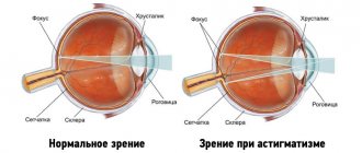
The human eye is a complex optical system whose main function is to focus images on the retina to transmit visual information to the brain. Moreover, on the retina the object is reflected upside down, but in the brain as it really is.
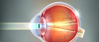
To ideally convey the image of an object, all layers of the eye must be of a uniform spherical shape; if a bulge or depression forms in one place of the optical system, then the ideal shape on the retina is no longer obtained. A person sees an object consisting of both clear fragments and blurred ones. This condition is called astigmatism, that is, the lack of focusing of an object visible in space.
There is nothing ideal in nature at all, and in most people the cornea cannot have a clearly defined spherical shape. Therefore, minor astigmatism is normally allowed, which does not affect visual acuity. This option is not a reason for correction; it does not lead to vision deterioration. When the distortion of the optical elements of the eye becomes the source of poor vision, then correction is required.
How to make a sliding screen
The sliding structure can also be made of plastic. For this you will need a plastic sheet. The guides along which the doors will move must be made of a special aluminum profile. First, a metal frame is made, as described above, then guides are fixed at the top and bottom along the entire length of the profile.
The required length and width of the doors are measured, their dimensions are transferred to the sheet and cut out. The doors are fixed on the sides of the screen in the first groove of the profile. A central fixed part is installed in the middle groove.
You can stick a plain or colored film on plastic doors in accordance with the overall interior of the bathtub
If you want the doors to move along the entire length of the bathtub, then two plastic pieces are cut out. Their size is calculated in such a way that when closed they overlap each other. The first door is installed in one groove, and the second in the other. Finally, furniture handles are placed on the doors.
Causes of acquired corneal astigmatism
The reason for the occurrence of astigmatism is different for each patient, but in most cases a hereditary predisposition is suspected, although such a concept as a family disease has not yet been officially introduced. The leading cause of the development of acquired vision defect is deformation of the cornea as a result of its healing after injury or disease. The injury can be not only spontaneous, but also surgical, for example, when removing a cloudy lens - a cataract, an incision is made in the cornea, which can heal, although not visible to the naked eye, but for such a thin matter as a rough scar.
With age, the cause of the pathology is uneven thinning of the cornea due to dystrophic processes, when it becomes like an uneven cone; this eye disease is called keratoconus.
Correction
To eliminate articulation defects, gymnastics, breathing exercises, and logorhythmics are used. The type of violation must be taken into account. It takes more time to correct dental and lateral sigmatism than to normalize phonation in interdental and nasal sigmatism.
Articulation gymnastics
The following articulation exercises are recommended:
- Fence
We make a wide smile, teeth are visible. We fix the position of the lips for a few seconds. Then we repeat the exercise 2-3 times.
- Mustache
The child holds flat, light objects (strips of paper, tubes, pen) with his lips.
- Inflating the balloons
The cheeks are involved. The child inflates and deflates his cheeks alternately.
- Rent a pencil
The kid blows on a pencil that lies on the table.
- Imitation of chewing
- Singing syllables and sounds.
Massage
- Spot
We vibrate with our fingers under the chin, under the tragus of the ears. Open and close your mouth alternately during the massage.
- Soft palate massage
We knead the palate with our fingers until we get a pharyngeal effect.
Exercises
Tongue strengthening exercises:
- Pancakes
Licking the mouth in wide, circular movements.
- Rain
Rhythmic slaps of the tongue on the lips.
- Biting the tip, back, sides of the tongue.
Tongue spreading exercises:
- Baking pies
Mechanical assistance to the tongue will be needed. Take the tongue with two fingers and fold it into a pie.
- Sleds
- Railway
Staging sounds
During speech therapy classes on sound production [C], the imitation method, working with a mirror, and the mechanical method are used.
Causes of lens astigmatism
Science cannot name the exact reasons, but a connection has been found between astigmatism and some chronic diseases. Thus, diabetes mellitus leads to clouding of the lens due to disturbances in the microvascular bed, and the frequency of astigmatism also increases.
Prolonged high pressure is also unfavorable for the transparency of the lens and also becomes a reason for the formation of this pathology, therefore, with uncontrolled pressure, there is a high probability of developing eye disease.
The preventive ophthalmological examination carried out at the Medicine 24/7 Clinic is based on world experience, which states that many paths lead to the disease, and all of them can and should be detected so that vision from a happy gift of nature does not become a big problem. Sign up for a consultation
Testing visual acuity using special tables helps make a diagnosis, which is beautifully called visometry, but literally means simply measuring vision.
How the membranes of the eye refract light is determined by a refractometer, and examination in a dark room with special lenses is used - skiascopy or shadow test. The cause of the pathology may well be uneven transparency of the cornea or lens.
We will call you back
leave your phone number
Today, using computer methods, it is possible to map the surface of the cornea, like a geographical map, with all the bulges, depressions and opacities; this is keratotopography.
Ophthalmometry in combination with ultrasound gives the most complete picture of changes in the parts of the eye and the shape of the eyeball, which is always present with astigmatism.
Biomicroscopy allows you to identify inflammatory eye diseases that cause symptoms of astigmatism.
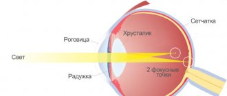
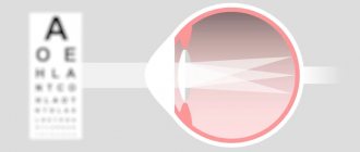
Symptoms of astigmatism in adults are similar to other eye diseases, because with farsightedness the focus of the image is located in front of the surface of the retina, with myopia - behind the retina, with astigmatism the image is focused in pieces to the retina and behind it.
Any eye pathology is detected exclusively by an ophthalmologist; it cannot be detected by eye; the Medicine 24/7 Clinic has installed the most modern equipment that will not allow astigmatism to be missed.
Astigmatism is a disturbance in the clarity of vision as a result of uneven changes in the structures of the eye, when vision is distorted not along the entire curvature, as with myopia or farsightedness, but locally. Astigmatism spoils vision, as if in pieces: here it’s good, in this place it’s bad. For example, the vertical lines are clearly and clearly visible in the drawing, but the horizontal ones are blurred.
Classification
Ophthalmologists classify astigmatism according to various parameters. For example, the disease can be lens, cornea, depending on the location of the pathological area.
According to the method of refraction in the main meridians, there are 3 types of disease:
- Direct (genetic form of the disease). Deviations appear along vertical meridians;
- With oblique axes (usually an acquired disease);
- Reverse astigmatism. Increased refraction occurs horizontally.
There are physiological and pathological astigmatism. The first form of the disease is characterized by dysfunction in the area of one diopter; in the second, the patient experiences discomfort all the time, objects do not look sharp, with blurred edges.
Astigmatism can be either independent or accompanied by farsightedness or myopia. It is divided into the following types:
- Simple, when one meridian has normal vision, and the second has farsightedness or myopia.
- Complex, with deviations in 2 meridians at the same time and is accompanied by farsightedness or myopia.
- Mixed - on one meridian there is farsightedness, and on the second there is nearsightedness.
Based on how people with astigmatism see, there are 5 types of dysfunction:
- Simple myopic (M). A combination of myopia in the 1st meridian with correct vision in the other.
- Simple hypermetropic (H). In one meridian there is farsightedness, in the other there is clear vision.
- Complex hypermetropic (CH). Farsightedness on the main meridians of the eyeball.
- Complex myopic (CM). Accompanied by myopia in the horizontal and vertical main meridians.
- Mixed (NM and MN). It is distinguished by farsightedness in one meridian and myopia in the other.
Types of astigmatism
Light passes to the retina through the cornea, lens and then the vitreous body, which has no blood vessels and is a dense jelly riddled with fibers. The vitreous body is not involved in the formation of the irregular image. An irregular cornea and a cloudy or deformed lens can spoil vision. In the preparation of astigmatism, the leading role is given to the cornea; it is exposed much more than the lens, so corneal astigmatism prevails over the lens.
You can be born with such an eye pathology; it will be a congenital or functional variant, which is detected in early childhood and corrected depending on its severity with glasses. They prefer to do operations after reaching adulthood.
Acquired astigmatism is most often caused by rough scars on the cornea as a result of injury or severe inflammation of the eye, often of herpetic etiology. With age, degenerative changes begin in the eye, as elsewhere in the body - keratoconus, which also leads to this vision pathology. Anomalies in the development of teeth and the jaw system are also included in the spectrum of causes leading to visual impairment.
Prevention
If the change is at the level of a weak degree of astigmatism (up to 1 D), then prevention plays a big role in preventing the development of such a disease.
One of the methods involves the use of vitamin complexes. When the body has sufficient amounts of vitamins, macro- and microelements, the load on the cornea of the eye is much less. In addition, vitamins can generally improve human health.
You should not neglect the recommendations for doing eye exercises, especially if your work involves constant contact with a computer monitor. Exercises must be performed at least 2 times a day. Gymnastics will help prevent not only the development of astigmatism, but also myopia and farsightedness. These are the simplest rotations of the eyes, lowering and raising them. And of course, stick to a healthy lifestyle, be active and take care of yourself.
Treatment of astigmatism without surgery
There are three main methods for correcting eye astigmatism:
- The most common way of not so much treating as correcting a vision defect is correction using special glasses. They select so-called “complex glasses”; they are not easy to wear, because the glasses cannot be perfectly adjusted to the individual deformation of the eye shape. For the most acceptable result, it is better to select them in several stages. This treatment simultaneously prevents the progression of astigmatism.
- Contact lenses can be tailored to accommodate changes in corneal curvature and defects. It is more convenient and aesthetically pleasing, and if you follow the instructions for wearing lenses and hygiene, it is quite safe. However, increasingly, clinical studies note among the adverse events when using contact lenses, scar changes in the cornea, which themselves can cause astigmatism.
- Surgical correction. Surgical treatment is quite radical and consists of applying laser incisions to the cornea, which help reduce its tension and level the surface. During the operation, the degree of refraction of the rays is corrected; it reaches the retina as close as possible to normal. And laser resurfacing of the deep layers of the cornea increases its transparency.
Any eye pathology needs to be treated and it is better to do this earlier, when vision is not yet very bad.
The correct diagnosis is half the battle, but in ophthalmology only adequate vision correction helps to get rid of the feeling of personal inferiority and inferiority. At the Medicine 24/7 Clinic we will do everything that can restore vision; if this is not possible completely, then we will offer the optimal correction
Classification
Astigmatism can be correct or incorrect, it depends on whether the main meridians in the structure of the eye are perpendicular or not.
The correct type in its orientation can be direct, reverse and with oblique axes. With direct astigmatism, the refraction of the beam occurs along a vertical plane. It is physiological and is compensated by convergence, has diopters from 0.5 to 0.75, and this does not affect vision.
Refraction of the beam with reverse astigmatism occurs along the horizontal plane; this type is compensated by an additional mechanism of accommodation.
In ophthalmology, astigmatism is distinguished by its structure:
- lenticular;
- corneal;
- general;
- the other is internal.
This depends on which part of the eye has a changed shape; the general one combines several types, for example, the corneal and lens.
According to the type of changes in refraction, astigmatism can be simple and complex, simple and complex myopic, mixed.
Myopic astigmatism is a combination of myopia and astigmatism. To correct such vision, an ophthalmologist selects special glasses or contact lenses, focusing on diopters and an angle that corresponds to the curvature of the surface of the eye.
With simple astigmatism, changes in refraction develop in one of the meridians. With a complex type, the image is formed behind the retina (farsightedness).
In simple myopic astigmatism, the normal focus is located on one meridian, and on the other meridian it is in front of the retina.
Complex myopic is characterized by different degrees of refractive error, which predominate on different meridians.
With mixed astigmatism, two types of refractive error are combined, the image in the eye is focused twice: in front of the retina and behind it, the person suffers from both myopia and farsightedness.
Important! According to the degree of severity, astigmatism can be weak, medium and high.
A weak degree is characterized by refractive index within 3 diopters. This degree lends itself well to correction; it is the most common. In addition, with the help of hard contact lenses, you can get rid of astigmatism up to 1.5 diopters.
The average degree is a difference in refraction of 3 to 6 diopters, it can be corrected through surgery or wearing contact lenses.
More than 6 diopters is already considered a high or severe degree; in its presence, photophobia occurs, objects are very blurred, and surgical intervention is required.

