What is strabismus?
Astigmatism is an eye disease.
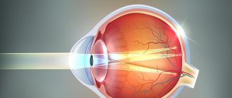
in which the structures responsible for refraction and focusing of light rays (lens or cornea) are affected (deformed). As a result of this, a person loses the ability to clearly see objects, which over time can cause the development of strabismus and other serious complications. In order to understand the essence of this pathology and the associated mechanisms of visual impairment. certain knowledge is required from the field of ophthalmology (the science that studies the organ of vision). The human eye is a complex organ, the main sensitive element of which is the retina. The retina of the eye is located on the back wall of the eyeball and is a huge number of neurons (nerve cells) that have the ability to capture light particles ( photons) and convert them into nerve impulses, which are then transmitted to certain parts of the brain and are perceived by humans as images.
However, before reaching the retina, light waves must pass through the refractive system of the eye, causing them to be focused directly into the center of the retina, which contains the maximum number of sensory neurons.
The presence of a refractive system allows you to create a clearer image of surrounding objects located at different distances from the eyes (this phenomenon is called accommodation).
- The cornea is the most convex part of the front surface of the eye, resembling half a ball in shape.
- The lens is a transparent elastic formation, shaped like a biconvex lens and located directly opposite the pupil.
- The vitreous body is a transparent substance that fills the space between the lens and the retina of the eye.
- Aqueous humor is a small amount of liquid located in the chambers of the eye (in front and behind the pupil).
The lens and cornea are of greatest importance in the refractive system of the eye, while the refractive ability of the vitreous body and aqueous humor is less pronounced. It is also worth noting that the refractive power of the cornea is relatively constant and is about 40 diopters (a diopter is a unit of measurement for the refractive power of a lens).
At the same time, the refractive power of the lens can vary from 19 to 33 diopters (depending on the distance from the eye the object on which the person focuses vision is located).
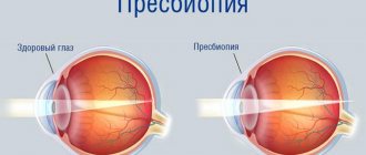
If a person looks at a nearby object, the muscles and ligaments that fix the lens tighten, as a result of which its refractive power increases. If a person looks into the distance, the above-mentioned structures relax, the lens flattens and its refractive power decreases.
Under normal, physiological conditions, all surfaces of the lens and cornea are ideally even and smooth. It is thanks to this that all light rays passing through each specific point of these structures are focused directly on the retina.
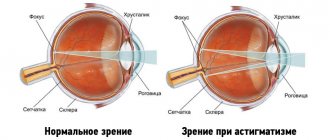
The essence of astigmatism is that with this pathology, the surfaces of the refractive structures of the eye are curved, that is, in some places dimples or bulges appear on them. As a result of this, certain light waves, after passing through them, will not be located in the central zone of the retina (as is normal), but in front of or behind it.
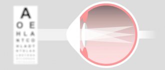
As a result, a person will not be able to focus his vision on any point, and images of surrounding structures will be unclear and blurry.
As mentioned earlier, the main link in the development of astigmatism is damage to the lens or cornea. The vitreous body and aqueous humor have little refractive power, so damage to them (which is relatively rare) does not lead to the development of astigmatism.
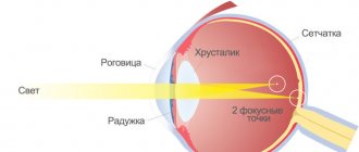
During normal operation of the optical system of the eye, the reflection of the object in question is focused at one point on the surface of the retina. With developing astigmatism, due to the presence of curvature of the cornea or lens, several foci and meridians are formed along which the light flux moves.
There are vertical and horizontal meridians. Astigmatism, if it is more pronounced on the vertical meridian, is called direct.
If the changes mostly affect the horizontal meridian, then this type is called reverse astigmatism.
The disease, which develops in combination with myopia, can be of two types - corneal and lenticular. The degree of development of disorders is established based on a comparison of refractive indices between two meridians.
There are two types of myopic astigmatism - simple and complex.
Simple myopic astigmatism is characterized by normal focus on one meridian and myopic focus on the other. The second focus is located in front of the retina, not on it. Complex myopic astigmatism is characterized by the presence of myopic disorders on both meridians.
In this case, both focuses are located in front of the retina and at different distances from it. Complex direct myopic astigmatism is characterized by the fact that on the vertical meridian of the eye the refraction of rays is stronger than on the horizontal meridian.
Unlike nearsightedness and farsightedness, astigmatism is characterized by a partially or completely blurred image when focusing on an object. The problem is the irregular shape of the eye's cornea or lens. Light rays are distorted with astigmatism and have several points on the retina, when with healthy vision there is only one focusing point on the retina.
With corneal astigmatism, a person sees a partially blurred image. This is all due to the high refractive power of the curved cornea of the eye.
With lenticular astigmatism, vision impairment is caused by an irregularly shaped lens, which causes the image to be poorly focused on the retina.
With astigmatism, as a rule, a person sees a blurry, unclear, sometimes ambiguous image. Usually the disease is partly associated with nearsightedness or farsightedness.
Astigmatism is a disease in which one of the lenses of the eye - the cornea - refracts light incorrectly, the focus on the retina is scattered, and the child's brain receives an unclear, blurry image.
The cornea is the front clear layer of the eye and is the strongest lens in the eye. Normally, the cornea should have an ideal spherical shape and also have the same refractive power in any area.
With astigmatism, the cornea distorts due to its “non-ideal” shape.
Congenital astigmatism is inherited and does not progress throughout life. To be more precise, the child inherits the shape of the cornea from the parents; just like, for example, the shape of the nose or the color of the hair.
Manifestations of congenital astigmatism depend on its degree and configuration: the more distortions the cornea gives, the worse the vision. Conversely, vision can be 100%, but with slight double vision in the form of “shadows” near letters or numbers.
Strabismus is a physiological deviation in the position of the eyeballs.
Most often it happens to both eyes at once. There is a distortion of the shape of one eye when looking straight ahead.
During a deviation of this kind, the binocular image does not merge in the cortical regions. In this regard, the human nervous system, in order to protect the brain from splitting the image visible to the eyes, excludes one of the parts of the image. Therefore, the squinting eye turns out to be blind during some periods of work.
Classification
There are many classifications of astigmatism. They are classified according to various parameters (aspects) of the disease.
According to the course of the pathology, they are distinguished:
- Correct astigmatism. Innate is considered correct. Congenital is a disease that is passed on from parents to a child. This type occurs in a mild form. Amenable to optical correction (glasses, lenses).
- Irregular astigmatism. Appears as a result of errors during ophthalmological operations, injuries, inflammation, infections. Difficult to correct. It is treated with laser correction and replacement of a biological lens. Irregular shape cannot be corrected by optics.
Based on the state of vision, there are 3 types:
- Myopic - the eye is affected by so-called myopia. The lesion extends to one or both eyes. Objects located at a distance blur. Up close, a person sees clearly.
- Hyperopic - one or both eyes are farsighted. Objects located at a distance are visible clearly, without distortion.
- Mixed. The mixed type is distinguished by blurriness and hazyness of the image at any distance. Farsightedness and nearsightedness can develop both in one eye at once and in different ones. For example, in the right the patient has myopia, in the left - farsightedness.
The meridians of the eye are located perpendicular (90 degree angle) to each other. Each meridian refracts incoming rays differently. One is stronger, the other is weaker. Based on the refraction of rays by meridians, the following types are distinguished:
- Direct astigmatism. The vertical meridian refracts incoming rays more strongly. Horizontal is passive and refracts rays weaker.
- Back. The horizontal meridian refracts rays more strongly, the vertical - less.
- With oblique meridians. The meridians are not located perpendicular to each other, as in the 2 types above, but at an offset angle.
According to the damage to the eye organ, they are distinguished:
- Lenticular. Occurs when the lens is irregularly shaped, subluxated, or damaged.
- Corneal - when the shape of the cornea changes, as a result of improper ophthalmological intervention, mechanical damage, inflammation and infections.
There are 3 degrees of development of pathology:
- weak – up to 3 diopters;
- average – 3-6 diopters;
- high – more than 6 diopters.
The higher the degree of development, the less a person sees a clear image.
Pathology with a degree below 0.4 diopters does not need adjustment. Astigmatism of this degree does not need to be corrected, since it does not cause discomfort to a person. In some cases, rapid fatigue and general discomfort are possible, then the person is prescribed glasses.
Visual acuity with astigmatism depends on the form, type of pathology, causes of occurrence, “neglect” and complexity of the course.
How do cataracts form and manifest?
Cataract is a clouding of the eye lens that causes visual disturbances of varying degrees. This pathology belongs to the category of the most common eye diseases. And it tends to progress gradually throughout a person’s life.
The main factor contributing to the development of cataracts is a person’s age. More than 50% of the entire human population of the planet over the age of 65 has this visual defect. In addition to the age factor, the causes of cataract formation are also often endocrine diseases, the use of steroid hormones and negative habits (in particular, smoking).
Depending on the degree of change in the transparency of the lens, this disease manifests itself:
- double visibility, when the unaffected organ of vision is covered;
- blurred visual perception that cannot be eliminated by optical instruments;
- the appearance of signs of myopia;
- the appearance of light glare during visual viewing at night;
- painful sensitivity of the eyes to light at night;
- decreased quality of color vision.
A visual defect such as a cataract must be corrected. If its development is not stopped in a timely manner, it can ultimately lead to complete loss of vision.
Causes and symptoms of strabismus
The most common cause of myopic astigmatism is the presence of congenital deformation of the cornea.
Deformation of the cornea, as a rule, is a disorder that is inherited. This is precisely the reason why regular examination of the visual organs of children whose parents have been diagnosed with this visual impairment is required.
Myopic astigmatism can also develop as an acquired disorder, which appears as a result of various injuries to the cornea of the eye or a person undergoing diseases or eye surgeries.
The result of the operations is the appearance of a surgical scar on the cornea, which leads to a change in the curvature of this element of the optical system of the eye. Less commonly, the cause of the disorder is an abnormal change in the shape of the lens of the eye.
- Weak degree - the pathology proceeds unnoticed, since with such a degree of development of the disorder it is difficult to identify deviations from the norm in the functioning of the visual organ. Violations with a weak degree of development of the disease can be determined by a specialist.
- The average degree of development is characterized by the appearance of significant distortions in the functioning of the visual organ, which require the use of corrective measures.
- Congenital strabismus
- Strabismus with impaired refraction
- Amblyopia
- Some other reasons
Strabismus can be recognized visually. It manifests itself as a deviation from the central axis of one or both eyes.
The disease is caused by uncoordinated work of the eye muscles. In the normal position, the eyes focus on one object, and the optic nerve, comparing two pictures, creates one three-dimensional image.
With strabismus, the focus of the gaze on the object in question is disrupted, and, as a result, the interpretation of two images into one does not occur. In this case, in order to avoid double vision, the nervous system analyzes the signal from the healthy eye and does not take into account the signal from the squinting eye.
According to statistics, one in fifty children suffers from strabismus. As a rule, this disease develops in childhood during the formation of coordinated work of the muscles of both eyes. Malfunctions in the functioning of the eye muscles appear in a child by the age of two to five years, together with visual impairments such as myopia, farsightedness, and astigmatism.
Brain diseases, head injuries, mental disorders, and eye surgeries lead to strabismus.
Also, strabismus can begin to progress after severe fright, stress, or as a result of infectious diseases: influenza, diphtheria, scarlet fever. measles
Symptoms of strabismus
There are concomitant and paralytic forms of strabismus.
With friendly, the right or left eye squints, while the degree of deviation from the central axis is approximately the same. Most often, the disease is inherited, manifests itself mainly in children and is associated with specific features of the eye structure.
In the case of paralytic strabismus, the healthy eye is squinted. In a situation where the movements of the diseased eye are complicated due to atrophy of one of the eye muscles, the second one has to do the work of both eyes, deviating to a larger angle. The causes are damage to the motor muscles of the eye or diseases of the visual-nervous tract.
There are convergent, divergent and vertical strabismus. Convergent is characterized by deviation of one of the eyes towards the nose. Often accompanied by farsightedness. Divergent is diagnosed as deviation of one of the eyes towards the temple. Accompanied by myopia. With vertical strabismus, the eye squints upward or downward.
In addition to asymmetrical eye function, the following symptoms are observed with strabismus: tilted or turned head, squinting, double vision. Moreover, adults will complain about double vision. In children, good adaptive abilities of the brain can compensate for this symptom.
Found an error in the text? Select it and a few more words, press Ctrl Enter
Treatment of strabismus
When choosing a method of treating strabismus, many factors should be taken into account: the age of the patient, the sides and degrees of strabismus, the causes of the disease. Accordingly, treatment methods can be: wearing glasses, sealing one of the glasses, exercises for the eye muscles, surgery.
Strabismus does not go away on its own, and its treatment is a long process that takes months, or even years. It is most effective to take action as soon as a problem is discovered.
The child's eye has great adaptive properties. This means that in a short period of time, the squinting eye partially or completely ceases to participate in the visual process, since the brain blocks its signal, and the healthy eye takes over all functions.
This condition is called amblyopia. If amblyopia develops, glasses with the left or right lens sealed are prescribed.
Moreover, the worse-seeing eye is left uncovered. Constant loads will serve as training for weakened eye muscles.
Another important part of the treatment is special eye exercises. They are also aimed at creating additional stress on the muscles of the unhealthy eye.
Strabismus is accompanied by other visual abnormalities, so its treatment must be approached comprehensively, combining it with the correction of astigmatism, myopia or farsightedness. For this purpose, the patient is prescribed glasses to wear constantly.
Glasses can completely restore vision. If a visible result cannot be achieved, surgical intervention is resorted to.
Most often, the operation is prescribed for children from three to six years old. For children under 14 years of age it is performed under general anesthesia, for adolescents and adults - under local anesthesia.
The operation is performed on the muscles of the eye and is aimed at restoring the balance between the muscles that rotate the eyeballs in the socket. If both eyes are operated on, then, as a rule, one muscle is strengthened and the other is weakened.
Recovery takes from a week to ten days. After surgery, it is necessary to resume a course of exercises for the eye muscles, and also regularly visit an ophthalmologist.
The ultimate goal of strabismus treatment is to achieve one hundred percent vision without glasses, symmetrical eye position and the formation of three-dimensional stereoscopic vision.
In adulthood, the disease does not have pronounced symptoms, and minor dysfunction of the visual system does not cause noticeable inconvenience.
You can recognize the disease yourself only by several indirect signs:
Congenital strabismus is called when a child was born with such a defect or he received it before the age of six months. During the uterine period of development or during childbirth, microscopic hemorrhages can occur, which lead to strabismus.
If you consult an ophthalmologist and neurologist in time, the child can be cured, and quite quickly. Sometimes parents' fears may be exaggerated, because after birth, most children's eyes visually diverge or converge, but this is absolutely not the case.
Therefore, in this case, the child does not need treatment. At the same time, if you do not pay attention to the child’s strabismus in time, it turns into pathology.
Strabismus is also associated with visual impairments such as nearsightedness, farsightedness and astigmatism. However, it must be remembered that all children at birth have farsightedness, which is associated with the age-related features of the eye design, and the eyeball in babies is not as round as in adults, but flattened.
Due to these features, doctors consider mild farsightedness to be the norm for children up to at least seven years of age.
Complications and prognosis
The main complications of visual anomaly manifest themselves as:
Amblyopia is a pathological condition that causes decreased visual acuity. This disorder, also called “lazy eye,” occurs as a result of the lack of quality treatment, long-term progression of the disease, leading to significant distortion of surrounding objects.
Strabismus in astigmatists is also associated with the prolonged course of the disease and the lack of adequate therapy. In some cases, the disorder may occur even after the underlying pathology has been eliminated.
A complication of surgical treatment may be a relapse caused by restoration of the original shape of the cornea and the return of symptoms of the disease.
A positive prognosis remains in adult patients who contacted an ophthalmologist at an early stage of the disease and underwent timely, high-quality treatment. If the problem is ignored or medical care is neglected, the pathology progresses quite quickly, leads to the development of complications, and becomes the cause of inferior social and professional activity.
Astigmatism is a serious disease
, which can negatively affect the functions of the visual system and significantly reduce the quality of life.
A frivolous attitude to the treatment of an illness can cause consequences such as strabismus, lazy eye syndrome
and even
complete blindness
.
Timely vision correction
will help not only stop the development of the disease, but also maintain eye health for many years.
How does a person with astigmatism see?
The refraction of light occurs with different strengths, as a result, some outlines of objects look sharp, others blurry. Some lines are clear, others are curved. Double vision is possible.
What happens with astigmatism?
Due to the fact that the lines converge not at one point on the retina, but at several before or after it, a blurry spot (circle of light scattering) appears, which a person sees, and through it the rest of the fuzzy world.
Why is it dangerous?
Causes severe headaches and pain in the eyes. In difficult cases it leads to strabismus, amblyopia, and blindness. Plus, a distorted picture is in itself bad and uncomfortable.
How to recognize?
Astigmatism negatively affects the ability to clearly and correctly distinguish objects both far and near. The ability to see in the twilight and examine details sharply decreases. Sometimes this persists, but is accompanied by severe blurring of peripheral vision. There are simple tests on the Internet to check for defocus, but only an ophthalmologist should confirm or refute fears with the help of in-depth diagnostics.
Types of astigmatism
Determining the type and form of astigmatism is of great importance, since the effectiveness of vision correction and treatment of the disease entirely depends on this.
From a geometric point of view, the eye is a sphere, the anterior pole of which is the cornea, and the posterior pole is the retina. Through this sphere it is possible to draw many meridians (circles) passing through its front and rear poles.
Two meridians perpendicular to each other (vertical and horizontal), having the most different refractive power, are usually called the main ones. It is the deviations (deformations) of the main meridians that determine the type of astigmatism.
Correct astigmatism
Correct astigmatism is said to occur if one of the main meridians refracts light most strongly, and the other most weakly, but both meridians have an even shape throughout their entire length.
Simple astigmatism is most often observed with a congenital disorder of the development of the cornea or lens, and they are not round (as is normal), but slightly flattened (in the form of an oval, ellipse).
In this case, rays that pass through the “longer” meridian (drawn through the longer axis of the oval) will be refracted the least strongly, while rays passing through the “short” meridian will be refracted as strongly as possible.
As mentioned earlier, during normal functioning of the refractive system of the eye, images of surrounding objects are projected directly onto the retina. In various diseases, the focusing of the image may occur not on the retina, but in front of it (in this case we are talking about myopia.
that is, about myopia) or beyond it (this condition is called hyperopia, that is, farsightedness). If the area of the cornea or lens affected by astigmatism increases the refractive power of the eye, we are talking about the myopic form of the disease, but if it decreases, it is a hypermetropic form.
Irregular astigmatism
Irregular astigmatism is characterized not only by different curvature of the main meridians, but also by different refractive power in different parts of the same meridian. This deformation usually develops with acquired astigmatism after injury, after surgery or after inflammation of the cornea, with keratoconus, and so on.
Physiological astigmatism
Under normal conditions, a healthy person may experience a slight difference in the refractive power of the main meridians of the cornea. Physiological is considered to be correct astigmatism, in which this difference does not exceed 0.5 diopters.
This deviation occurs in more than half of the world's population and is not a pathology, since it practically does not affect visual acuity and does not lead to the development of any complications.
First of all, astigmatism is a visual impairment. which can be restored with proper treatment and timely contact with an ophthalmologist. World statistics show that every 10th person has the initial stage of astigmatism, and at the same time a person may not even be aware of it.
Prevention
Prevention of this disease includes a careful and attentive attitude to the health of the visual organs, as well as timely diagnosis of astigmatism.
It is recommended to alternate visual stress with special eye exercises. Exercise will help relieve tension and fatigue.
You should avoid corneal injuries and promptly treat inflammatory eye diseases. To detect congenital astigmatism in children, it is necessary to undergo routine preventive examinations. If a child has been diagnosed with this disease, he will be registered with an ophthalmologist.
To prevent the development of complications of astigmatism, it is necessary to carry out its timely optical correction.
Refractive lens replacement for astigmatism - lensectomy
Lensectomy is a procedure that allows you to correct vision to almost ideal levels. During this procedure, the curved lens is removed and replaced with an intraocular lens (IOL, artificial lens). Hundreds of thousands of artificial lens implantation operations are successfully performed every year around the world.
The lensectomy procedure is usually prescribed for the following indications:
Correcting farsightedness with astigmatism is a fairly common combination in which lensectomy is used in most cases where laser correction is not possible. It is prescribed for high degree hypermetropia, as well as when the ability of the lens to accommodate has been lost.
There are also contraindications for refractive lens replacement:
- inflammatory diseases of the eyes;
- recent stroke or heart attack;
- diseases of the retina.
- An operation may be prescribed if interfering factors are eliminated after a certain period.
Postoperative period
The cataract removal procedure and toric intraocular lens implantation are performed on the same day. Visual function is restored in the operating room.
The very next day after the examination by an ophthalmologist, you are allowed to do your usual activities, provided that you follow the doctor’s recommendations and control your visual stress.
If you do not violate the ophthalmologist’s instructions, then the likelihood of complications is virtually eliminated. Dagaev Adam Huseinovich
Symptoms of strabismus
An objective symptom of any type of strabismus is the asymmetrical position of the iris and pupil in relation to the palpebral fissure.
With paralytic strabismus, the mobility of the deviated eye towards the paralyzed muscle is limited or absent. There is diplopia and dizziness, which disappear when one eye is closed, and the inability to correctly assess the location of an object.
In paralytic strabismus, the angle of primary deviation (squinting eye) is less than the angle of secondary deviation (healthy eye), i.e. when trying to fix a point with a squinting eye, the healthy eye deviates to a much larger angle.
A patient with paralytic strabismus is forced to turn or tilt his head to the side in order to compensate for visual impairment. This adaptation mechanism contributes to the passive transfer of the image of an object to the central fovea of the retina, thereby eliminating double vision and providing less than perfect binocular vision.
Forced tilting and turning of the head with paralytic strabismus should be distinguished from that with torticollis. otitis
The main manifestation of astigmatism is visual impairment, but over time, other symptoms from the central nervous system and other systems and organs may develop.
Symptoms of astigmatism may be minor and will become more severe over time. It is important to detect this disease in time and take the necessary medical measures - first of all, consult an ophthalmologist.
Methods of correction and treatment
Astigmatism is one of the common eye diseases; it is often combined with farsightedness and myopia. A healthy eye has a regular, even surface, but when abnormalities appear, the shape of the cornea changes noticeably (becomes bumpy).
With such violations, the refractive power and the integral image are distorted, and the person sees fuzzy or blurry pictures, while straight lines are distorted.
The main methods of correction and treatment of astigmatism are: laser correction, glasses, keratoplasty and contact lenses. Before using various correction methods, you need to visit an ophthalmologist who will help you make a choice and explain how to properly get rid of astigmatism. He may recommend wearing suitable glasses.
How can astigmatism be corrected?
If this visual impairment is not treated, it can eventually cause strabismus or contribute to significant visual impairment.
In childhood and elementary school age, glasses are used, middle and high school students can choose to wear glasses or toric contact lenses, and when a person turns 18, surgery can be performed to correct astigmatism. The following methods are used for this:
- laser correction (for corneal astigmatism);
- lensectomy (refractive replacement of the lens with an artificial one, recommended for high degrees of lens astigmatism);
- keratotomy (used for mixed or myopic astigmatism);
- thermokeratocoagulation (cauterization of the peripheral area of the cornea with a metal needle, used for farsightedness mixed with astigmatism);
- coagulation with a laser beam.
- Each of the described methods has certain advantages and indications, and which one to use for a particular patient is decided by the ophthalmologist after a thorough diagnosis and depending on the type of disorder in the patient. We will talk about lensectomy - one of the most effective ways to correct refraction for astigmatism.
