It happens that a person begins to see poorly in the distance, goes to an optician or clinic, finds out that he has myopia or astigmatism (or maybe both at the same time), puts on prescribed glasses or lenses, and uses them for some time. Meanwhile, his vision continues to deteriorate. The day comes when a person goes to an ophthalmologist in a specialized clinic, sometimes even with the goal of laser vision correction, and then an unpleasant surprise awaits him: he finds out that he has keratoconus. And it’s good if this is a latent or early stage of the disease - in this case there is a chance to save vision and your own cornea. If this is a developed process, we will talk about surgical treatment. In recent years, there seems to be an increase in patients with keratoconus. But I think this is not because someone began to get sick more often, but because diagnostic capabilities have improved, and now the conical cornea can be caught in the initial stages.
In general, keratoconus is a genetic pathology of the cornea. As the patient gets older, the cornea thins in the center and stretches due to pressure from inside the eye. The outer capsule of the eye also loses its elasticity. The first manifestation is irregular astigmatism. The patient begins to change glasses frequently because the axis and degree of astigmatism changes quite quickly.
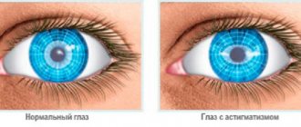
Where does astigmatism come from? The fact is that the protruding cornea changes the properties of the “lens system” of the eye, and the new “front lens” does not correspond to the projection of the pupil. The cornea with astigmatism is uneven, but regularly uneven, symmetrically uneven. A characteristic “butterfly” is visible on a diagnostic scan.
History of the discovery of the disease
The first mention of keratoconus dates back to 1748 in the writings of the German physician Burchard Mauchart. John Nottingham described this phenomenon in detail and identified it as a separate group in 1854. A few years later, British surgeon William Bowen performed the first corneal surgery for keratoconus. The surgical intervention was performed on a young woman, which resulted in a very good result, namely improved vision in an 18-year-old patient. At that time, German ophthalmologist Albert von Graefe developed methods for treating keratoconus. To restore the original shape of the cornea, a silver nitrate solution was used to cauterize it, then a bandage soaked in a special solution was applied to induce mitosis. The invention of contact lenses in 1888 did not ignore this disease. A special glass scleral shell was used to compress the surface of the cornea.
How to improve vision with keratoconus?
At the initial stage of development of keratoconus, high visual acuity can be achieved with the help of glasses that correct low myopia and astigmatism.
However, with further development of keratoconus, glasses can no longer help. In this case, contact lenses are used, as well as methods such as implantation of intracorneal rings (Intacs), corneal cross-linking, or corneal transplant surgery.
Contact lenses
Contact lenses are often the best way to achieve satisfactory vision for keratoconus. Contact lenses, covering the eye, smooth out all the irregularities of the cornea and form a more regular optical surface in front of the eye.
However, contact lenses do NOT treat keratoconus . They simply improve vision, which has become worse due to keratoconus. Wearing contact lenses does NOT stop or slow the progression of keratoconus.
Various types of contact lenses are used to correct vision in keratoconus: rigid gas permeable lenses (RGP), soft ones, a combination of “rigid lens + soft” (Piggyback), hybrid lenses and scleral lenses.
Rigid gas permeable contact lenses
Rigid gas permeable lenses are the most common method of contact vision correction for keratoconus . Thanks to their “rigidity”, the lenses perfectly smooth out all the irregularities of the cornea and create a perfectly correct anterior optical surface for the eye. The optical system “eye + ZGP lens” is capable of focusing all light rays passing through the cornea exactly onto the retina of the eye.
It is the “rigidity” of this type of contact lenses that makes it possible to correct the incorrect refraction of light rays in keratoconus. This is the main advantage of ZGP lenses compared to soft lenses, which “fit” the cornea, although they smooth out its irregularities to a certain extent, but still repeat its shape and therefore cannot provide an ideal spherical shape for the anterior optical surface of the “eye + lens” system "
However, the rigidity of this type of lens is also associated with the main disadvantage of all rigid gas permeable lenses - significantly less comfort, especially at the stage of getting used to them, compared to soft contact lenses.
Advantages of rigid gas permeable lenses:
— allow you to correct myopia and astigmatism of the highest degrees accompanying keratoconus,
- this is a very “healthy” type of contact lenses, since ZGP lenses perfectly transmit oxygen to the cornea of the eye, which is so necessary for it to maintain its health,
- they are easy to put on and take off, easy to care for,
- they can be made to individual measurements, i.e. for a specific cornea shape.
Soft contact lenses
Custom-made soft contact lenses can be used in cases where discomfort prevents the patient from wearing GP lenses. However, for the reasons stated above, soft lenses often provide significantly less visual acuity than hard lenses.
Combination “hard lens + soft” (Piggyback)
This technique consists of putting a soft contact lens on the eye, on top of which is a ZGP lens. The advantage of this combination is the combination of the comfort of soft lenses with the good visual acuity of hard lenses.
Hybrid lenses
Hybrid lenses are lenses in which the central part is a rigid gas permeable lens, and the peripheral zone (the so-called “skirt”) is made of a soft hydrogel polymer. Hybrid lenses provide users with a better combination of comfort and vision quality than soft and hard lenses. However, this type of lens is difficult to manufacture. When wearing the first hybrid lenses, problems arose due to the detachment of the hydrogel “skirt” from the zone. The latest generation of hybrid lenses offered today, such as SynergEyes lenses, have successfully solved this problem.
Scleral lenses
Scleral lenses are PGP lenses that rest, at least partially, on the sclera (the white part of the front surface of the eye) to reduce pressure on the cornea with keratoconus and not traumatize it. Let us remind you that ordinary soft and hard lenses “lie” on the cornea. The mechanical impact of the lens on keratoconus is traumatic and can lead to scar formation. In order to reduce or completely eliminate lens pressure on the cornea, the diameter of scleral lenses must be larger than the diameter of the cornea. In this case, the lens will rest partially or completely on the area of the eye (sclera) that lies outside the cornea. Scleral lenses are divided into 2 categories based on diameter: semi-scleral and scleral. Semi-scleral lenses have a smaller diameter (up to 15-16 mm) compared to scleral lenses (from 16 to 25 mm). Semi-scleral lenses rest partly on the cornea and partly on the sclera (hence why they are sometimes called corneoscleral lenses).
In severe cases of keratoconus, large diameter scleral lenses are the only alternative to surgery to achieve acceptable visual acuity.
What types of contact lenses are selected in Russia for keratoconus
Soft contact lenses used to correct vision for mild keratoconus can be selected in any contact correction office. However, this type of contact lens is not effective for “true” keratoconus, which requires other methods of correction. ZhGP lenses are not selected in every contact correction office. The selection of these lenses, especially in the case of keratoconus, requires extensive experience and professionalism of the ophthalmologist. However, in many cities in Russia there are contact correction rooms where experienced specialists successfully work with ZGP lenses.
They do not work with Piggyback and hybrid lenses in Russia. Unfortunately, we have very few specialists with experience working with scleral lenses, but they still exist. For example, in some contact vision correction offices in Moscow they successfully select corneoscleral lenses, which are manufactured here in Moscow using high-precision foreign equipment.
Advantages of treating keratoconus at the MGK clinic
Thanks to diagnostic equipment, the clinic’s specialists can diagnose keratoconus at the earliest stages of the disease, which allows for easier treatment methods and minimizing costs. After a detailed diagnosis, the doctor draws up a complete picture of the state of the visual system and determines the indications for a specific treatment method, and can also predict the outcome of treatment.
If cross-linking is indicated, the procedure is performed on an outpatient basis on a one-day basis. At the same time, according to indications, laser vision correction can be performed to eliminate refractive errors (myopia and astigmatism). The installation of stromal rings is also performed on an outpatient basis, under local drip anesthesia. After the operation, patients receive detailed recommendations to speed up the rehabilitation period; the doctor also draws up a schedule of follow-up visits for preventative control examinations.
The clinic employs Professor Alexey Yurievich Slonimsky, a recognized leader in the field of end-to-end corneal transplantation for keratoconus, the author of a monograph on keratoconus. Professor Slonimsky Yu.B. - Member of the Board of the Russian Society of Ophthalmologists. Among fellow ophthalmologists, Yuri Borisovich is a recognized authority in the field of treatment of patients with severe corneal pathology.
The operations are also carried out by a surgeon of the highest category, the chief physician of the clinic, Fomenko Natalya Ivanovna, who has successfully performed more than 12 thousand operations of various categories of complexity.
The operations of cross-linking and installation of stromal rings (kera-ring) are carried out by Yuri Ivanovich Kishkin, Candidate of Medical Sciences, and other clinic specialists.
Acute keratoconus
- Treatment
- Conservative therapy
- Surgical methods
Keratoconus is a non-inflammatory, progressive, bilateral dystrophic disease of the cornea, characterized by protrusion of the cornea anteriorly, changes in optical properties and thinning in the area of its apex. As the disease progresses, in approximately 30% of cases, acute keratoconus (hydrops corneae) may occur, characterized by painful swelling of the corneal stroma due to rupture of Descemet's membrane and penetration of anterior chamber moisture into the stroma.
Acute keratoconus, as a rule, occurs suddenly and is accompanied by severe pain and severe corneal edema. The area of edema and stromal opacification can be different - from local opacification in the central or paracentral zone to total corneal edema. If left untreated, the process lasts about 3-6 weeks and ends with the formation of a rough scar.
Acute keratoconus develops during a progressive course in patients with an advanced stage of the disease. Acute keratoconus is somewhat more common in young men. Keratoectasia can be primary (keratoconus, pellucid marginal corneal degeneration, keratoglobus) and secondary (iatrogenic). Hydrops corneae can also occur with other severe keratoectasias, such as pellucid marginal corneal degeneration, keratoglobus, Therrien marginal corneal degeneration, and posterior keratoconus.
With untreated corneal hydrops, which occurs without complications, corneal edema spontaneously resolves within 2 to 5 months. Over time, the endothelial cells enlarge and gradually close the gap, restoring the integrity of Descemet's membrane. After stopping the process in acute keratoconus, some flattening of the cornea occurs as a result of scarring with the formation of opacification and local closure of the zone of rupture of Descemet's membrane. As a result of corneal flattening, patients note a slight improvement in vision and in some cases are able to use contact correction again.
The time required for complete resolution of edema can vary widely depending on the size of the tear in Descemet's membrane. An extensive cataract, especially a vascularized one, occurs extremely rarely, and perforation of the cornea, even with its threatening thinning, occurs infrequently.
Patients with acute corneal hydrops are a special and burdened group of patients. They often have systemic allergic diseases and have a habit of vigorous rubbing of the eyes (2-3%); and the progression of keratoectasia occurs at a faster pace.
In acute keratoconus, the area of the resulting corneal edema is of great importance (partial - 6 mm or less, subtotal - 7-9 mm and total - 10 mm or more).
The most common misdiagnosis for corneal hydrops is discoid herpetic keratitis with the prescription of drug treatment, which can lead to worsening of the eye condition. Acute bacterial keratitis is often diagnosed.
In the English-language ophthalmological literature, the biomicroscopic picture of a rupture of Descemet’s membrane in the outcome of acute keratoconus is designated as a “fish mouth” symptom.
Treatment
When acute keratoconus occurs, both conservative and surgical treatment methods are used.
Conservative therapy
- Frequent instillations of glucocorticoid drugs (dexamethasone 6 times a day) in combination with broad-spectrum antibiotics. It is possible to use combination drugs (Tobradex, Combinil Duo, Maxitrol, etc.) 6 times a day - 7 days, then 4 times a day for 1 month, then 3 times a day for 2 weeks, then gradual withdrawal of drugs .
- Subconjunctival injections into the sub-Tenon's space of glucocorticoids (dexamethasone) or long-acting drugs (Diprospan).
- Instillation of antihypertensive drugs - if necessary (diacarb orally).
- Instillation of non-steroidal anti-inflammatory drugs - Indocollir 4 times a day for 2 weeks.
- The use of epithelializing and keratoprotective drugs (Korneregel, Hiloparin Komod, Vit-A-Pos, etc.)
- In case of aggravated allergic background - instillation of the drug Opatanol 4 times a day for 1 month + Erius, 1 tablet. 5 days.
We would like to point out that many ophthalmologists are unreasonably afraid to prescribe steroids for acute keratoconus, for fear of causing deterioration of the cornea, which can lead to perforation, as well as an increase in IOP. The use of NSAIDs in the treatment regimen also has its own characteristics. It is known that NSAIDs in various pathologies of the cornea (and after penetrating keratoplasty) can in many cases lead to disruption of the normal course of epithelization, the occurrence of epitheliopathy and trophic changes in the cornea (or corneal graft), especially in patients who are characterized by a slowdown in reparative processes. We do not recommend using diclofenac 0.1% (Diclo-F, etc.). Its use in acute keratoconus may cause corneal perforation. According to indications (ciliary pain) from the NSAID group, we recommend using indomethacin 0.1% (Indocollir).
Surgical methods
Surgical methods include injection of autoplasma into the anterior chamber, cryoapplication method with puncture of the anterior chamber. To achieve the effect of tamponade and prevent saturation of the corneal stroma and the increase in edema, Myata et al. (2002) proposed introducing filtered air into the anterior chamber. As a result, conditions are created for faster healing of microcracks in the Descemet membrane. A number of other authors use gases such as hexafluoride - SF6, perfluoropropane - C3F8.
Percutaneous transplantation of a donor cornea, first performed for keratoconus in 1936 by Castroviejo, is a radical method of treating the disease. The problem is not only the shortage of donor material. In case of total acute keratoconus, penetrating keratoplasty is technically impossible.
The epiceratoplasty method allows you to stop the acute process, prevent relapses of the disease, and also simultaneously correct the myopia and high degree of astigmatism accompanying keratoconus. The advantages of this method are relative simplicity in technical terms, preservation of the native cornea, and the possibility of removing the graft if necessary. Due to constant compression on the recipient's cornea, the surface graft ensures comparison and fusion of the edges of the ruptured Descemet's membrane, restoring its integrity, and performs bandage functions, strengthening and thickening the cornea, which prevents relapses of acute keratoconus. The therapeutic effect of the transplant is manifested by the disappearance of corneal edema, relief of corneal syndrome, restoration of the transparency of the recipient's cornea and, accordingly, an increase in visual acuity. Unlike penetrating keratoplasty, the innervation of the native cornea is not disrupted. The operation is extraocular, which significantly reduces the risk of intra- and postoperative complications. Thanks to the suturing of negative biolenses and compression of the ectopic cornea, in the process of applying fixing sutures, a decrease in the refractive power of the cornea is achieved.
Summing the epigraft in patients with a rupture of Descemet's membrane and deep lesions of the corneal stroma, both in the form of edema and hydration, and already formed scars, leads to an almost complete restoration of the transparency of the cornea. The phenomenon we discovered refutes the established opinion that deep stromal scars and opacities do not undergo resorption, and their treatment, other than penetrating keratoplasty, is futile. Our experience has shown that this is not the case. Epikeratoplasty brings the patient's ectopic cornea to the spherical shape inherent in it by nature, and the epigraft, made from a “living” non-preserved donor cornea, is a biostimulator of reparative processes and improves the trophism of the pathologically altered cornea. All this ultimately leads to the resorption of corneal scars. It is likely that the structure and composition of the cornea affected by ectatic disease is such that, if factors contributing to ectasia are eliminated, they are capable of restoring normal characteristics, the main one being transparency.
Causes of keratoconus
As a rule, keratoconus occurs after 14-15 years, sometimes a little later. A person should be wary if myopia or astigmatism suddenly appears at a young age for no apparent reason. If this happens, he should immediately consult an ophthalmologist and undergo a thorough special examination to exclude a disease such as keratoconus.
The tendency towards the appearance of keratoconus is determined by a number of factors. Presumably it is genetics, corneal injuries, constant stress, environmental influences. Recently, there has been an increase in keratoconus diseases due to the large number of operations to correct abnormal refraction using laser technologies.
What is keratoconus
Keratoconus is an eye disease that results in progressive thinning of the cornea. It usually occurs during puberty and results in blurred vision, double vision, myopia, astigmatism, and light sensitivity. Affects both eyeballs at the same time. Keratoconus occurs infrequently, occurring in one in 2,000 cases.
This disease does not lead to complete loss of vision, but remains with the person for life.
Keratoconus is manifested by a convexity of the cornea.
The disease is manifested by a strong cone-shaped convexity of the cornea. This organ is responsible for clarity of vision, since light rays are refracted and focused on the retina. Changing the shape of the cornea leads to defocus and subsequent changes in vision. Patients with keratoconus feel discomfort when reading, working on a computer and watching TV, and scuba diving is strictly contraindicated for them.
The development of keratoconus in children begins during puberty. The reasons for this are not fully understood. It is assumed that the mechanism of pathology formation is triggered by sex hormones. The influence of environmental factors and heredity is also noted.
Keratoconus - video
Which contact lenses are suitable for vision correction for keratoconus - all options
Contact correction for the ophthalmological pathology in question is not capable of leading to complete recovery; the disease will continue to develop. However, contact lenses can improve vision - and thereby affect the patient's quality of life.
Today, the medical market offers several types of contact lenses that can be used for keratoconus.
There is no absolute leader - the choice of a particular product depends on the individual characteristics of a particular person.
Let's take a closer look at the existing types of contact lenses.
Soft
They are relevant for small and medium refractive defects; in more advanced conditions they will be useless. These optical structures are quite flexible - they take the shape of a modified cornea, fitting tightly to it - thereby leaving no space for tear fluid.
This type of lenses is used in cases where wearing rigid models is impossible due to constant discomfort in the cornea, as well as persistent pain.
According to their characteristics, soft lenses are divided into 2 groups:
- Axisymmetric. Due to the fact that the center of such models is thickened, they are used to correct disorders that are more pronounced in the lateral sections of the cornea. They are comfortable to wear, but they cannot significantly improve vision. In addition, these optical designs are not able to combat the manifestations of astigmatism.
- Toric. They have proven themselves to be excellent as a treatment for astigmatism. Often, such lenses are used in the absence of deformations on the cornea, or after surgical procedures for the treatment of keratoconus.
Rigid gas permeable contact lenses
They received a similar name due to the type of optical design, as well as the properties of the material from which they are made. Due to gas permeability, oxygen can freely flow to the cornea.
At the moment, the type of lenses in question is the main means of correcting visual abilities for keratoconus: with their help, even complex refractive defects .
Video: Hard contact lenses for keratoconus
Many experts are inclined to believe in the need to use hard lenses in the initial stages of pathology, while others insist on their use as keratoconus progresses.
Soft lenses are more gentle - patients adapt to them quickly and can wear them constantly. With hard products, things are more complicated - when wearing them, you get the feeling of having a foreign body in the eye, which makes it difficult to concentrate on everyday concerns.
The type of contact lenses under consideration, based on its parameters, is divided into 2 groups:
- Corneal . Their diameter does not exceed 10 mm, and they are the most popular in the treatment of keratoconus symptoms. In the office of almost every ophthalmologist there are 3 different sets of corneal optical glasses - you can choose the right product without much effort.
- Scleral. They can reach up to 25 mm in diameter. Based on this parameter, this type of lens can be semi-scleral or corneoscleral (13.5-16 mm) - and scleral (16-25 mm). The main point of support when wearing such models is the sclera - this makes it possible to minimize pressure on the periphery of the cornea and protect it from injury. They are often considered as an alternative to surgery in cases of very poor visual acuity. In addition, such lenses protect the eye from dust and dirt getting into it. The negative side of the design under consideration is the difficulty in accurately “fitting” to the surface of the eyeball: their selection takes a lot of time.
Rigid contact lenses can slow the progression of keratoconus, but they cannot achieve remission of the disease.
Hybrid
The creators of these products tried to combine the positive aspects of hard and soft contact lenses in one model.
The central part of the lens is rigid. With its help, complete vision correction is provided. Along the perimeter, such a lens is framed by a soft silicone hydrogel “skirt”. Thus, it is possible to improve the patient’s visual abilities without causing him much discomfort.
However, this model also has its drawbacks. The tight fit of the hydrogel polymer limits wetting of the cornea with tear fluid. This leads to its drying out and the formation of microcracks, which only aggravates the course of keratoconus.
To avoid such exacerbations, patients must strictly adhere to the wearing periods that were established by the doctor.
Double layer Piggyback lenses
If it is impossible to wear hard lenses due to the poor condition of the corneal layers, or in case of a hypersensitive reaction to gas-permeable material, patients are prescribed another type of contact correction. First, a soft lens is placed on the surface of the eye, and a hard lens is placed on top of it.
This combination makes it possible to achieve improved visual acuity without feeling discomfort when wearing the optical structure.
However, such models cannot be worn for a long time: the area under them is poorly wetted by tears and is also poorly supplied with oxygen. These phenomena can provoke a number of complications.
Symptoms
The initial signs of the disease are a blurry image of objects, a gradual decrease in the refraction of the eye, which over time is less and less amenable to glasses correction
If you do not consult a doctor in a timely manner, vision gradually deteriorates, myopia increases, astigmatism increases and the patient begins to see everything “in the fog.” The eye begins to become inflamed. As the disease progresses, the cornea becomes more cone-shaped, and vision becomes even more blurry and distorted. Doubling of objects may occur, which increases over time.
Diagnostic methods
Before starting the examination, the ophthalmologist interviews the patient. The anamnesis consists of objective and subjective symptoms, complaints of decreased vision. The doctor must find out whether there have been recent injuries or exacerbation of chronic diseases that could trigger the development of keratoconus.
Important! The doctor must examine allergic history . Keratoconus often occurs in combination with bronchial asthma, eczema and other allergies.
Instrumental methods are used to diagnose keratoconus:
- Slit lamp - using this method, you can easily detect the classic sign of keratoconus - Feischer's ring . It is detected in 50% of patients.
- Examination of the cornea with a keratoscope - the study helps the ophthalmologist assess the degree of its curvature.
- Topographic map of the cornea - the computer determines the topology of the surface of the stratum corneum - scars , severity of curvature, and irregularities are clearly visible in the picture. The method is considered especially informative at the onset of the disease and helps to track its dynamics.
- Biomicroscopic research method - allows you to identify cellular and histological changes - opacities, degeneration, membrane cracks.
- Ophthalmoscopy - assessment of the condition of the fundus, retina and optic nerve.
- Pachymetry is carried out when an exact value of the uneven thickness of the stratum corneum is needed.
- Skiascopy - determination of the degree of retraction.
- Refractometry - to detect astigmatism and myopia.
Diagnosis of keratoconus
Diagnosis of keratoconus is carried out in specialized ophthalmological institutions, and it is very important to identify this disease in the early stages. During this period, it is very difficult to determine keratoconus; patients are often diagnosed with myopia and myopic astigmatism, which is successfully corrected with glasses or soft contact lenses. If such a patient decides to improve his vision with the help of operations such as laser vision correction LASIK or scleroplasty, then keratoconus begins to progress very quickly after these interventions.
Diagnostics may consist of a number of stages:
— First, a survey is carried out, a visual examination and a check of the client’s visual acuity. — Next, refractometry is done to determine myopia and astigmatism. - Biomicroscopy allows you to detect the degree of thinning of the horny surface, its turbidity, Vogt's stripes, hemisiderin deposits and “Munsen's signs.” - Using keratoscopy, the curvature of the cornea is determined. - With skiatoscopy, you can determine the “scissors symptom” characteristic of keratoconus. The fundus of the eye is examined with an ophthalmoscope. — The thickness, configuration, refraction and sphericity of the cornea are determined using computer keratopography. — An ultrasound of the eye is also performed.
You may be interested in Photophobia (photophobia): symptoms, treatment
Treatment of keratoconus
Treatment methods for keratoconus depend on the stage of the disease, taking into account the existing pathological changes in the cornea. Therapeutic and surgical treatment methods are used.
Therapeutic methods for treating keratoconus include the use of hard contact lenses and cross-linking, and surgical methods include implantation of corneal stromal rings and corneal transplantation (keratoplasty).
Contact lenses for keratoconus
The selection of contact lenses for the correction of keratoconus can only be made by the attending physician, taking into account the characteristics of the disease in an individual patient, as well as his tolerability of different types of contact lenses. To correct visual impairment in patients with keratoconus, hard gas-permeable contact lenses or a combined option are used: the simultaneous use of hard and soft lenses. Strictly speaking, contact lenses can improve vision in patients, but do not cure keratoconus.
Crosslinking is a treatment technique aimed at strengthening corneal tissue in order to prevent the progression of keratoconus symptoms. During crosslinking, a riboflavin solution is applied to the cornea of the affected eye, and then a laser is applied to the surface of the cornea. The formation of new collagen fibers is stimulated, which increases the density of the cornea, prevents its thinning and prevents deformation. More about crosslinking>>>
Surgeries for keratoconus
The installation of stromal rings is carried out a month and a half after the cross-linking procedure. Stromal rings (segments) are made of biologically inert material (polymethyl methacrylate). During implantation, a shallow round incision is made along the periphery of the cornea into which corneal rings are placed. Over time, the rings can be replaced, moved or removed, i.e. the technique is a reversible procedure. Stromal rings make it possible to change and stabilize the surface of the cornea, so it is possible to compensate for the astigmatism present in keratoconus and improve the patient’s vision, preventing the progression of the disease. More about stromal rings>>>
In advanced stages, corneal transplant surgery (keratoplasty) . During the operation, the damaged area of the cornea is replaced with a donor graft. Depending on the indications, the graft is transplanted into the thickness of the cornea, to its entire thickness (penetrating keratoplasty) or to replace the superficial layers of the cornea (epiceratophakia). Read more about keratoplasty>>>
Keratoconus and the army
“Do they accept people with keratoconus into the army?” - the question is very important and has a great social aspect, since, as mentioned earlier, keratoconus is a disease of the young, and its first signs may appear shortly before conscription. It should be noted right away that people with such a disease are not accepted into the army. Moreover, if there is a suspicion of keratoconus of the eye, then the young man receives a deferment from conscription for six months. After a specified period of time, the diagnosis must either be confirmed or refuted.
In this situation, you need to clearly understand that any military registration and enlistment office has its own medical commission, which evaluates the health of the conscript, and only it has the right to decide whether the patient is fit for military service or not. Ordinary ophthalmologists cannot make such decisions on commission.
Classification of keratoconus
Keratoconus is classified according to its stages:
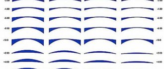
— Stage I is the initial stage and is characterized by visual acuity of 0.1-0.5. At this stage, glasses and contact lenses for astigmatism help. — Stage II already has a visual acuity value of 0.1-0.4. The cornea begins to thin out. It can also be corrected with glasses with cylindrical lenses and contact lenses. — At stage III, protrusion and thinning of the cornea occurs. Visual acuity is 0.1-0.12. Glasses for correction become unsuitable. Rigid gas-permeable contact lenses are used for this purpose. — Stage IV is clinical. Visual acuity is 0.01-0.02 and cannot be corrected.
By type, keratoconus is divided into:
— anterior (true), — posterior; - acute (dropsy).
Thanks to the development of modern equipment, it is no longer difficult to determine, using special functions in measuring instruments, keratoconus at the initial stage, as well as a type such as posterior keratoconus.
Keratoconus: main symptoms
At the initial stage of the disease, its symptoms are practically no different from the signs of myopia, farsightedness or astigmatism. The patient complains of blurred vision; he sees objects unclearly, blurred and, in some cases, distorted. This is due to the fact that at different points of the shell, light rays are refracted unevenly due to its cone-shaped shape. The main difference is that this progressive corneal disease cannot be corrected with glasses or soft contact lenses. In some cases, in the early stages, keratoconus is mistaken for other refractive errors due to similar symptoms. Only a qualified ophthalmologist can reliably identify the cause of visual impairment when conducting a thorough examination of the visual system using modern diagnostic equipment. Over time, as the deformity increases, the patient experiences various phantom images. Ophthalmologists characterize this phenomenon as monocular polyopia. This effect is most often observed when the patient looks at objects with high contrast for a long time, for example, while looking at dark dots on a light background. Instead of one point, a person observes a picture with various chaotic images. In the evening and at night, halos and glare may appear, which causes discomfort. In the final stages, the pathology of the eye with keratoconus becomes visually noticeable: a cone-shaped protrusion of the cornea is formed.
Keratoconus of the cornea: main signs:
- blurred vision (similar to myopia and farsightedness);
- distorted image perception (similar to astigmatism);
- ineffective use of standard optical aids (glasses and soft contact lenses);
- progressive decrease in visual acuity;
- glare and halos in the evening;
- photophobia and double vision;
- phantom images when looking at contrasting objects for a long time;
- cone-shaped protrusion of the cornea, visible to the naked eye (in the final stages).
Symptoms of corneal thinning: features of the latent form
The main symptoms of the pathology are progressive myopia and irregular astigmatism. The disease often occurs latently; over the course of several years, a person may experience slight visual disturbances. In the initial stages, the patient notices a slight blurred vision and comes to the ophthalmologist with a request to prescribe lenses or reading glasses. At this time, keratoconus has similar symptoms to other eye refractive errors. As the disease progresses, vision quickly deteriorates. Sharpness drops at any distance, the patient ceases to distinguish objects in twilight lighting.
In some cases, one eye sees better than the other. Photophobia and itching appear; when reading, a person squints. The classic symptom of keratoconus is the appearance of “ghostly” forms in the field of vision. Monocular polyopia occurs (the appearance of many virtual images). It is clearly visible in a field with high contrast, such as a point of light on a dark background. The patient sees not one object, but several, located in a chaotic order. When looking at a light source, a person notices banding and distortion, sometimes rhythmically moving spots appear.
Manifestations of keratoconus depending on the stage - table
| Stage | Characteristic |
| Stage I |
|
| Stage II |
|
| Stage III |
|
| Stage IV |
|
Video about operations for hydrops of the cornea (hydropsis)
In the absence of treatment for acute keratoconus, swelling of the cornea goes away on its own within 4–5 months. Upon completion of the relief of the pathological process in acute keratoconus, the cornea becomes somewhat flattened due to the formation of turbidity and local closure of the area of rupture of Descemet's membrane. As a result of this flattening of the cornea, patients' vision improves slightly, and contact correction is often available again.
It must be said that acute keratoconus is often misdiagnosed as keratitis. In this case, drug treatment is prescribed, which can aggravate the patient’s eye condition!
After relief, relapses of acute keratoconus usually do not occur. In the available ophthalmological literature there is no mention of recurrence of the disease in one eye. However, we had to deal with the recurrence of acute keratoconus twice in our clinical practice: in the first case, in a patient with Down syndrome, after three years, and in the second case, after 25 years (the patient’s discharge summary recorded acute keratoconus suffered for the first time ).
Treatment
Treatment and correction of keratoconus consists of taking measures and interventions that prevent and slow down its development, as well as correction of visual acuity.
— In the initial stages of the disease, the patient is prescribed glasses with cylindrical lenses, as for the correction of astigmatism.
- In later stages, rigid gas permeable contact lenses are used. On the one hand, they increase visual acuity, on the other hand, they mechanically prevent further protrusion of the cornea. Correction with these lenses does not stop the disease. Therefore, wearing these lenses cannot be considered a complete treatment for keratoconus. Currently, the trend of selecting LCL for patients is decreasing. The very wearing of these lenses in most cases causes both inflammatory diseases, ranging from conjunctivitis, ending with keratitis and other inflammatory diseases of the deep structures of the eye, and there is intolerance of GCL by patients. However, contact correction to improve vision in patients with keratoconus remains one of the main correction methods.
Double-layer lenses are also used to a lesser extent. They consist of two parts, hard and soft. Hybrid lenses are JGCLs that have a hydrogel layer as part of the material. Scleral lenses, which, due to their larger radius, unlike other types of lenses, are located on the sclera rather than lying on the cornea.
— If it is impossible to wear liquid contact lenses, if there are complications caused by wearing these lenses, as well as with low visual acuity and thinning of the cornea, penetrating or layered keratoplasty is used, i.e. corneal replacement. It should be noted that, if previously only penetrating keratoplasty was performed, now, with the advent of the femtosecond laser, layer-by-layer deep corneal transplantation is performed. Those. Only the stroma and outer layers are transplanted. A number of advantages of layer-by-layer corneal transplantation include a low risk of developing possible postoperative complications, as well as a higher functional result in the postoperative period.
You may be interested in Retinal dystrophy: causes, symptoms, treatment
Keratoconus - symptoms and treatment
There is no single algorithm for managing patients with keratoconus yet. Modern literature presents treatment methods that are focused on the stage of the disease.
The main task of the specialist who has diagnosed keratoconus is to correctly inform the patient about the choice of treatment and rehabilitation method. At the initial stage of keratoconus, spectacle or contact correction is performed, and in the case of severe keratoconus, contact vision correction (scleral or rigid corneal lenses) or penetrating/layered keratoplasty is performed.
What does ophthalmology have in its arsenal today?
Crosslinking
Crosslinking is the treatment of keratoconus using ultraviolet light and riboflavin (a special vitamin). This new method was proposed by the German ophthalmologist Theo Seiler in 1997, and the first successful clinical trials of this treatment were published in 2003 [11]. Since then, corneal crosslinking has entered clinical practice and has been widely used for more than 10 years in various European countries and more than five years in different countries around the world. However, permission to perform the procedure in the United States was issued only in 2016 [12].
The method is based on strengthening the biomechanical properties of the cornea - collagen fibers, the connections between them, as well as their spatial structural arrangement. “Cross-linking” with ultraviolet light under the influence of a sensitizer—0.1% riboflavin dissolved in 20% dextran—inhibits metabolic processes in stromal collagen, thereby increasing the biomechanical stability of the cornea [12].
Many different corneal crosslinking techniques have been developed. Numerous studies are still being conducted on the effectiveness of techniques and the percentage of riboflavin used.
The cross-linking procedure is minimally traumatic, is performed under local anesthesia and is usually uncomfortable only in the first few days after the intervention. After treatment, patients note an increase in visual acuity.
In 95% of cases, cross-linking stops the progression of keratoconus. The percentage of surgical risk is about 1% (infection, scarring, slow healing, etc.) [13].
Crossing is performed even in adolescence, but its effectiveness is higher in the early stages of the disease. In case of advanced keratoconus, the procedure is no longer possible.
Implantation of corneal segments
This treatment method involves strengthening the side of the cornea opposite the apex of the stretch using special segments - rings. The introduction of such segments into the cornea improves visual functions (for example, improves visual acuity), but does not solve the problem of keratoconus progression [15]. The disadvantages also include the economic factor: the operation is very expensive.
The operation of introducing segments is not technically difficult. Mandatory conditions for implantation are transparency of the central zone of the cornea and sufficient central thickness.
This treatment method is indicated in the early refractive stages. Other benefits of implantation include:
- no risk of rejection;
- flattening of the cornea without invading its central zone;
- significant persistent decrease in refraction;
- reversibility of the procedure: in case of complications, the segments are removed, while the ophthalmometric characteristics that were before the operation are preserved.
Keratoplasty
There are two options for keratoplasty - layer-by-layer and penetrating corneal transplantation.
The apotheosis of the struggle for vision remains penetrating keratoplasty. Despite the use of a femtosecond laser, this surgery is high risk. Its success depends on the patient’s age and the condition of his body.
Despite the widespread use of keratoplasty in the treatment of keratoconus, this operation has disadvantages:
- the risk of transplant rejection and its limited “life”;
- residual postoperative myopia and astigmatism;
- long-term risks of infections;
- problems with donor material;
- high cost of the operation [14].
Another unpleasant disadvantage of keratoplasty is the formation of new blood vessels in the cornea. Without blood vessels in the cornea, the immune system does not have direct access to its top layer. In this case, all immune processes proceed slowly and carefully. When blood vessels appear, the immune system begins to react sharply to the transplant.
But, probably, the biggest disappointment of patients after keratoplasty is low visual acuity. This condition is associated with surgical astigmatism, which is almost impossible to avoid. According to Boston professor Perry Rosenthal, more than 40% of patients after keratoplasty use correction, and their visual acuity averages 20/50 (0.4) [15].
Deep lamellar keratoplasty for keratoconus is just entering surgical practice. Its significant difference from penetrating keratoplasty is the surgical correction of keratoconus while preserving the patient’s corneal endothelium. Due to this, the risk of transplant rejection is reduced.
Optical correction
Progression of keratoconus is a good reason to consult a surgeon about surgical intervention: cross-linking or implantation of stromal segments. But if progression is not observed, or it is highly questionable, then surgery is not justified. What to do? Select adequate optical correction and observe dynamically.
Today in Russia there are several types of contact correction:
- soft individual lenses for keratoconus (several options);
- hybrid lenses (hybrid of soft and hard lenses);
- rigid gas permeable corneal lenses and rigid corneoscleral lenses;
- rigid gas permeable scleral lenses.
One popular way to compensate for visual acuity is gas permeable rigid lenses. They fit closely to the outer part of the eye, smooth out all the imperfections of the cornea and form the most correct optical surface.
Scleral gas permeable lenses are quite specific. They rest on the sclera and do not contact the cornea and eyelids. They have a stable fit throughout the day, provide comfort and high visual acuity. Such lenses change the geometry of the cornea due to the tear film and fluid under the lens, which allows them to compensate for irregular astigmatism.
Optical correction can be used in combination with surgery: lens selection after installation of an intrastromal segment or corneal transplantation. This allows patients to see the world in a better way.
Diagnostics
Keratoconus is diagnosed by an ophthalmologist during a visual examination. The doctor will definitely check whether there are cases of the disease in the family. If necessary, the patient is sent for additional examinations:
- Refraction measurement. The doctor asks the patient to look into a device containing lenses of different sizes (phoropters). The examination allows us to draw conclusions about the stage of the disease.
The ophthalmologist will determine the stage of the disease
- Slit lamp. In this test, a vertical beam of light is directed into the patient's eye. The doctor uses a microscope to examine the organ, evaluate the shape of the cornea, and look for other potential problems.
- Keratometry. A point of light is focused onto the cornea. The reflection is measured, and based on the data obtained, the shape of the cornea is determined.
Keratometry is one of the methods for diagnosing keratoconus
- Computer research. A series of high resolution photographs are taken. From these, a topographic map of the surface of the eyeball is compiled. This method shows the thickness of the cornea.
Cross stitching of the cornea
This procedure helps strengthen the structure of the cornea, which is weakened by keratoconus. To strengthen the cornea, riboflavin (vitamin B2) and exposure to ultraviolet light in the UVA range are used. Riboflavin is instilled onto the cornea, and then the area is irradiated with UVA rays. Activated by ultraviolet rays, riboflavin binds to collagen fibers of the cornea. As a result of this process, the number of cross-links between the collagen fibers of the cornea increases, which helps strengthen the corneal tissue and stop the further development of keratoconus.
This technique does not eliminate changes in the shape of the cornea that have already occurred in keratoconus (cone-shaped protrusion). Cross-linking the cornea stops further progression of keratoconus. This procedure can be used in conjunction with Intacs, which flatten the cone shape of the cornea itself.
The ability to strengthen the structure of the cornea for a long time and stop the progression of keratoconus is a huge advantage of this method. The technique is already used in some clinics in the country.
Causes
Experts still do not fully understand how keratoconus of the eye occurs and the reasons for its appearance. It is currently believed that this pathology is observed in the following cases:
- in people with characteristic structural features of the cornea;
- with hereditary predisposition;
- after diseases or injuries of the cornea;
- due to prolonged use of corticosteroid drugs;
- as a result of the negative impact of unfavorable environmental conditions.
Treatment of keratoconus with folk remedies
In the treatment of keratoconus, folk remedies are used as general strengthening, as well as preventive, to avoid complications of the disease, such as corneal rupture. For itching, burning and rapid eye fatigue, rinsing and lotions with decoctions of medicinal herbs such as chamomile, sage, coltsfoot are recommended. Medicinal herbs can also be used as teas for mild immune correction, for example, tea from the leaves of Echinacea purpurea.
Another folk remedy for treating keratoconus is honey and other bee products. In this case, honey and propolis are used both topically, in the form of aqueous solutions, and in food, as a general strengthening and immunostimulating agent.
Prognosis and complications
If you notice the disease in time and consult a doctor about it, the prognosis is favorable: ophthalmology knows many methods of diagnosis and treatment, and uses them successfully. The only exception is advanced keratoconus at the last stage. It’s difficult to cure, and sometimes even modern medicine can’t cope.
The main complication - and at the same time the result - of keratoconus is blindness, and irreversible. However, if you follow all the doctor’s instructions, the chances that it will reach her are small.
Classification
Keratoconus can be unilateral, when only one eye is affected, or bilateral. Statistics show that about 95% of all cases of the disease occur in both eyes.
Due to appearance:
- Primary, caused by genetic factors;
- Secondary, arising as a result of adverse environmental influences, surgical operations on the organs of vision, and the consequences of injuries.
According to the form of the disease:
- Acute;
- Chronic;
- With variable current.
Deviation of vision from the norm with keratoconus is divided into 3 stages:
- Weak, with a value of up to 40 diopters. During this period, slight morphological changes occur in the cornea of the eye, signs of astigmatism appear;
- Average degree, no more than 55 diopters. Cracks in the Descemet's membrane of the cornea form, allowing moisture to pass through from the anterior chamber. The apex of the cone takes on a cloudy outline, and the patient practically ceases to see in the dark;
- Severe, with a diopter value of more than 55. Visually, the unnatural shape of the cornea is already noticeable; it can become completely cloudy. Vision deteriorates sharply, the person sees poorly even during the day. If there is a high probability of corneal rupture, urgent surgery is required.
The level of corneal dystrophy can be:
- Moderate, with its thickness close to 0.5 mm;
- Medium, with a value of 0.4–0.5 mm;
- Over-permissible or close to rupture, with a thickness of less than 0.4 mm.
According to the degree of corneal deformation, the following forms are distinguished:
- Point dome. The pathology has a diameter of about 5 mm and is located in the center of the cornea;
- Oval dome shape. The cornea protrudes up to 6 mm, its deformation is localized below the center and sags;
- Ball-shaped deformation. The cone is more than 6 mm, the disease covers up to 70% of the cornea.