Causes of blurred vision
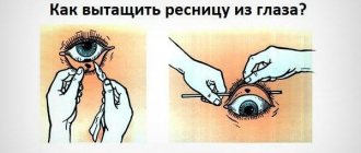
Transient blurred vision in one eye often indicates an internal infection or mechanical irritation. For example, if something gets into the eye, it starts to itch. At the same time, scratching the eye too intensively can damage the mucous membrane. This often results in the release of mucus, which, when it comes into contact with the pupil, causes defects in the perception of visual information.
In this case, the surest way is to rinse the eye with clean running water and give it a good rest. No reading, working at close range and at the computer, no watching TV, etc. In a word, the eye needs to be given rest for the entire next day, protecting it from any stress.
Causes of poor vision at close distances
The risk of occurrence and development of the disease concerns patients over forty years old, but the problem can also occur in children. Let's look at the main causes of poor near vision:
Physiological characteristics
If a small child has difficulty seeing close up, this problem is most often caused by features associated with the formation of the tissues of the eyeball. With age, such disturbances usually go away and vision returns to normal.
Eye muscles don't work well
This problem occurs among people leading a sedentary lifestyle, among office workers who need to concentrate attention on objects that are at a constant distance from the eyes (drawing, working in front of a computer monitor, with documentation, journalism and writing articles, etc.)
Therefore, the curvature of the lens rarely changes, the muscles lose tone and the ability to work effectively. It is very important to take breaks and “force the eye muscles to function”, concentrating on distant objects and distracting from the monotony of the activity.
Changes in the retina associated with aging
The light-sensitive pigment that allows us to see is gradually destroyed. Image clarity is lost and visual acuity decreases. You can slow down the aging process with a diet that includes all the necessary vitamins and microelements.
Age-related hardening and sclerosis of the lens
As a result of which a person loses the ability to focus the eyes on objects, a lens that has lost its elasticity cannot provide a clear image of things that are located close to the eyes.
Hereditary predisposition of some people to such pathologies
There are ethnic groups that are characterized by refractive errors and the development of similar diseases. The culprit for this is the miniature size of the eyeball and limited optical power.
| In such cases, vision correction is recommended; it is impossible to improve the patient’s condition without medical intervention. |
Disorders of blood flow in the vessels and nutrition of the retina
Poor nutrition, alcohol abuse and addiction to tobacco products can lead to deterioration of blood circulation and the condition of the blood vessels of the eyes. In these cases, doctors recommend sticking to a diet and limiting visits to the hammam and sauna.
Severe visual tension
It is associated with poor or constantly changing lighting, working in bright light. Ophthalmologists draw patients' attention to the importance of wearing sunglasses, avoiding reading in transport and in low light indoors, and using “chameleon” glasses while working at the computer.
Return to contents
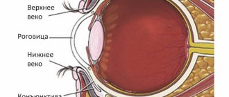
Infection
This is a very common cause of blurred vision, which goes away over time. For example, inflammation of the conjunctiva. In this case, the eye begins to see poorly due to incessant lacrimation, swelling and pus discharge. It is the pus that gets on the pupil that causes blurriness of visible objects.
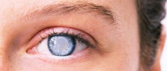
In the case of stye, the enlarging pustule covers the area of the pupil and the eye also sees with interference.
Treatment of infections is prescribed according to the stage of the disease. In this case, it is necessary to use antibacterial drugs prescribed by the doctor. All bacterial infections are treated with antibiotics, but only a doctor can determine the type of pathogen and prescribe a drug to which this pathogen is especially sensitive.
Normal vision is restored literally a few days after complete recovery. There is no need to worry about this, especially when the diagnosis is made correctly and the results of treatment are already visible.
Why does it happen?
The causes of the disease are divided into two groups:
- congenital;
- acquired.
More common are congenital forms of anisometropia, which are hereditary in nature. In infants, the disease does not manifest itself externally and is initially asymptomatic. But with age, the manifestations become brighter, and the disease progresses. The degree of development depends on the timeliness of diagnosis and correct correction.
The causes of the acquired form of the disease are divided into 3 groups:
- cataracts that are progressive and cannot be treated;
- complication after surgery;
- disturbance of accommodation in old age.
The disease can be stopped if you choose the right treatment in a timely manner and do corrective exercises.
Acquired forms develop faster than congenital ones, which contributes to early diagnosis, correct diagnosis and corrective measures. In most cases, the congenital form is genetic in nature, so they talk about the hereditary nature of the pathology. If you start proper eye care in time and select corrective exercises, the development of the disease can be slowed down or even stopped.
Separately, there is an idiopathic form of different vision in the eyes, in which it is impossible to identify the causes of the pathology. The diagnosis can only be made after a full examination by an ophthalmologist and a geneticist, who will help rule out other causes. According to statistics, the idiopathic form is very rare.
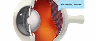
Retinal disinsertion
Retinal detachment is considered a serious disorder of eye function, which can cause not only a temporary deterioration in vision, but also its complete loss. Such a condition must be prevented whenever possible, and if this is not possible, eliminated literally at the initial stage.
The causes of retinal detachment may be age-related changes that affect the strength of the tissue; various diseases that make the connective layer thinner, which causes detachment; various injuries leading to retinal tears. Therefore, it is especially important, in the event of an injury, to consult a doctor, even when the blow was not severe, and the result of the impact was only a small bruise.
Useful video
Clouding of the image in one eye may go away on its own and does not require specific therapy. The presence of a long-term decrease in the quality of vision is the basis for contacting a doctor.
Author's rating
Author of the article
Alexandrova O.M.
Articles written
2031
about the author
Was the article helpful?
Rate the material on a five-point scale!
( 9 ratings, average: 4.89 out of 5)
If you have any questions or want to share your opinion or experience, write a comment below.
Taking vitamins
Every person needs vitamins throughout his life. Therefore, it is especially important to choose the right combination of elements for a particular individual. Taking fish oil will probably be the simplest - everyone always needs it.
Fish oil is rich in retinol, i.e. vitamin A, which plays a very important role in ensuring normal visual function. In addition, taking blueberry extract (anthocyanins) and lutein is very beneficial for vision. Thanks to the last element, the carotenoid lutein, age-related degeneration of the retina, which can lead to retinal ruptures and detachment, slows down.
What to do?
The doctor will conduct an examination and try to select lenses.
If visual function is impaired, you should contact an ophthalmologist. Only he is able to determine the method and method of correction. The first thing the doctor will do after diagnosis is to try to choose glasses with such lenses that each eye is comfortable. The lenses will differ from each other, since each has its own optical power. It is important that the doctor selects the lenses, because inadequately applied refractive power can damage vision even more.
Another conservative method is the use of corrective lenses. The method allows you to get rid of the need to wear glasses. The selection of lenses occurs only individually with each patient, but it should be remembered that during the installation of lenses the cornea and the epithelial layer of the eye shell may be damaged, and this is fraught with complications. The use of this corrective method is accompanied by gradual adaptation, periodic examinations by an ophthalmologist and proper eye hygiene.
Glasses are safer for patients with anisometropia.
Surgical treatment
One comes to the surgical method of vision correction when conservative therapy is powerless and the visual acuity progressively decreases. Laser surgery is used to restore the normal refractive power of light rays. This method is difficult to implement and has a long and difficult rehabilitation period. Clarity of vision in the eyes is restored gradually and requires the implementation of strict rules. After the operation, it is necessary for the organs to be provided with 1-2 weeks of complete calm. Only then can you do eye exercises and apply other development methods. Heavy physical activity and eye strain are contraindicated for six months after surgery.
Gymnastics
Eye exercises can not only prevent certain diseases, but also actually improve vision. They help activate local blood circulation, which is very important. At the same time, if you take some well-known techniques as a guide, your vision will be restored literally after a couple of weeks of active exercise.
And in conclusion, I would like to remind everyone once again that if your eyes begin to see blurredly, you should immediately go to an ophthalmologist. Only a specialist can help you choose the right vitamins for your eyes, recommend the most effective sets of exercises, and, if necessary, prescribe medication for the disease.
In the medical department, everyone can undergo examination using the most modern diagnostic equipment, and based on the results, receive advice from a highly qualified specialist. The clinic provides consultations to children from 4 years old. We are open seven days a week and work daily from 9 a.m. to 9 p.m. Our specialists will help identify the cause of vision loss and provide competent treatment for identified pathologies.
You can find out the cost of a particular procedure or make an appointment at the Moscow Eye Clinic by calling 8 (800) 777-38-81 (daily from 9:00 to 21:00, free for mobile phones and regions of the Russian Federation) or using the form online entries.
Fomenko Natalia Ivanovna
Treatment methods
Depending on the degree of impairment, the doctor chooses what to do in a particular case if a person has difficulty seeing nearby objects.
Main methods of treatment:
- optics for vision correction (glasses, contact lenses);
- software-computer methods of treatment;
- vitamins;
- special exercises for the eyes;
- surgical treatment.
A slight decrease in vision clarity can be improved with eye exercises and the use of reading glasses. In case of more severe impairments (plus 3 or more), a person must wear lenses or glasses constantly in order to see both near and far. The complex prescribes treatment using computer programs.
In most cases, it is enough for children to perform eye exercises, and their vision gradually improves.
It will be useful to take eye vitamins for myopia: “Blueberry-Forte”, “Aevit”, “Okuwait Lutein Forte”.
If treatment is ineffective or at the request of the person, surgical treatment is performed. Types of operations:
- laser correction;
- laser thermokeratoplasty (thermal effect on the cornea to change refractive power);
- replacement of the affected lens with an IOL;
- keratoplasty (failed cornea is replaced with a healthy one);
- lens implantation;
- radial keratotomy (correction of the refractive function of the cornea by applying incisions);
- thermokeratocoagulation (point temperature effect on the cornea).
Additionally, we invite you to look at several ways to improve vision at home:
Diagnostics
Only an ophthalmologist can diagnose the exact cause of the appearance of a veil before the eyes. The main methods for diagnosing blurred vision are:
- visual inspection of the affected area using a slit lamp;
- tonometry – determination of intraocular pressure indicators;
- fundus examination;
- Ultrasound examination of the eyeballs.