Types of injuries
Bladder injuries are classified based on a number of criteria:
- Rupture in relation to the abdominal cavity:
- extraperitoneal;
- intraperitoneal;
- combined;
- By type of bladder injury:
- open (with violation of skin integrity);
- closed (without violating the integrity of the skin);
- By severity of injury:
- bruise (the integrity of the organ is not compromised);
- incomplete rupture of the bladder wall;
- complete rupture of the bladder wall.
- Based on the presence of damage to other organs:
- isolated injury;
- combined trauma (in combination with damage to other abdominal organs).
BLADDER DAMAGE
There are two types of bladder injuries: extraperitoneal and intraperitoneal. The mechanism of closed injuries to the bladder: 1) for an extraperitoneal rupture - a fracture of the pelvic bones, traction of the walls of the bladder by the ligaments fixing it or perforation of the bladder wall with bone fragments, 2) for an intraperitoneal rupture - a direct blow to the stomach with a full bladder, which leads to a sudden increase in intravesical pressure .
Rupture of the bladder occurs more easily when its wall is thinned (due to long-term bladder outlet obstruction and overdistension of the bladder).
There is no typical clinical picture for bladder ruptures. The main symptoms are disturbance or cessation of urination, pain in the lower abdomen, painful urge to urinate, and blood in the urine.
With intraperitoneal rupture of the bladder, tenesmus may be absent if urine flows freely into the abdominal cavity. But in this case, signs of peritoneal irritation appear, and 10-12 hours after the injury, peritonitis develops.
Since a rupture of the bladder often accompanies a fracture of the pelvic bones, along with the symptoms listed above, symptoms of a pelvic fracture are noted. For all pelvic fractures, it is necessary to determine whether there is simultaneous damage to the bladder or urethra.
The simplest way to diagnose a ruptured bladder is the Zeldovich test. 300 ml of liquid is injected into the bladder through a catheter and then the volume of liquid released through the same catheter is accurately measured. When the bubble ruptures, the volume of liquid released is significantly less than the volume introduced. However, sometimes with intraperitoneal rupture of the bladder, the amount of fluid released through the catheter is 2-3 times greater than the amount injected. This happens when the catheter penetrates through a defect in the wall of the bladder into the abdominal cavity and the catheter begins to release urine that previously entered the peritoneal cavity. This situation also argues in favor of bladder rupture.
We combine the Zeldovich test with ultrasound examination of the abdominal cavity and bladder. Moreover, after filling the bubble, in the event of a rupture of its wall, the liquid in the peri-vesical space is clearly visible, and sometimes it is even visible how the liquid flows outside the bubble. If the bladder ruptures intraperitoneally, an abdominal ultrasound will show the presence of fluid in the abdominal cavity.
If damage to the abdominal organs is suspected, laparoscopy may be performed. During laparoscopy, a bladder rupture can also be detected by the significant content of hemorrhagic contents in the abdominal cavity.
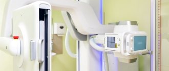
Retrograde cystography remains the main test used to document bladder rupture. The study allows you to establish the fact of bladder rupture, determine the type of rupture (intra- or extraperitoneal) and the location of urinary leaks. It is necessary to strictly follow the rules for performing cystography in patients with suspected bladder rupture:
1) a panoramic photograph of the abdominal cavity is performed with maximum coverage of the pelvis; 2) a concentrated solution of a contrast agent (at least 35%) and in a volume of at least 300 ml must be injected into the bladder; 3) photographs must be taken in frontal and lateral projections; 4) after the bladder has emptied, another photo must be taken.
Cystoscopy for suspected bladder rupture can be performed if it is not possible to perform ultrasound or cystography. If these studies can be performed, cystoscopy in cases of suspected bladder rupture is not indicated.
If a bladder rupture is diagnosed, emergency surgery is necessary.
In case of intraperitoneal rupture of the bladder, it is necessary to perform a laparotomy with revision of the abdominal cavity and evacuation of the contents. The bladder is sutured with 2-row catgut or vicryl sutures, leaving an extraperitoneal epicystostomy. In this case, it is necessary to inspect the bladder either through a rupture in its wall or, if the rupture is small, through an additional cystotomy hole on its anterior wall. Inspection of the bladder is mandatory, since we have encountered cases of combined multiple bladder ruptures: extra- and intraperitoneal. In such cases, an extraperitoneal (second) rupture is diagnosed only by careful examination of the inner surface of the bladder.
With extraperitoneal rupture of the bladder, the need for laparotomy arises in cases of suspected damage to the abdominal organs. The bladder is sutured and drained in a similar way, but in addition it is necessary to drain the paravesical space through the anterior abdominal wall or through the obturator foramina on the right and left (according to McWhorter-Buyalsky).
Fractures of the pelvic bones, combined with a rupture of the bladder, are not a contraindication to drainage of the peri-vesical tissue. The fear that this type of drainage converts a closed pelvic fracture into an open one is unfounded. Drainage of pelvic urohematoma, on the contrary, sharply reduces the percentage of pelvic osteomyelitis.
In all cases of rupture of the bladder in combination with a fracture of the pelvic bones, treatment measures should be aimed not only at eliminating the rupture of the bladder, but also at treating the fracture of the pelvic bones.
In the event of a rupture of the symphysis pubis, it is advisable to stitch it with nylon braid, loose bone fragments should be removed, the sharp ends of bone fragments should be bitten off with pliers to prevent damage to the pelvic organs, and primary metal osteosynthesis is performed according to indications. To stop intense bleeding from sites of pelvic bone fractures, bilateral ligation of the internal iliac arteries can be performed.
Our doctors
Perepechay Dmitry Leonidovich
Urologist, Candidate of Medical Sciences, doctor of the highest category
40 years of experience
Make an appointment
Khromov Danil Vladimirovich
Urologist, Candidate of Medical Sciences, doctor of the highest category
35 years of experience
Make an appointment
Mukhin Vitaly Borisovich
Urologist, Head of the Department of Urology, Candidate of Medical Sciences
34 years of experience
Make an appointment
Kochetov Sergey Anatolievich
Urologist, Candidate of Medical Sciences, doctor of the highest category
34 years of experience
Make an appointment
Complications after bladder rupture
There are no complications with timely and correct treatment. However, if treatment is carried out later than the recommended period, the following additional difficulties may arise:
- peritonitis;
- intestinal obstruction;
- abscesses;
- bloating;
- uroascites;
- incontinence;
- general disturbance of urination;
- fistula;
- urinary tract infections;
- persistent hematuria and more.
But such complications rarely occur with simple bladder ruptures. They usually accompany more complex cases of tissue damage. In any case, the prognosis for the treatment of these conditions is optimistic. And if you seek medical help in a timely manner, such conditions can be avoided.
This article is posted for educational purposes only and does not constitute scientific material or professional medical advice.
Answers to questions on the topic: Bladder rupture
Publications in the media
Injuries to the bladder with timely assistance are compatible with life, however, severe combined injuries and their complications can lead to death. Frequency. Complete or partial disruption of the bladder wall accounts for 5–15% of all traumatic injuries. In children - 4.4–11.5% of injuries to internal organs. Combined injuries account for more than 60% of all cases of bladder injuries. Classification • Closed, open; isolated and combined (in combination with fractures of the pelvic bones, external genitalia, abdominal organs, rectum, etc.) • Intraperitoneal, extraperitoneal and combined • Non-penetrating and penetrating.
Causes • Injuries of the abdominal wall (penetrating, non-penetrating) • Blunt injuries • Labor efforts, heavy lifting (with a full bladder) • Iatrogenic factors. The clinical picture is a combination of signs of injury to the bladder itself, shock, internal bleeding and the abdominal organs, pelvis and other bones bordering the bladder. At the initial stages, the clinical picture is dominated by symptoms of a violation of the patient’s general condition and injuries to other organs. • With incomplete ruptures of the bladder, urination is often preserved, the only sign being hematuria. • Early manifestation of extraperitoneal injuries is the occurrence of false urge to urinate. • With intraperitoneal ruptures of the bladder, the earliest and most common symptom is abdominal pain •• As the circumvesical urohematoma increases, the pain in the lower abdomen intensifies, the tension of the abdominal wall above the pubis increases •• Percussion - dullness without clear boundaries •• Swelling of tissues in the pubic, inguinal areas or perineum •• Symptom of false urge to urinate •• Sometimes urination is preserved due to tamponade of the bladder wall defect with a loop of intestine or omentum •• Symptoms of peritoneal irritation increase gradually, which complicates diagnosis. • With combined damage to the bladder and pelvic bones, the patient is pale, covered in cold sweat, and blood pressure is reduced. • Open injury - urine leakage from the wound canal (symptom observed in 11% of cases). Special studies • Catheterization of the bladder - urine is not released or flows out in a weak stream and contains an admixture of blood; with intraperitoneal injury, you can get a large amount of turbid bloody fluid (Zoldovich's sign) • Cystoscopy is only possible for incomplete or very small injuries, when it is possible to fill the bladder for examination • Excretory urography and descending cystography • Ascending cystography • Tests with dyes (ingestion of methylene blue, intravenous administration of indigo carmine) confirm the release of urine from the wound with open injuries to the bladder.
TREATMENT. The main method is surgical. Surgical treatment. Types of operations: • For closed intraperitoneal injuries - laparotomy, revision of the abdominal organs (ruptures of parenchymal organs are sutured, then interventions are performed on the gastrointestinal tract). The bladder wound is sutured with a double-row suture. The abdominal cavity is drained. A permanent catheter is left in the bladder for 5–7 days • For closed extraperitoneal injuries, a median suprapubic approach is used. The peri-vesical urohematoma is emptied and loose bone fragments are removed. Bladder ruptures are sutured, preferably with a double-row catgut suture. An epicystostomy is applied. If necessary, the cellular spaces of the small pelvis are drained. Conservative treatment is indicated for bruises and incomplete ruptures of the bladder. In stationary conditions - complete rest, cold on the stomach. Hemostatic, anti-inflammatory, and painkillers are prescribed. In rare cases, a permanent urinary catheter is installed for 3–5 days or intermittent catheterization is performed 3–4 times.
The prognosis depends on the severity of the injury and the timeliness of surgical treatment. For incomplete ruptures of the bladder - favorable.
ICD-10 • S37.2 Bladder injury
conclusions
Anastomotic repair in a patient with extensive damage to the urethra was performed on the 10th day from the moment of injury. Satisfactory results of the operation can, to one degree or another, be due to adequate surgical sanitation of the urethral bed with complete removal of softened tissue, as well as adherence to the principles of microsurgical technique at the reconstructive stage. However, despite the results obtained, the operation was of the nature of desperation and such tactics in no way pretend to change the established canons in the treatment of patients with urethral injuries and can only be applied in exceptional, forced situations by surgeons who know the principles of urethral surgery.
Types of urethral ruptures
Urethral ruptures are divided into several types depending on the severity of the injury. So, the injury can be open or closed.
And, accordingly, the gaps are divided into:
- non-penetrating (internal ruptures of muscle fibers and mucous membrane): partial ruptures and complete ruptures;
- penetrating (complete ruptures of the mucous membranes of the urethra).
If the urethra is completely ruptured, urine may leak into the surrounding soft tissue. This process is the main cause of further complications.
Also, damage to the urethra, depending on the severity, is divided into degrees:
- bruises;
- sprains;
- partial urethral ruptures;
- complete rupture with a distance between the edges of the urethra less than 2 cm;
- a complete rupture with a distance between the edges of the urethra greater than 2 cm.
With different types of urethral ruptures, the symptoms may differ.
You can make an appointment with a venereologist from our consultants by calling +7 (495) 125-49-50
Prices for venereologist services Addresses of clinics Pain during urination Smear from the urethra Ultrasound of the appendages Urologist at home


