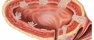
The bladder is a hollow organ in which urine accumulates. This organ, like others, is susceptible to various inflammatory processes and ailments that manifest themselves regardless of the gender or age of the patient. One of the most common ailments is bladder bloating. This pathological process is an abnormal increase in urinary output due to disturbances in the outflow of urine.
Violation of the outflow of biological fluid can be caused by various reasons. These include:
- mechanical obstacle;
- neuromuscular disorders;
- biological disorders.
In addition to the above reasons for urine outflow disorder, there is another one that occurs as a result of taking certain medications as a side effect.
In most cases, men who have passed the age of forty are susceptible to bladder bloating. However, the likelihood that this pathology can occur in young men, adolescents, and women is also quite high.
Causes
Urinary bloating can be caused by various factors and pathological processes developing in the body.
Problems observed from the endocrine system. A disease such as diabetes mellitus causes disorders in the autonomic nervous system. This pathological process, in turn, causes a disruption in the outflow of biological fluid, the urinary tract becomes overfilled and swells.
Inflammatory processes of the genitourinary area. For example, an inflammatory process that occurs in the prostate gland and occurs in a particularly acute form causes mechanical urinary retention and provokes bloating. In addition, urethritis, cystitis, pyelonephritis and many other ailments can also cause a violation of the outflow of urine.
Disorders arising from the nervous system. For example, during multiple sclerosis, the patient cannot control his bladder, which causes urinary stagnation.
Urolithiasis disease. This disease is manifested by the formation of stones in the urinary cavity or urethra. However, the presence of stones alone does not cause the development of urinary bloating. This pathological process occurs only if the stones cause a disruption in the outflow of biological fluid.
Basically, blockage of the outflow of urine occurs when there are stones in the urethral canal, since its diameter is small. Stones enter this area from the kidneys and bladder or arise against the background of diseases such as urethritis or prostatitis, which have become chronic. As a result, all of the above factors provoke bloating.
Tumor in the urethra, prostate or urinary area. New growths that increase in size can provoke the development of problems with the secretion of biological fluid, which, in turn, causes bloating.
Urethral stricture. In most cases, this disease affects the male population, but there are cases, albeit rare, of its development in women. Urethral stricture can be either congenital or acquired. Congenital development of pathology in medicine is very rare and occurs due to anterior valve narrowing of the urethra. The acquired structure of the urethra occurs after injury, surgery or acute inflammatory processes.
Urinary catheterization. Urinary bloating can be caused by medical procedures, in particular, improper installation of a catheter. If the health care provider is not competent in catheterizing a patient, catheter obstruction may occur, causing urinary stasis. In addition, in some patients, after removal of this medical instrument, an inflammatory process in the walls of the urinary tract may develop, as well as their outflow, which will cause a disruption in the outflow of urine.
Use of medications. A course of taking certain types of medications can cause retention of biological fluid in the bladder. In most cases, this pathological process is provoked by sedatives and painkillers, as well as anesthetics and opiates.
Ultrasound diagnosis of squamous cell carcinoma of the bladder
Ultrasound machine RS85
Revolutionary changes in expert diagnostics.
Impeccable image quality, lightning-fast operating speed, a new generation of visualization technologies and quantitative analysis of ultrasound scanning data.
Introduction
The incidence of bladder cancer around the world is showing a steady upward trend. According to the World Health Organization, bladder cancer accounts for about 3% of all malignant neoplasms. In terms of prevalence, it is second only to tumors of the stomach, esophagus, lungs and larynx. Among all oncological urological diseases, bladder neoplasms occupy the second place in incidence after prostate cancer. More than 150 thousand new cases are registered annually in the world. In terms of prevalence in Europe, bladder cancer ranks fifth in men and 11 in women among all forms of this disease [1]. In 1999, 11,267 cases of bladder cancer were first detected in Russia, of which only 2.1% were detected during preventive examinations [2]. Of all morphological forms, transitional cell carcinoma is the most common, accounting for up to 90%. Less than 10% are adenocarcinoma, squamous cell carcinoma and squamous cell carcinoma.
It has been established that the carcinogenic agent is present in the urine and that the epithelium of the bladder mucosa is predisposed to proliferation. Under the influence of certain types of irritation, the epithelium undergoes changes both morphologically and biologically, which ultimately can lead to neoplasm [4]. More often it occurs in the area of the triangle and the neck of the bladder, which differ in structure from the rest of it.
Among the main etiological factors leading to the appearance of bladder tumors are chemical irritants, mainly aniline products, functional liver disorders, viruses, impaired metabolism of microelements (copper, silver, zinc, manganese, etc.), previous chronic diseases of the bladder (interstitial cystitis, granular cystitis, ulcers, bladder leukoplakia, stones, diverticula, etc., as well as chronic cystitis caused by parasites, in particular schistomatosis), smoking, stagnation of urine, high activity of lactate dehydrogenase [4,5].
At the very beginning of the disease, the clinical manifestations of bladder cancer are scanty and largely depend on the stage of the disease, the presence of complications, and concomitant diseases. The main symptoms of epithelial bladder tumors are hematuria (70%) and dysuria (15-37%). As the tumor process progresses, patients experience pain in the suprapubic region, which is constant. The pain intensifies at the end of urination. The intensity of pain depends on the location and nature of tumor growth. Exophytic neoplasms can reach large sizes without causing pain. Endophytic growth is accompanied by constant, dull pain above the womb and in the pelvic cavity. If the tumor grows into the bladder wall and spreads to the paravesical tissue and neighboring organs, symptoms of pelvic compression may occur, manifested by swelling of the lower extremities, scrotum, phlebitis, pain in the perineum, lumbar region, and genitals.
Descriptions of cases of ultrasound diagnosis of squamous cell carcinoma of the bladder are extremely rare in the literature. That is why in this observation we want to share our experience.
Description of observation
Patient A., born in 1930, was referred by a urologist for an ultrasound scan of the kidneys, bladder and prostate gland with a preliminary diagnosis of prostate adenoma and chronic pyelonephritis. From the anamnesis it is known that over the past 5-6 months. He noted dysuria (frequent urge to urinate, accompanied by a burning sensation when urinating, pollakiuria). Later, the process of urination became painful, pain appeared in the suprapubic and left lumbar regions. On examination: condition is satisfactory. The physique is asthenic. The skin and visible mucous membranes are in satisfactory condition. The physique is asthenic. The skin and visible mucous membranes are pale. Breathing is vesicular, no wheezing. Heart sounds are muffled. Pulse 82 beats per minute, satisfactory filling. BP=140/85 mmHg. The tongue is moist and covered with a white coating. Pasternatsky's sign is weakly positive on the left. In a general urine test taken on the day of the study: specific gravity 1025, dark orange color, cloudy urine, acidic reaction, protein 1.12 g/l, leukocytes 7-8 in p.s., red blood cells 15-20 in p.s. sp., mucus, bacteria in moderate quantities.
Ultrasound revealed the following picture: the right kidney is bean-shaped, with a smooth, clear contour, dimensions 110x55 mm, parenchyma thickness 13 mm, single dilated calyces up to 8 mm are located. The left kidney is oval in shape, with a smooth, clear contour, dimensions 115x58 mm, parenchyma thickness 11 mm, the collecting-pelvis system is expanded, calyces up to 12 mm, pelvis 25x12 mm. The sinuses of both kidneys have unevenly increased echogenicity, corticomedullary differentiation is difficult, the parenchyma has small echo-positive inclusions up to 2 mm without an acoustic shadow. After emptying the bladder, the ultrasound picture of the FLS of both kidneys was unchanged.
Bladder: anterior-posterior size 8 cm, transverse - 7 cm, superior-inferior - 7 cm, volume 188 cm³, wall - 4 mm, contents anechoic. On the left side wall an echo-positive formation of irregular shape is visualized, with uneven, bumpy contours, heterogeneous structure, with areas of higher echogenicity along the contour facing the bladder cavity, measuring 52x35x36 mm. The wall of the bladder closer to the mouth of the left ureter is not clearly differentiated and is blurred. The residual volume of the bladder is 102 ml. (Fig. 1 a, b). Prostate gland: oval, symmetrical, with an even, unclear contour, increased echogenicity, anterior-posterior size 48 mm, transverse - 35 mm, superior-inferior - 38 mm, heterogeneous structure, with small areas of reduced and increased echogenicity without clear contours, with echo-positive areas up to 3 mm without an acoustic shadow and with a slight acoustic shadow. Ultrasound of the inguinal lymph nodes: on the right - without features; on the left — a single hypoechoic formation of an oval shape, with clear contours, homogeneous structure, dimensions 15x7x8 mm; retroperitoneal lymph nodes - without features. Conclusion: diffuse changes in the parenchyma and sinuses of the kidneys. Pyeelectasia on the left. Ultrasound picture of a bladder mass with signs of wall infiltration. Increased volume of residual urine. To clarify the diagnosis, cystoscopy is recommended. Diffuse changes in the prostate gland. A single enlarged lymph node in the groin area on the left.
Rice. 1.
Echogram of bladder cancer.
A)
Transverse scanning.
b)
Longitudinal oblique scanning.
Cystoscopy performed a day later revealed that the bladder mucosa was hyperemic and slightly swollen. On the left side wall, a bright pink, lumpy formation is determined, with a coating of fibrin, isolated foci of necrosis and incrustation with salts. During biopsy, bleeding is increased, the tissue is rigid, dense, and cannot be displaced. Conclusion: Bladder tumor; germination cannot be ruled out. In the material obtained from the biopsy: squamous epithelium of varying degrees of differentiation, histological picture of squamous cell carcinoma.
Conclusion
The presented observation proves that ultrasound is a highly informative method for diagnosing space-occupying formations of the bladder; in addition, it is painless and harmless to the patient. Ultrasonography allows you to identify a tumor, preliminary assess the degree of infiltration of the bladder wall, the spread of the tumor process in the bladder and beyond, as well as the degree of disruption of the passage of urine from the kidneys and the condition of the parenchyma, regional and retroperitoneal lymph nodes.
Literature
- Pereverzev A.S., Petrov S.B. Bladder tumors. Kharkov, 2002.
- Haeashi K., Nagashima M., Iwata M. Et al. Hiperthernochemotherapy, which intraarterial infusion by vascular access device in uterine cervicalcancer //Y.upn.Sos.Cancer. 1991/-Vol/26.-Pp 760-773.
- Kaprin A.D., Kostin A.A. // Attending physician. 2003.-N7.-C 40-44.
- Clinical oncourology // Ed. E.B.Marinbakh. M: Medicine, 1975.
- Shipilov V.I. Bladder cancer. M: Medicine, 1983.
Ultrasound machine RS85
Revolutionary changes in expert diagnostics.
Impeccable image quality, lightning-fast operating speed, a new generation of visualization technologies and quantitative analysis of ultrasound scanning data.
Symptoms of bloating
It is quite difficult to determine bladder bloating on your own, since everything depends on the factors that provoked it. If the bloating was caused by urolithiasis, the patient will feel slight discomfort.
During the inflammatory process that occurs in the prostate gland, a man feels acute pain in the abdominal area.
Source: Megamedportal.ru
If the pathology is provoked by cystitis or urethritis, then the symptoms will be similar to these diseases:
- burning pain when urinating;
- the frequency of the urge to emit biological fluid increases, while its volume decreases significantly;
- the feeling of not emptying the bladder immediately after emptying it;
- discomfort and pain in the lower abdomen.
If the cause is a urethral stricture, the patient will experience clear signs of difficulty voiding.
The appearance of tumors in the prostate, urinary cavity or urethra also has its own symptoms. With prostate adenoma, a man will feel pain in the perineum and a feeling of incomplete urinary emptying; constipation and hematuria may bother him. With cancer of the urinary tract and urethra, the frequency of urination may increase, and vomiting, diarrhea, or, conversely, constipation may occur. In addition, as the size of the tumor increases, acute pain in the abdominal area will occur more often.
In most cases, the pathological process occurs without pronounced symptoms. The only thing the patient should pay attention to is swelling of the abdomen in its lower part. If such a phenomenon is observed, you should contact a specialist to determine the type of pathological process.
How long do men with bladder cancer live?
The prognosis for bladder cancer in men is determined by the stage and degree of aggressiveness of the tumor, age, health status, and concomitant diseases. A special indicator is used - five-year survival rate - it indicates the percentage of patients surviving for 5 years.
The five-year survival rate for bladder cancer in men at different stages is:
- At stage 0: 98%.
- Stage I: 88%.
- At stage II: 63%.
- At stage III: 46%.
- At stage IV: 15%.
Diagnosis and treatment
As soon as the patient contacts a specialist, a number of measures are taken to identify the pathology. First of all, the doctor examines the area of the swollen abdomen using palpation, then prescribes tests:
- blood and urine, which reveal signs of an inflammatory process in the body;
- Ultrasound examination of the urinary tract. Ultrasound helps determine the presence of urolithiasis, neoplasms or various inflammations; Urinary pressure is measured.
If the patient has problems with the endocrine system, a consultation with a specialist qualified in this field is scheduled. You will definitely need to visit a neurologist to rule out problems with the nervous system. If the doctor doubts the diagnosis, he may prescribe an MRI or CT scan.
Treatment is carried out based on the pathology identified by doctors. If urinary bloating occurs slowly and the patient does not feel any strong changes occurring in his body, the doctor prescribes special massages and warm baths.
In addition, the patient needs to temporarily reduce fluid intake. If the bladder fills too quickly, internal pressure may rise. In this case, emergency measures are required: catheterization, which allows the release of biological fluid from the urinary tract. If these measures are not taken promptly, serious consequences may occur: the urinary lining will become thinner and rupture will occur.
Urinary bloating is a serious pathology that requires careful attention.
As you already understand, there are a lot of reasons that can cause this process, so a person cannot cope on his own. Share:


