Anatomy of the urinary system
Basic knowledge of the structure will help you more easily navigate the processes that result in the formation, accumulation and removal of urine from the body.
- The kidneys are a paired organ of the abdominal cavity. It includes large veins and arteries through which blood enters the filtration system. During the day, this process is repeated completely 300 times; a total of 1500 - 2000 liters of blood passes through the kidneys in 24 hours. The smallest structure of this organ is the nephron; it is a glomerulus with a capsule surrounded by a network of capillaries. The tubules of the nephrons form branched connections, they exit into the ducts and then pass into the calyces, which make up the renal pelvis.
- The ureters are paired hollow tubes that deliver finished urine, formed in the kidneys, to the bladder. In a healthy person, the ureter can hold up to 700 ml of fluid.
- The bladder is a large hollow sac with a volume of up to 0.5 liters in women, 0.7 liters in men. Urine accumulates in it until a sufficient amount is collected for the urge to appear (200 - 250 ml).
- The urethral canal is a narrow hollow tube with 2 sphincters (internal and external) inside, through which the collected fluid is released out. Its structure depends on gender - in women the urethra is wide and short (4 - 6 cm), in men it is narrower and much longer (up to 16 cm). The structural features of the reproductive system play a role in this.
Note! This characteristic applies to adults. Children only need 100 ml of accumulated urine to feel the urge to visit the restroom. The bladder capacity of a newborn is only 30–50 ml and reaches adult size by 10–12 years.
Nephrons
The structural and functional unit of the kidney is the nephron, which consists of a glomerular capsule, shaped like a double-walled glass, and tubules. The capsule covers the glomerular capillary network, resulting in the formation of the renal (Malpighian) corpuscle.
The glomerular capsule continues into the proximal convoluted tubule. It is followed by a nephron loop, consisting of a descending and ascending part. The nephron loop passes into the distal convoluted tubule, which flows into the collecting duct. The collecting ducts continue into the papillary ducts. Throughout their entire length, the nephron tubules are surrounded by adjacent blood capillaries.
How urine is formed
Instructions
Excretory organs include the lungs, skin and kidneys. In this case, the kidneys, ureters, bladder and urethra, which carry out urination, play a major role. The main function of the excretory organs is to maintain a constant internal environment. Blood enters the kidneys through the renal arteries. Here it is cleared of excess substances and returned back to the bloodstream through the renal veins. Harmful substances filtered by the kidneys form urine, which goes through the ureters to the bladder. At the moment of urination, the circular muscle (sphincter), which closes the exit to the urethra, relaxes, the walls of the bladder contract, and urine is pushed out. The kidney is a paired bean-shaped organ. The concave part facing the spine is called the renal hilum. The renal artery entering them carries unpurified blood. The renal veins and ureter emerge from the hilum of the kidney. Through the veins, “clean” blood goes to the inferior vena cava of the systemic circulation, and through the ureter, the released waste products enter the bladder.
The kidney consists of an outer cortex and an inner medulla. The latter is differentiated into renal pyramids, their bases adjacent to the cortex, and their apices directed into the renal pelvis. The renal pelvis is a reservoir that collects urine before it exits into the ureter.
The microscopic structural and functional unit of the kidney is the nephron. There are about a million of them in each kidney, and it is in them that the blood plasma is filtered. The nephron consists of a capsule that extends into a long convoluted tubule. The capsules and the initial part of the tubules are located in the renal cortex, and their continuation is in the medulla.
Blood plasma penetrates in portions through the thin wall of the blood vessel into the gap in the nephron capsule. Formed elements (erythrocytes, leukocytes, platelets) and proteins remain in the arterioles. Decay products, water and nutrients enter the nephron tubule. Together they make up primary urine. About 150 liters of primary urine are formed per day, and all the blood (5 liters on average) passes through the kidneys about 300 times.
Primary urine moves further along the convoluted tubule. Here, the necessary substances and most of the water are reabsorbed into the blood, and “waste” that is unnecessary for the body remains in the tubule itself. This is how secondary, final urine is formed - a concentrated solution of urea and salts of oxalic, uric, phosphoric and other acids. The convoluted tubules are followed by collecting tubules, which direct fluid into the renal pelvis. 1.5-2 liters of secondary urine are produced per day.
Video on the topic
www.kakprosto.ru
Evolution of the excretory system
In the process of evolution, excretory products and the mechanisms of their removal from the body have changed greatly.
With the complication of organization and the transition to new habitats, along with the skin and kidneys, other excretory organs appeared or the excretory function began to be performed secondarily by existing organs. Excretory processes in animals are associated with the activation of their metabolism, as well as much more complex life processes. Protozoa are released by diffusion through the membrane. To remove excess water, protozoa have contractile vacuoles. Sponges and coelenterates - metabolic products are also removed by diffusion. The first excretory organs of the simplest structure appear in flatworms and nemerteans. They are called protonephridia, or flame cells. Annelids have a pair of specialized excretory organs, metanephridia, in each body segment. The excretory organs of crustaceans are green glands located at the base of the antennae. Urine accumulates in the bladder and then comes out. Insects have Malpighian tubules that open into the digestive tract. The excretory system of all vertebrates is basically the same: it consists of renal corpuscles - nephrons, with the help of which metabolic products are removed from the blood. In birds and mammals, in the process of evolution, a third type of kidney was developed - metanephros, the tubules of which have two highly convoluted sections (like in humans) and a long loop of Henle. In long sections of the renal tubule, water is reabsorbed, which allows animals to successfully adapt to life on land and use water sparingly.
Thus, in different groups of living organisms one can observe different excretory organs that adapt these organisms to their chosen habitat. The different structure of the excretory organs leads to differences in the quantity and type of metabolic products released. The most common excretory products of all organisms are ammonia, urea and uric acid. Not all metabolic products are excreted from the body. Many of them are useful and are part of the cells of this organism.
The process of urine formation
Urine is formed from blood plasma during the process of its “distillation” through the kidney structures in several stages, including filtration, reabsorption and secretion. As a result of the sequential operation of these stages, purified blood returns to the organs, and urine entering the excretory tract is removed from the body.
Filtration
Due to the difference in pressure in the vessels and renal capsules, blood penetrates through the walls of the latter. The pores in them are very small (they represent a kind of filter), they retain large molecules - proteins, formed elements (platelets, erythrocytes, leukocytes). After blood passes through this filter, primary urine is formed, which contains water and small molecules of enzymes, and may also contain particles of albumin and hemoglobin. Primary urine is formed in the amount of 130 - 180 liters per day. Of course, not all of this volume is discharged outside.
Reabsorption
Primary urine contains many useful elements that should be returned back to the vascular bed. Therefore, secondary urine is formed, which begins with the absorption of the necessary molecules into the blood in the kidney tubules. Initially, the tubular walls transport proteins and glucose, which consumes a large amount of chemical energy. Salts and water, under the influence of the laws of osmosis, passively flow into a low-density environment behind proteins and glucose.
Interesting! When secondary urine is filtered and formed, up to 90% of the water and substances found in the primary urine end up in the blood system.
Secretion
At this stage, the formation of urine is completed. It contains substances that need to be removed from the body (products of the transformation of nitrogen, drugs, bacterial toxins, dyes). Secondary urine becomes more concentrated due to the accumulation of toxins, salts, and ammonia. Its characteristic feature is its specific smell and color, which distinguishes it from primary urine.
Pathways for excreting metabolic products
As a result of metabolism, simpler end products are formed: water, carbon dioxide, urea, uric acid, etc. they, as well as excess mineral salts, are removed from the body. Carbon dioxide and some water are eliminated in the form of steam through the lungs. The main amount of water (about 2 liters) with urea, sodium chloride and other inorganic salts dissolved in it is excreted through the kidneys and in smaller quantities through the sweat glands of the skin. The liver also performs the excretion function to some extent. Salts of heavy metals (copper, lead), which accidentally entered the intestines with food and are strong poisons, as well as rotting products are absorbed from the intestines into the blood and enter the liver. Here they are neutralized - they combine with organic substances, while losing toxicity and the ability to be absorbed into the blood - and are excreted with bile through the intestines, lungs and skin, the final products of dissimilation, harmful substances, excess water and inorganic substances are removed from the body and the constancy of the internal environment is maintained .
The influence of the concentration of substances circulating in the blood on the degree of filtration in the kidneys
- Threshold - amino acids, vitamins, various ions, glucose. They are not eliminated along with the urine until their quantity exceeds a certain level in the blood plasma. Presence of pain.
- Non-threshold - urea, sulfates. They are released during ultrafiltration into primary urine (regardless of their quantity), without being reabsorbed.
The detection of an excess of threshold substances in secondary urine tests may indicate a violation of the reabsorption mechanism, or may signal a disruption in the functioning of the body.
DETAILS: How much urine is needed for a urine test? Required volume of urine for analysis
Excretion in plants
Plants, unlike animals, excrete only small amounts of nitrogenous products, which are excreted in the form of ammonia by diffusion. Aquatic plants release metabolic products by diffusion into the environment. Land plants accumulate unnecessary substances (salts and organic substances - acids) in the leaves - and are released from them during leaf fall or accumulate them in stems and leaves, which die in the fall. Due to changes in turgor pressure in cells, plants can tolerate even significant shifts in the osmotic concentration of the surrounding fluid as long as it remains below the osmotic concentration inside the cells. If the concentration of dissolved substances in the surrounding fluid is higher than inside the cells, then plasmolysis and cell death occur.
Composition of urine
Primary urine differs from secondary urine in volume, place of production and chemical properties. If the first is, rather, purified blood, then the second is the final product, which should not be retained in the body.
This table will tell you what is the difference between primary and secondary urine.
| Comparison of urine properties | |
| Primary composition: water - 99%, urea, glucose, amino acids, chlorine, calcium, magnesium, potassium and sodium ions, hormones, vitamins, small protein molecules - hemoglobin, albumin. | Secondary composition: water - 95%, sulfates, urea, magnesium, sodium, chlorine and potassium salts in concentrations exceeding the content in the blood, bile pigments, ammonia, harmful metabolic products, excess medicinal substances. |
| It is formed in the nephron capsule in a large volume and consists of harmful and beneficial elements. | Formed in loop tubules, the final amount is a thousand times less than the primary fluid, contains excess nutrients and toxic metabolic products. |
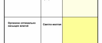
| Colorless or barely colored. | It has a bright yellow, straw color and a characteristic odor. |
The chemical composition of the secreted fluid can change during illness, increased physical activity, pregnancy and other conditions with changes in hormonal levels. Short-term differences due to dietary habits and medications often pass without a trace. A change in the formation of primary urine may indicate a systemic pathology and should be regularly monitored.
Kidney functions
The kidneys are the main excretory organ. They perform many different functions in the body.
| Function | |
| excretory | The kidneys remove excess water, organic and inorganic substances, and nitrogen metabolism products from the body. |
| Regulation of water balance | Allows you to control the volume of blood, lymph and intracellular fluid by changing the volume of water excreted in the urine. |
| Regulation of the constancy of osmotic pressure of liquids (osmoregulation) | Occurs due to a change in the amount of osmotically active substances excreted. |
| Regulation of the ionic composition of liquids | It is caused by the possibility of selective changes in the intensity of excretion of various ions in the urine. It also affects the acid-base state through the excretion of hydrogen ions. |
| Formation and release of physiologically active substances into the bloodstream | Hormones, vitamins, enzymes. |
| Regulation | Regulation of blood pressure by changing the volume of blood circulating in the body. |
| Regulation of erythropoiesis | The released hormone erythropoietin affects the activity of division of red bone marrow stem cells, thereby changing the amount of formed elements (erythrocytes, platelets, leukocytes) in the blood. |
| Formation of humoral factors | Blood coagulation (thromboblastin, thromboxane), as well as participation in the metabolism of the physiological anticoagulant heparin. |
| Metabolistic | They take part in the metabolism of proteins, lipids and carbohydrates. |
| Protective | They ensure the release of various toxic compounds from the body. |
Dependence of urine composition on external factors
The composition of urine directly depends on the following factors:
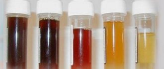
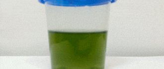
- Colors (normally straw yellow), but when taking a number of foods or medications, the urine turns orange, this is not considered a deviation from the norm. If a red tint and the color of meat slops appear, a hemolytic crisis or glomerulonephritis should be suspected. When a black tint appears - alkaptonuria, black-brown - jaundice, hepatitis, and a greenish tint - an inflammatory process in the intestines.
- Odor – urine normally does not smell. But if the smell of ammonia appears, you should think about the appearance of mucus in the urine, suppuration in the urinary cavities, or the development of cystitis. When the smell of decomposing fish appears, trimethylaminuria develops, the smell of sweat - fistula, suppuration in the urinary tract.
- Protein - normally, doctors do not observe it and the urine comes out clear. If the permissible amount is exceeded, the urine begins to foam, and when a bacterial infection is attached, it becomes cloudy and leaves with sediment.
Additional factors affecting the condition of urine:
- Acidity is normally 5–7 pH. When levels decrease, diarrhea, lactic acidosis, and ketoacidosis develop. If the indicators increase above 7 - pyelonephritis, cystitis, hyperkalemia, hyperfunction of the thyroid gland, and other kidney diseases.
- Protein - the norm is 33 mg/l of urine. In children and infants up to 300 mg/l. When protein appears above 30 mg/l, one should speak of microalbuminuria or kidney damage. Although for pregnant women, an amount not exceeding 300 mg/l does not indicate the development of kidney diseases.
- Leukocytes and red blood cells: present in the fluid in the form of 13 mm/g urine. When the amount is small, microhematuria develops; when the amount is higher than normal, macrohematuria develops. The normal leukocyte count in women is 10 mg per sample, in men – 12 mg. When exceeding 60 mg/l, urine becomes yellow-green in color and comes out with a putrid odor. Normal urine should not contain epithelial particles. Otherwise, this indicates the development of urethritis or an inflammatory process in urine.
- Salts – the main portion of urine includes inorganic salts that precipitate. But normally their amount should not exceed 5 mg/l of urine. If there is an excessive accumulation of urate, gout should be suspected when a pinkish-brick sediment appears. When oxalates appear, there is an inflammatory process, the development of colitis, pyelonephritis, and diabetes.
- Sugar - glucose is not present in normal urine, but detection of sugar up to 3 mmol/l in a daily dose is not considered pathological. Deviation from the norm indicates diabetes mellitus, diseases of the liver, pancreas and kidneys. At the same time, for pregnant women, 60 mmol/l is not considered a deviation from the norm.
- Bilirubin - the permissible value in the composition of the liquid must be insignificant. Deviations indicate diseases of the gallbladder, the development of cirrhosis of the liver, jaundice, hepatitis B, when foamy brown urine begins to pass.
Chemical composition of primary urine.
Primary urine is the liquid part of blood plasma containing a small amount of low molecular weight protein. The composition of primary urine depends entirely on the composition of blood plasma. Only those substances contained in the blood plasma enter the primary urine, therefore a number of parameters of the primary urine coincide with those in the blood plasma. Thus, primary urine has the same pH (about 7.4) and the same osmolarity (about 300 mOsm/L) as blood plasma.
Osmolarity is an important indicator for both blood and urine. However, while blood plasma osmolarity can, in most cases, be calculated with a reasonable degree of reliability, urine osmolarity is almost impossible to calculate, and osmometers are often unavailable, so laboratories usually use a measurement that has a high correlation coefficient with osmolarity. This indicator is the density of urine. Density is the mass per unit volume of a substance and is measured in g/l (for example, 1018 g/l), or in g/ml (for example, 1.018 g/ml).
There is a formula for moving from density to osmolality:
osmolality = 33275 x density - 33270
The ultrafiltrate also contains a significant amount of substances necessary for the body, including glucose, amino acids, protein, electrolytes, etc.
1) Cytolytic syndrome (cytolysis) occurs due to disruption of the structure of liver cells, increased membrane permeability, usually due to increased processes of lipid peroxidation (LPO) and the release of enzymes into the blood. With cytolytic syndrome, both cytoplasmic and mitochondrial components of enzymes enter the blood. Cytolysis mainly accompanies acute liver diseases and increases with exacerbation of chronic ones. The main available markers of cytolysis in acute hepatitis are alanine (AlAT) and aspartic (AST) transaminases, gamma-glutamyl transpeptidase (GGTP), lactate dehydrogenase (LDH). An increase in the activity of ALT and AST is observed in 88-97% of patients, depending on the type of hepatitis. Maximum activity is typical for the 2-3rd week of the disease, and a return to normal at 5-6 weeks. Exceeding the period for normalization of activity is an unfavorable factor. AlAT activity > AST, which is associated with the distribution of AST between the cytoplasm and mitochondria. The predominant increase in AST is associated with mitochondrial damage and is observed with more severe liver damage, especially alcoholic ones. The activity of transaminases increases moderately (2-5 times) in chronic liver diseases, more often in the acute phase, and liver tumors. Increased transaminase activity is not typical for liver cirrhosis. AST increases in acute hepatitis. A moderate increase is observed in obstructive jaundice, in patients with liver metastases and cirrhosis. The de Ritis coefficient, that is, the AST/ALAT ratio (normally equal to 1.33) for liver diseases is lower than this value, and for heart diseases - higher. The level of alanine amine transferase (ALAT) activity is normally 7-41 IU/l. In acute hepatitis, regardless of its etiology, the activity of aminotransferases increases in all patients. The activity of ALT contained in the cytoplasm especially changes, which promotes its rapid exit from the cell and entry into the bloodstream, therefore ALT is a more sensitive test for the early diagnosis of acute hepatitis than AST. In acute viral hepatitis, ALT and AST increase 10-15 days before the onset of jaundice with hepatitis A, and many weeks with hepatitis B, and they increase simultaneously, but ALT - much more. In the typical course of viral hepatitis, ALT activity reaches its maximum at 2-3 weeks of illness. If its course is favorable, ALT activity normalizes after 30-40 days, AST activity after 25-35 days. A repeated or progressive increase in aminotransferase activity indicates new necrosis or relapse of the disease. Prolongation of the period of increased aminotransferase activity is often an unfavorable sign, because may indicate the transition of an acute process to a chronic one. Chronic hepatitis is characterized by moderate and moderate hyperenzymeemia. Increased activity of ALT and AST can also be detected in practically healthy carriers of the hepatitis B surface antigen, which indicates the presence of apparently asymptomatic active processes in the liver. Gamma-glutamyl transpeptidase is found in the cytoplasm (low molecular weight isoform) and is associated with the membranes of the biliary pole (high molecular weight isoform). An increase in its activity may be associated with cytolysis, cholestasis, alcohol or drug intoxication, tumor growth, therefore, an increase in GGTP activity is not specific for a particular disease, but to a certain extent universal or screening for liver diseases, although presumptive requires additional searches for the cause of the disease. The enzyme is more sensitive to disorders in liver cells, ALT, AST, alkaline phosphatase, succinate dehydrogenase, glutamate dehydrogenase, etc. Lactate dehydrogenase (LDH) is elevated in many diseases. The diagnostic value is limited to the determination to exclude tumor and hemolytic processes, as well as for the differential diagnosis of Gilbert's syndrome (normal) and chronic hemolysis (increased). For the diagnosis of liver diseases, the assessment of the hepatic isoenzyme LDH - LDH5 - is more significant. In acute viral hepatitis, LDH activity in the blood serum is increased in the first days of the icteric period, and in mild and moderate forms of the disease it quickly returns to normal levels. Severe forms of viral hepatitis, and especially the development of liver failure, are accompanied by a pronounced and longer-lasting increase in LDH. In obstructive jaundice, in the first stages of blockage of the bile ducts, LDH activity is normal; in later stages, an increase in activity is observed due to secondary liver damage. In liver carcinomas or metastases of cancer to the liver, an increase may occur
29) Proteinograms characteristic of diseases associated with acute inflammatory processes. This type is characterized by a decrease in albumin. At the same time, characteristic of this type of electropherogram is an increase in the levels of fractions α1 and α2 - globulins, with which the main groups of proteins of the so-called acute phase are associated. In later stages, an increase in the content of γ-globulins may be recorded. The content of β-globulins may be increased. This type of proteinogram is typical for the initial stages of pneumonia, acute polyarthritis, exudative tuberculosis, acute infectious diseases, sepsis, extensive myocardial infarction, etc. Proteinograms are characteristic of chronic inflammatory processes. The proteinogram of such diseases shows a moderate decrease in the albumin fraction, with an increase in the peaks of α2 and γ - globulins, because when the disease becomes chronic, the formation of a number of acute phase protein fractions decreases, which leads to a pronounced redistribution of this type of protein on the electropherogram. β-globulins may be increased. This type of protein distribution corresponds to the late stage of pneumonia, chronic pulmonary tuberculosis, chronic endocarditis, cholecystitis, cystitis, etc. Proteinograms characteristic of hepatitis reflect a moderate decrease in albumin content, an increase in the level of γ - globulins and a less pronounced β - globulins. This type of electropherogram occurs in conditions of toxic liver damage, hepatitis, hemolytic processes, and possibly in malignant neoplasms and dermatoses. Proteinograms consistent with liver cirrhosis. There is a significant decrease in albumin content with a strong increase in the γ-globulin fraction; this type of proteinogram is detected in liver cirrhosis, sepsis, and in some forms of polyarthritis and collagenosis. Proteinograms characteristic of obstructive jaundice. Differs in the complex of changes; a decrease in the level of albumin and a moderate increase in the content of α2, γ and β - globulins. This type of electropherogram is characteristic of obstructive jaundice, as well as jaundice caused by the development of cancer of the biliary tract and the head of the pancreas. Proteinograms characteristic of impaired renal filter function. A significant decrease in albumin content is recorded against the background of an increase in the concentration of α2 and β - globulins with a moderate decrease in the level of γ - globulins. This type of proteinogram is characteristic of genuine or lipoid nephrosis, nephritis, cachexia, toxicosis of pregnancy, as well as a number of other diseases. Proteinograms corresponding to malignant neoplasms. A sharp decrease in albumin content is detected with a significant increase in all globulin fractions. The level of γ-globulins reaches the greatest peak of rise. This type of proteinogram accompanies metastatic neoplasms with different localizations of the primary tumor. Proteinograms characteristic of γ-globulin plasmacytomas. They are distinguished by a significant decrease in the content of albumin α2 and β - globulins with an increase in the concentration of γ - globulins. They are typical for macroglobulinemia and some reticuloses.
49) Hyperproteinemia - increased protein content relative to the norm;
Hypoproteinemia - a decrease in protein content relative to the norm; dysproteinemia is a change in the ratio between the main fractions of blood proteins, which is clearly visible from the results obtained by electrophoresis.
Dysproteinemia can be recorded when the level of total protein in the blood remains unchanged.
The cause of a decrease in the concentration of blood proteins - hypoproteinemia - may be:
- impaired protein synthesis due to enzyme deficiency / fasting, enteritis, pancreatitis, liver disease, including parenchymal hepatitis, long-term treatment with corticosteroids, alcoholism; eating food poor in animal proteins/;
— insufficient intake of proteins /pyloric ulcer, starvation/;
— insufficiency of digestion and absorption /dysentery, gastroenteritis/;
- acute and chronic blood loss of protein /glomerulonephritis and other kidney pathologies, diabetes mellitus, transudates and exudates, ascites, peritonitis, pleurisy, burns, bleeding/;
- increased protein breakdown /thyrotoxicosis, prolonged physical activity, prolonged fever, trauma, malignant tumors, sepsis/; - hyperdihydration;
- a decrease in the protein content in the blood plasma can be recorded in women during lactation and in late pregnancy.
Dysproteinemia - a violation of the ratios of the main fractions of blood proteins almost always occurs during the development of a pathological process.
Dysproteinemia can be due to a large group of proteins - globulins, or due to an individual protein, the change in which is specific to the pathological process. Electrophoresis of blood proteins makes it possible to determine the presence of dysproteinemia and makes it possible to determine the group of proteins that cause this condition
Dysproteinemia may be present with hypo- and hyperproteinemia due to simultaneous changes in the number of individual groups of proteins or accompany these changes in the blood when the balance of protein synthesis and breakdown is disturbed and a pronounced deviation of protein metabolism from the norm.
A similar picture occurs when protein is lost in the urine due to kidney pathology or through the gastrointestinal tract, as well as during fasting, cachexia, and chronic liver damage affecting the protein-synthesizing apparatus.
Deviations from the norm in the content of individual protein fractions may be due to hereditary metabolic defects associated with disruption of processes at the genetic level.
Changes in the concentration of blood proteins can be both relative and absolute. The relative change in protein content is recorded with a change in plasma /blood volume. It is known that hydremia leads to relative hypoproteinemia, and dehydration leads to relative hyperproteinemia. Dehydration hides absolute hypoproteinemia, since with this combination the protein concentration in the blood plasma is not always different from the norm. To assess absolute and relative changes in plasma protein content, it is necessary to determine either the plasma volume or the hematocrit.
51)NT-proBNP is a precursor of brain natriuretic peptide (BNP).
The heart normally produces small amounts of the protein proBNP, which are cleaved to produce the active hormone, brain natriuretic peptide (BNP), and the inactive fragment, N-terminal pro-brain natriuretic peptide (NT-proBNP). The function of the BNP is to regulate the volume of blood circulating throughout the body and, accordingly, the force that the heart must apply to pump this blood throughout the body.
BNP and NT-proBNP are mainly formed only in the left ventricle of the heart - this is the strongest “chamber” that does the most work of “pumping” blood. If fluid is retained in the body, the left ventricle works with greater load and its walls stretch. As a result, the concentration of BNP and NT-proBNP in the blood increases significantly, which leads to increased excretion of sodium, along with which excess fluid leaves. Most often, such changes occur in heart failure.
How is kidney function determined?
The efficiency of urine formation is shown by such an indicator as the rate of fluid filtration in the glomeruli of nephrons. GFR is an important diagnostic criterion that helps clarify the cause of the disease or indicate the direction to search for it. To determine how much liquid the urinary system is able to filter in 1 day, a daily portion of urine is examined. Knowing the total amount of urine, laboratory technicians can determine how many ml pass through the kidneys in 1 minute. GFR is influenced by the condition of the kidneys: the number of healthy nephrons, the total area of working capillaries and the speed of blood flow in the incoming and outgoing vessels.
Next is a comparison of the result with gender and age norms. If the value is significantly different, up or down, it is recommended to re-run the analysis. The diagnosis can be clarified using various formulas, the calculation of which requires collecting urine and donating blood in a certain order. The purpose of the sample data is to find out how much creatinine the body fluids contain.
In a healthy person, GFR is: up to 110 ml/min in women, up to 135 in men, it falls in old age. In newborns, plasma is filtered at a rate of 40 ml/min, reaching 130 by 10 years.
If urine formation does not occur properly, then too few or many cells penetrate from the blood, elements do not return to the vessels during the process of reabsorption, and urination becomes difficult. Regardless of the cause of the disorder, toxic substances accumulate in the body and kidney failure develops.
Causes of disturbances in urine production
Any problems associated with the kidneys are immediately reflected in the appearance, consistency or smell of a straw-yellow (normal) liquid.
- Protein is an element that is absent in the urine of a healthy person. When it appears in large quantities, the urine resembles beer. If it becomes cloudy, a sediment appears in it, this is a clear indication of the presence of infection.
- Smell. Usually there is none. Ammonia odor is a typical symptom of cystitis or a purulent process in the urinary system. If the toilet suddenly smells of spoiled fish, then you can be sure of “fishy odor syndrome” - trimethylaminuria. This is the name given to the body's inability to eliminate tertiary amines.
- Color. Glomerulonephritis (damage to the kidney glomeruli) and hemolytic crisis (massive destruction of red blood cells) give it the color of meat slop. A greenish tint is typical for inflammation that begins in the intestines. Hepatitis turns urine blackish-brown. A rare congenital disease - alkaptonuria (impaired protein metabolism) is rightly called “black urine disease” because of its shade.
- Normal kidney function is assessed by one important indicator - glomerular filtration rate. In men, 125 ml of primary urine should be produced in one minute, normal GFR for women is 110 ml/min.
Normal kidney function is assessed by one important indicator - glomerular filtration rate. In men, 125 ml of primary urine should be produced in one minute, normal GFR for women is 110 ml/min. Serious kidney disorders are provoked by:
- amyloidosis, systemic lupus erythematosus, glomerulonephritis, diabetes, tumors, rheumatism, scleroderma;
- poisoning with dyes, toxins, drugs;
- injuries leading to large blood loss.
To avoid kidney problems, if you suspect a problem, you should go to a urologist or nephrologist. After taking the tests, you need to follow the doctor’s recommendations, carefully monitor your diet and drinking regime, and leave bad habits in the past.
Causes of decreased kidney function
Diseases and conditions that affect GFR:
- Chronic kidney failure - the amount of creatinine and urea in the blood increases. The nephron glomeruli are damaged and filtration efficiency decreases. The same thing happens with hydronephrosis (dropsy), amyloidosis, and malignant kidney tumors. Nephropathy (disorder of the urinary system) is indicated when the value is less than 89 ml/min. If it falls below 20, the patient may enter a terminal state of uremia. The blood completely stops being purified, and a large amount of harmful decay products enters the organs. This is an indication for artificial dialysis, intensive care or kidney transplantation.
- Infections of the urinary apparatus - glomerulo- and pyelonephritis. Inflammation affects the glomerular and tubular system. In response to this, the rate at which liquid passes through natural filters decreases.
- Severe hypotension - due to the lack of pressure difference in the vessels and renal nephrons, blood cannot diffuse through the walls of the capillaries. A similar process occurs in chronic heart failure. The cause of severe hypotension may be a progressive malignant tumor, gastric ulcer, endocrine disorders, inflammation of the heart muscle (myocraditis, endocarditis), acute blood loss (as a result of injury, surgery).
- Violation of the production of hormones - substances that regulate the activity of the entire body. If the values of adrenal, thyroid, and pituitary products differ from the norm, kidney function may deteriorate. For example, in diabetes mellitus there is an excess accumulation of glucose in the blood. It penetrates through the pores of the glomerular walls, and the filtered liquid differs significantly from the primary urine of a healthy person. In the future, this disrupts the 2nd and 3rd stages of urine formation.
- Malignant neoplasms - uncontrolled tissue formation destroys adjacent cells. Decomposition products are toxic to the body and must be eliminated. Dying cells negatively affect the performance of the kidney filter. A similar mechanism of damage occurs with massive chemical and thermal burns.
- Poisoning - food, poisonous plants and mushrooms, bacterial and viral toxins, chemical dyes, heavy metal salts, overdose of medications (you must strictly follow the dosage and read the descriptions of the medications before starting therapy). Harmful substances travel through the blood to the kidneys, where an insoluble complex is formed. Toxins can damage the structures of the filtration system or “not pass” through the smallest pores, remaining in the body for a long time.
- Transfusion of incompatible blood, which causes life-threatening phenomena - shock, acute kidney failure, intravascular coagulation and breakdown of blood components.
- Rheumatic lesions - scleroderma, systemic vasculitis, rheumatism. They increase the load on the kidneys and change GFR.
- Arterial hypertension - due to the narrowing of the vascular lumen, blood is pumped into the urinary organs under increased pressure. More substances penetrate into the lumen of the glomeruli, and the final composition of urine changes significantly.
Changes in GFR in diseases are accompanied by a decrease in the amount (oliguria - up to 0.5 l, anuria - practically no discharge) and color (dirty brown, dark brown) of urine.
Conclusion
In order not to get a deliberately false result, before you start collecting daily urine (in the morning), you should urinate in the toilet. It is advisable to store the container in a cool place throughout the day. In heat, bacteria multiply intensively, which accelerate the transition of creatinine to creatine and distort the analysis values. The urine is saved for 24 hours and, together with the morning portion, is sent to the laboratory. To make it easier for a specialist to compare answers, it is recommended to calculate the level of creatinine in the blood serum (biochemical analysis) in advance.
Gomutra - cow urine... Medicinal properties.
Gomutra - cow urine. Medicinal properties. In traditional Indian medicine, the cow is literally a moving treasure trove of remedies. WONDERS OF NATURE! “GOMUTRA is a great elixir. It is pleasant to the heart, gives strength to the mind and body, gives longevity, eliminates all disorders associated with blood circulation, balances bile, mucus and air, cures heart disease and eliminates the effects of poisons.” GOMUTRA - evaporated cow urine. The value of this remedy lies in its ability to balance the three doshas (Vata, Pitta, Kapha), which helps strengthen the immune system and improve health. It also strengthens cow urine to cleanse the liver and blood, normalize digestive tract processes, lower cholesterol and reduce body fat. Strengthens the mind and purifies the heart, bringing your consciousness to a higher level of goodness. Restores damaged cells and tissues. Antibacterial agent. Instructions: 2 tablespoons per (150 ml) water, can be mixed with honey to taste. Taken on an empty stomach once daily. Cow urine can help cure numerous diseases. “Atharva Veda” and other classical texts of Ayurveda, such as “Charak Samhita”, “Rajni Ghuntu”, “Amritashagar”, “Bhavprakash”, “Shushrut Samhita”, “Ashtanga Shangraha”, etc. describe the healing properties of cow urine as a universal remedy and cure for many diseases. It is known that cow urine has been used for many centuries not only in India but also in many countries around the world. Roman emperors, as well as many European doctors and Chinese traditional healers, used cow urine for medicinal purposes. Cow urine is also included in Ayurvedic medicines: Shiva Gutika, Panchagavya Ghrita, Maha Panchagavya Ghrita, Gomutra Haritaki Gomutra is not a toxic waste product of the body. 95% of it is water, 2.5% is urea, and the remaining 2.5% is a mixture of minerals, salts, hormones and enzymes. A large number of valuable elements vital for human health were found in cow urine, such as nitrogen, sulfur, phosphoric acid, sodium, manganese, carbolic acid, silicon, chlorine, magnesium, various salts, vitamins A, B, C, D , E, minerals, lactose, enzymes, creatine, hormones, important oxides and others. Cow urine also contains volatile salts, which are very beneficial for humans, as they reduce acidity in the body. Gomutra has the property of Rasayana Tattva, which is responsible for modulating various functions of the body, including immunity. Antimicrobial and bactericidal properties are due to the presence of urea, creatinine, gold hydroxide, carbolic acid, phenols, calcium and manganese; its anticancer effect is due to the antioxidant properties of uric acid and allantoin; immunity is enhanced by gold hydroxide; and also the presence of allantoin promotes wound healing. The health of the cardiovascular system is supported by a number of its components: kallikrein is a vasodilator; the urokinase enzyme acts as a fibrinolytic agent; nitrogen, uric acid, phosphates and hippuric acid act as diuretic agents; ammonia maintains the integrity of blood elements; the components nitrogen, sulfur, sodium and calcium act as cleaning agents; while iron and erythropoietin are stimulating factors in maintaining hemoglobin levels. Kidney health is maintained by nitrogen, which acts as a renal stimulant, and urinary components act as diuretics. Gold hydroxide and copper act as antidotes for various poisons in the body. Cow urine distillate increases the effectiveness of antibacterial, antifungal and antitumor drugs. The lack of nutrients that are less available through food is compensated by the nutrients present in the cow's urine. Cow urine (gomutra) can treat severe liver diseases. Food cooked on fire by burning dry cow dung is very beneficial for health. Her milk has the ability to cure almost all ailments. The cow is our mother because she manifested from Goddess Gayatri, the most revered goddess who is worshiped by most of the followers of Hinduism. It doesn't matter whether you worship Krishna, Shiva, Kali, Hanuman or anyone else. Gayatri is worshiped by everyone and therefore her deity form, the cow, is our mother and we should never harm her. Since urine is a destroyer of poisons in our body, it destroys those diseases that are caused by toxins. Cow urine helps strengthen the body's immune system, thereby increasing the ability to resist disease. Gomutra, being a natural tonic, eliminates dizziness, nervous tension, apathy, paralysis, colds, diseases of the brain, nerves, and joints. Today, thousands of patients in India and other countries use home therapy to treat diabetes, blood pressure, asthma, psoriasis, eczema, heart attacks, vascular blockages, seizures, cancer, AIDS, prostatitis, arthritis, migraines, thyroid diseases, ulcers, hyperacidity, constipation, gynecological problems, hearing problems and breathing problems, as well as many other diseases. Cows of the “Ukrainian Gray” breed are genetic relatives of the original “Zebu” breed. Vegetarian online store “Mother Farm”
If you found an error, highlight it and press Shift + Enter or click here to inform us.