Progressive myopia is an ophthalmological disease in which visual acuity decreases at a rate of more than 1 diopter per year. Most often it occurs in people whose activities involve working at close distances to objects and with strong visual loads of other kinds. The progressive form is fraught with significant complications, including complete blindness. Therefore, the problem cannot be ignored. Let's consider how you can cope with this pathology and what can provoke it.
Causes of myopia progression
Unlike congenital myopia, progressive myopia is a pathology, the development of which is caused not by any one, but by a whole set of different factors. It is based on genetic inheritance - if parents suffered from this disease, then their child also increases the likelihood of developing the problem. Read about lens replacement surgery for astigmatism here.
The following provoking factors are genetically determined:
- weakness of the eye muscles;
- scleral weakness;
- thickening of the cornea;
- thickening of the lens.
External factors that additionally influence the development of pathology include:
- some infectious diseases;
- prolonged sitting in front of a computer/TV;
- severe strain on vision;
- hormonal and immune system disorders;
- lack of a number of microelements;
- incorrect organization of work processes, including prolonged work at close range.
Myopia also directly depends on age. At a young age, before primary school, the chance of its occurrence is minimal, no more than 6%. By high school, this probability increases to 16%, and after 20 years, the risk of its development gradually decreases, again reaching minimum levels at a more mature age.
Glaucoma
Glaucoma is a progressive disease that leads to irreversible blindness.
Due to increased intraocular pressure in glaucoma, retinal cells are destroyed, the optic nerve atrophies, and visual signals stop entering the brain.
A person begins to see worse, peripheral vision is impaired, as a result of which the visibility area is limited.
Mentions of glaucoma (translated from Greek this word means “green color of the sea”) are found in the works of Hippocrates dating back to 400 BC. However, modern ideas about glaucoma began to take shape only in the middle of the 9th century.
Currently, glaucoma is understood as a fairly large group of diseases, often of different origins and with different courses. There is still no consensus on what causes the development of these ailments, but in the absence of treatment, their outcome is the same - optic nerve atrophy and blindness.
Glaucoma
Normal vision
“Risk groups” for glaucoma include:
- people over 60-70 years of age who do not even have eye complaints;
- people over 40 years of age who: intraocular pressure is in the upper limit of normal;
- the difference between the intraocular pressure of the right and left eyes is more than 5 mmHg. Art.;
- the difference between intraocular pressure measured in the morning and evening is more than 5 mmHg. Art.;
Glaucoma can occur at any age, but the disease most often develops in older people.
Age groups
Newborns
40-50 years
60-75 years
Disease frequency
There is 1 case of glaucoma in approximately 10,000 newborns.
Experts diagnose primary glaucoma in approximately 0.1% of the population.
In this age group, glaucoma occurs in approximately 1.5-2% of cases.
According to the World Health Organization, glaucoma is a major disease that, if not treated promptly, irreversibly causes blindness. More than 5 million people have lost their sight due to glaucoma, accounting for 13.5% of all blind people in the world.
Causes of glaucoma development
In a healthy eye, a certain pressure is constantly maintained (18-22 mm Hg) due to the balance of fluid inflow and outflow. With glaucoma, this circulation is disrupted, fluid accumulates, and intraocular pressure begins to rise.
The optic nerve and other structures of the eye experience increased stress, and the blood supply to the eye is disrupted. As a result, the optic nerve atrophies and visual signals stop reaching the brain.
A person begins to see worse, peripheral vision is impaired, as a result of which the area of visibility is limited - and eventually blindness may occur.
Glaucoma is an irreversible disease. Therefore, it is very important to start treatment on time.
The main symptoms of glaucoma are:
- pain, pain, feeling of heaviness in the eyes, narrowing of the field of vision;
- blurred vision, the appearance of a “mesh” before the eyes;
- when looking at a bright light, for example, a lamp, “rainbow circles” appear before the eyes;
- deterioration of vision in the evening and at night;
- feeling of eye moistening;
- minor pain around the eyes;
- redness of the eyes.
- Open angle glaucoma
- Angle-closure glaucoma
The open-angle form is diagnosed in more than 90% of cases of glaucoma. In open-angle glaucoma, access to the natural drainage system is open, but its functions are impaired. The result is a gradual increase in intraocular pressure. As a rule, open-angle glaucoma is characterized by an asymptomatic, almost imperceptible course of the disease. Since the field of vision narrows gradually (the process can continue for several years), a person sometimes accidentally discovers that he can see in only one eye. In some cases, one can identify complaints about the periodic appearance of rainbow circles when looking at a light source, “fogging,” and asthenopic complaints associated with weakened accommodation.
In angle-closure glaucoma, intraocular fluid accumulates due to the fact that there is no access to the natural drainage system of the eye - the iris blocks the angle of the anterior chamber. As a result, the pressure increases, and this can lead to an acute attack of glaucoma, which is accompanied by:
- sharp pain in the eye and the corresponding half of the head;
- obvious visual disturbances (blurred vision or its sharp decrease up to complete blindness);
- redness of the eye (dilation of the vessels of the anterior segment of the eyeball), corneal edema, decreased depth of the anterior chamber, dilation of the pupil and lack of its reaction to light;
- the appearance of halos around light sources.
Ophthalmologists draw attention to the fact that as a result of an acute attack of glaucoma, sudden loss of vision is possible.
Diagnosis of glaucoma
To detect the onset of the disease, simply measuring intraocular pressure is not enough. It is necessary to examine the fundus and optic nerve head in detail, as well as examine the visual fields, that is, conduct a thorough diagnostic examination.
In Excimer ophthalmology clinics, examinations are performed using a whole range of modern computerized equipment and include:
- study of the visual field (using a computer perimeter);
- measurement of refraction (the ability of the optical system of the eye to refract light rays);
- measurement of intraocular pressure;
- ultrasound examinations;
- determining the depth of the anterior chamber of the eye and the thickness of the lens (since high pressure is often caused by displacement or enlargement of the lens);
- Using gonioscopy, the structure of the anterior chamber angle, through which fluid outflows from the eye, is assessed.
Also, during the diagnosis, an examination is necessarily carried out on a computer perimeter and on a fundus analyzer - a unique device available in the equipment of a few Russian clinics. This makes it possible to identify the initial manifestations of glaucoma that occur before changes in the visual field, and to stop the onset of the pathological process in time.
Gudkov Yuri Alekseevich
Ophthalmologist of the highest category
Glaucoma is surrounded by a huge number of rumors and just as many misconceptions. Some believe that only older people are at risk, others are confident that it is impossible to miss the first symptoms of the disease, while others even try to treat themselves by prescribing drops to lower intraocular pressure or using folk remedies.
As a result, it turns out that the patient reaches the ophthalmologist in a state that is far from the initial stage of the pathological process.
The changes that occur in the patient’s eye before going to the clinic, unfortunately, can be irreversible.
That is why we recommend that every patient undergo regular eye examinations, regardless of whether he or she has complaints or visible symptoms of the disease.
How to deal with glaucoma?
Remember, without timely detection of the disease and timely treatment, vision is irretrievably lost! The Excimer Clinic offers its patients the most advanced and reliable methods for diagnosing and treating glaucoma. Don’t delay treatment, don’t risk the health of your eyes!
Ë
È
Please tell me, is it possible to cure glaucoma in the early stages, without surgery?
Glaucoma is a disease characterized by a constant or periodic increase in intraocular pressure due to impaired outflow of intraocular fluid. Left untreated, it can lead to optic nerve atrophy and blindness.
In Excimer clinics, treatment of glaucoma (depending on the degree of the disease) is carried out either by conservative methods or by surgery. The goal of any treatment for glaucoma is to reduce intraocular pressure.
However, it should be noted that the drugs used during the medical treatment of glaucoma do not restore the fluid balance in the eye, but only artificially maintain it. The decision on the appropriateness and method of treatment is made by the doctor after a complete diagnostic vision examination.
Ë
È
I heard that if cataracts are not operated on, glaucoma may develop. This is true?
At an advanced stage of cataract, the swollen lens begins to occupy most of the anterior chamber of the eye, thus disrupting the outflow of intraocular fluid, as a result of which secondary glaucoma can develop. This is very dangerous, because without surgical treatment, vision is irretrievably lost.
Ë
È
Is it possible to have cataract surgery and glaucoma at the same time?
After a comprehensive examination of vision in cases where cataracts are complicated by glaucoma, the doctor decides on the possibility of combined surgical intervention (phacoemulsification of cataracts with implantation of an intraocular lens + anti-glaucoma surgery) or (if intraocular pressure is compensated) performing the operation in stages: laser treatment of glaucoma before or after phacoemulsification of cataracts with implantation of an intraocular lens.
| Ask a Question | All questions |
Article rating: 4.9/5 (478 ratings)
Source: https://rnd.excimerclinic.ru/glaucoma/
How does sport help prevent myopia?
Physical activity plays an important role in the prevention of myopia. During breaks in visual work, it is useful to move, go outside, take a walk. Children are encouraged to take walks and play outdoors more often. Sports activities help strengthen the retina, prevent spasms of accommodation and the development of myopia. Ophthalmologists advise adults and children to engage in swimming, long-distance running, and cycling.
You need to be very careful when choosing a sport, since not all of them are equally useful for myopia. To avoid the development of myopia, you should avoid strength training and heavy physical activity, which can lead to traumatic brain injury, boxing, all types of wrestling, and weightlifting. When choosing a sport, it is recommended to agree on the possibility of practicing with an ophthalmologist.
Kinds
There are several approaches to classifying myopia, but the most common one is to distinguish between equality or inequality of eye refraction. Read about the treatment of false myopia in school-age children here.
There are anisometropic and isotropic types of myopia.
Isometric
With this type of myopia, its degree is approximately the same in both eyes. The refraction value is 1 diopter or more. This is not such a strong stage of myopia, although, undoubtedly, it also requires correction.
Anisometropic
With this type of myopia, its degree is different in both eyes. Myopia is called anisometropic when the difference exceeds 4-5 diopters. With this type of myopia, it is necessary to begin optical correction as quickly as possible and carry it out as competently as possible. This material will tell you about Indian eye drops Ujala.
Myopia and glaucoma
Description
Greater unanimity is expressed regarding the appropriateness of AGO in eyes suffering from myopia.
G. in myopes often occurs without the formation of excavation, but flows according to the type of malignant progressive myopia, with the formation of true staphyloma. The formation of staphyloma contributes only to visible compensation of IOP. This should always be remembered when encountering rapidly progressing myopia. It has been established that a decrease in visual function in patients with progressive myopia (M) is more often caused not by M., but by an unrecognized glaucomatous process; that they often go blind not from M-, but from G.
The following factors are to blame for this:
1. The difficulty of assessing the nature of the process, the nature of the excavation of the optic disc.
And normal IOP numbers, often obtained during tonometry due to reduced scleral rigidity in a myopic eye, further confuse the picture.
2. Lack of alertness of ophthalmologists regarding G.
in patients. And then the deterioration of visual functions in highly myopic people is associated with the progression of M. and G. is not suspected, and, therefore, is not examined in this direction.
Among glaucomatous patients of young age (up to 40 years), myopic refraction occurs from 51.4% to 57.3% of cases. At the same time, in elderly glaucoma patients, M. refraction was noted in only 7.4%. The predominance of high degrees of M. when combined with G. is clearly determined. Moreover, the degree of M. increases as the glaucomatous process progresses. As noted by A.Ya.Bunin and E.S.Avetisov (1982), these clinical features of G. in M. determine the delayed diagnosis of glaucoma in persons with myopia.
Studying the features of the hemodynamics of the eyes of the sixth G., combined with M., it was found that they have a pronounced decrease in hemodynamic parameters compared to G. in emmetropes.
Therefore, some researchers believe that primary G. of the myopic eye occupies a special place among the clinical forms of glaucomatous syndrome. It is characterized by a progressive course with a pronounced lack of blood supply to the eye and a slightly increased level of IOP.
Huth and Messin selected a completely random group of myopic people who went to an ophthalmologist for glasses at the age of 12-40 years. And in 25% of cases G. was detected! At the same time, in the control group of the same age, but without M., not a single case of G. was identified.
To clarify the connection between high myopia and the reaction to local use of corticosteroids was studied in individuals with high myopia. And in 88% of cases, a moderate to significant positive reaction was obtained to the local use of corticosteroids. Of these, 29% had an IOP rise above 31 mm Hg. Thus, the response to corticosteroids in patients with high M. is very close to those of OAG and differs significantly from the reaction of healthy individuals.
Difficulties in diagnosis, as well as the frequent combination of G. with M. determine the relevance of an in-depth study of this type of G. Based on a retrospective study of G. in M., some researchers believe that with the help of only conservative treatment, with the help of only miotics, it is possible to steadily compensate for IOP in myopic glaucomatics impossible. And with untimely surgical treatment of glaucoma, irreversible changes develop in the eyes. Therefore, some ophthalmologists recommend surgical treatment without attempting to prescribe drug antihypertensive therapy.
Patients with progressive myopia should be classified as a group with an increased risk of glaucomatous disease and should be examined in great detail and repeatedly to exclude or identify the glaucomatous process. And having identified glaucoma in a myopic patient, you should not delay surgical treatment.
RELATED ARTICLES: Restoring vision after surgery for glaucoma Glaucoma training Celandine glaucoma
What glasses should I wear?
If myopia is diagnosed, glasses with negative diopters are usually selected. In the early stages of myopia, it is possible to restore vision with the help of correctly selected glasses. But doctors recommend using them only when necessary, since constant wearing can lead to weakening of the eye muscles, and myopia will only progress.
If the stage of development of the disease is expressed in the range from 3.0 to 6.0 diopters, then it is no longer possible to do without glasses. This is an average degree of visual impairment; glasses are selected carefully, which will correct vision and relieve the person from discomfort.
A high degree of myopia – 6.0 diopters – requires a full vision diagnosis and the selection of glasses 1 diopter less. This approach is explained by ophthalmologists by the fact that strong negative lenses of glasses significantly reduce objects and objects, which disrupts the perception of reality. In addition, there is a chance to achieve a reduction in eye fatigue.
When choosing glasses, you should also consider the distance between the centers of the pupils. If the glasses have an inappropriate center distance, the person may experience dizziness and eye fatigue.
Preventive actions
High myopia in a patient requires regular visits to the ophthalmologist once every 6 months. Its prevention includes visual hygiene, adherence to a regimen, proper nutrition and regular intake of multivitamin complexes. To prevent complications of myopia, operations such as scleroplasty and laser coagulation are also used. These manipulations will avoid detachment and rupture of the retina, slow down the progression of degenerative changes and maintain normal binocular vision.
How to stop the progression of the disease
Before carrying out treatment, you need to make sure that you are dealing with myopathy, and not with its false variety, which is also called spasm of accommodation. Read about the treatment of progressive myopia in children and adults at this link.
Fighting accommodation spasm
Treatment tactics for the latter depend on the etiology of the disease.
Drug treatment
If the doctor chooses a medicinal approach, the patient takes a number of drugs that dilate his pupil.
Self-selection of medications is prohibited; you must follow only the instructions of the attending physician.
Optical correction
This method is used only in extreme cases and only for a short period of time. It consists in the fact that the patient wears minus glasses, as well as polydiaphragms or perforation glasses. The latter are a “screen” with small holes placed in front of the patient’s eyes. Due to this, the depth of focus increases and visual acuity improves.
Surgery
Accommodation spasms are often characterized by fairly low effectiveness of conservative therapy, so theoretically surgical treatment can also be prescribed, but at the moment this is not a common practice, but isolated exceptions.
Functional treatment
There are other treatment options for spasm of accommodation:
- optical-reflex training;
- physiotherapy;
- home training (for example, exercises using a mark on glass);
- hardware treatment (for example, local barotherapy).
In general, the prognosis for spasm of accommodation is not very positive - treatment may remain ineffective, and the likelihood of relapse is high. The severity of relapses may vary. Often the spasms resolve spontaneously even without major intervention.
Fighting myopia
Now let's look at the approaches to therapy used when a patient is diagnosed with progressive myopia.
Non-drug treatment
This treatment option includes:
- Nutrition balanced in vitamins and proteins.
- Physiotherapeutic procedures, such as electrical stimulation through the skin, which helps improve blood circulation in the retina.
- Eye exercises specially selected by your doctor.
- Targeted reduction of eye strain (for example, reducing time spent in front of the TV, computer, etc.).
- Frequent walks and exercise, including swimming.
- Eye exercises specially selected by your doctor.
Drug treatment
This method cannot guarantee that vision will return to previous levels, but it can stabilize the condition of the eyes.
Also, drug treatment stops the active progression of the disease.
The following categories of drugs are used:
- Drugs that have a positive effect on strengthening the sclera.
- Drugs that lower pressure inside the eye.
- Drugs that help activate metabolic processes in the retina and blood vessels of the eyes.
- Drugs that help restore the ciliary muscle.
Self-medication in this case is unacceptable; you can only take medications prescribed by your doctor. Their selection takes into account the individual characteristics of a person.
Non-surgical treatment
This is the name for therapy using glasses or contact lenses.
For myopia
Children use glasses in any case.
In adults, everything depends on the specific situation and stage at which the disease is located.
- Glasses. They are used for myopia of both stronger and weaker degrees. But with a weak degree, it is necessary to minimize the period of wearing them, since with prolonged use they can weaken the eye muscles. It is strongly recommended to use glasses when vision ranges from 3 to 6 diopters. It is recommended to remove glasses when reading or working with small objects.
- Contact lenses. This is a more useful option for the muscles - contact lenses, as it were, form a common optical system with the eye - and the muscles begin to work as healthy. As a result, in the early stages of myopia, a better effect is achieved than with glasses, but with a high degree of myopia, the effect will be just the opposite, since the lenses are very close to the eye.
There is a special type of lenses - orthokeratological. They are able to change the shape of the cornea - they are put on at night, taken off in the morning, after which a person sees better all day. The best option is in the range from 1.5 to 5 diopters. This article will tell you about vitamins for eyes for myopia.
Surgical
If myopia has reached six diopters, you can proceed to more drastic measures - surgical sclera-strengthening operations. There are, among other things, the following:
- Scleral strengthening injection. A special polymer substance is injected into the posterior part of the eye, where it enters the sclera and becomes a kind of framework that promotes the growth of connective tissue.
- Posterior scleroplasty. Small fragments of special scleroplastic tissue are inserted into the incisions in the back wall of the eye. It is used for myopia, which is caused by an increase in the size of the eye. A common indication is during pregnancy.
- Keratoplasty. An operation that involves replacing the cornea.
- Refractive lens replacement. It is performed for advanced myopia.
- Lensectomy. Replacing the lens with an implant.
High myopia
Myopia (nearsightedness) is the most common cause of visual impairment in people under 40 years of age.
In recent years, the number of patients with this disease has been continuously growing. Worldwide, studies show that in 2000, approximately 25% of the population had varying degrees of myopia. By 2050, doctors expect that about half the people on the planet will suffer from myopia.
High myopia is the most severe form of the disease, affecting 2% of people worldwide. At this stage, the pathology necessarily requires correction.
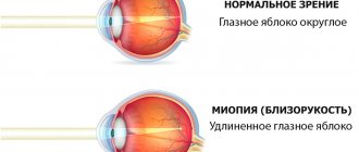
High myopia is a severe degree of myopia. The main reason for its occurrence is considered to be the irregular shape of the eyeball. It grows larger than it should and becomes very long.
There is also myopia, in which there is a high level of refraction of light rays, and the size of the eyeball is normal.
The development of myopia and its transition to the last degree is influenced by the following factors:
- genetic predisposition (the highest risk is when both parents have myopia);
- poor ability to accommodate;
- high intraocular pressure and weakening of scleral tissue;
- working at a computer, using gadgets;
- violation of diet, consumption of junk food.
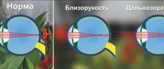
At this stage, myopia is characterized by a deviation from normal vision of more than 6 diopters. In advanced cases, this figure reaches 30 D. The main symptom is that a person sees objects clearly if they are close, but blurred if they are at a distance. Other signs of myopia include:
- wavy lines or opaque spots in the patient's field of vision;
- the need to squint to see distant objects;
- excessive eye strain;
- loss of visual acuity;
- headache.
Patients with high degrees of myopia require regular eye examinations to check for retinal damage. In many cases, myopia and its symptoms are not obvious.
What is the danger of the disease?
If left untreated, a high level of myopia can lead to the development of several dangerous ophthalmic pathologies:
- Cataract
. Patients whose treatment did not correct high levels of myopia have a 17% greater risk of developing cataracts than those in milder stages. - Glaucoma
. A systematic review of the researchers' available evidence found that people with high myopia are almost 50% more likely to develop glaucoma than those with low myopia. - Retinal detachment.
If myopia has reached the last degree, the patient experiences axial elongation of the eyes. This means that the retina is more stretched. Also, in people with myopia, the vitreous humor degenerates, which is more likely to collapse and separate from the retina. For this reason, there is a high risk of retinal tears, retinal detachment and degenerative changes. - Maculopathy
. The disease affects the central part of the retina (macula), which is responsible for the central vision of the eye. If myopia is pathological, myopic maculopathy develops. As a result, vision deteriorates and objects become blurrier.
To prevent a high degree of disease from leading to complications, regular examinations by an ophthalmologist and correction are necessary.
Methods for treating high myopia
In medical science, there are several methods of therapy that improve vision and, accordingly, the patient’s quality of life. The specific method of correction is selected by the ophthalmologist after examination. When choosing a treatment method for high myopia, various factors are taken into account: intraocular pressure, visual acuity, condition of the fundus, general anamnesis.
High myopia can be corrected in the following ways:
- Optical
. With high myopia up to 16 diopters, patients can wear glasses or contact lenses - constantly throughout the day. To make a person comfortable, the doctor selects lenses or glasses that correspond to the state of vision. - Laser correction
. Laser treatment is carried out if vision has dropped to -13 diopters. Sometimes laser correction is used at -15 D, but at higher degrees it will not be effective. The essence of the procedure is that the device makes a micro-incision in the cornea. Then the cells evaporate, as a result of which it acquires the correct shape, and vision returns to normal. There are several laser treatment options (LASIK, photorefractive keratectomy, and others). The specific option is selected by the ophthalmologist after examination. Patients aged 18-65 years are allowed for treatment. The laser correction procedure is irreversible. - Phakic correction.
The method is suitable for patients not only with extreme degrees of myopia (up to -18 diopters), but also with moderate degrees of myopia. Phakic intraocular lenses are made from a unique, highly biocompatible material for eye tissue - collamer, which includes collagen. They are implanted into the eye to reduce a person's need for glasses or contact lenses without removing the natural lens. An operation is performed for implantation. A small incision is made through which the phakic lens is inserted. This is an effective treatment method if myopia has reached the last degree, but the ability to accommodate remains. The phakic correction procedure is reversible. The lens can be replaced or removed if necessary.
Before choosing treatment, consult an ophthalmologist. It is necessary to undergo a detailed examination and accurately determine the degree of myopia in order to choose the appropriate technique. You can choose a clinic to visit here
Source: //xn--80aaa1adblkqd3ewb8g.xn--p1ai/company/miopiya-vysokoy-stepeni/
Proper nutrition to prevent myopia
When the diet is disrupted and there is a lack of vitamins, myopia develops quite often. The diet of children and adults should be enriched with foods that contain substances that are beneficial to the eyes. It is important to follow a routine - eat at the same time. Animal products will help develop visual acuity and avoid myopia: seafood, eggs, beef, turkey and other lean meats.
Vegetables and fruits are beneficial for the eyes. Carrots, white, red and cauliflower, bell peppers, pumpkin, and beets contain many useful vitamins. Blueberries, rowan, sea buckthorn, citrus fruits, and persimmons will also help resist the development of myopia. Among the drinks, freshly squeezed juices, blueberry juice, rosehip decoction, as well as milk, kefir, and fermented baked milk are very useful. It is better to give up coffee and give preference to green tea.
Maintaining the correct visual load regime
To maintain proper visual and physical activity, it is highly recommended to alternate eye strain (for example, reading or watching television) with active rest.
But if with myopia, which does not exceed 3 diopters, there are no restrictions on physical activity, then with 3 diopters and above, jumping, heavy lifting and a number of other activities are limited. It is also recommended to follow the tips below:
- Always maintain good lighting when reading or working on a computer.
- Do not read in moving vehicles, this increases eye strain.
- Do not sit closer than 40 cm from the computer.
- Every 55 minutes of working at the computer, take at least a 5-minute break.
Wearing glasses/lenses
If your doctor has prescribed you glasses or contacts (the selection principle is indicated above), it is important to take into account the specifics of the patient - where he spends more time, at the computer and reading - or in activities that involve looking into the distance.
Two pairs of glasses/lenses can be prescribed - for near and for distance, they need to be alternated depending on what type of activity is currently prevailing.
Visual gymnastics
There are many different eye exercises that help improve blood circulation to the eyes and train the ciliary muscle. Let's give a few examples.
- "Mark on the window." Place a two-centimeter mark on the window glass. Stand at a distance of 30 cm from her and look from her to some object at least 5 meters away - and back. Repeat 10 times.
- Sit down, close your eyes tightly for 5 seconds, then open them sharply for 5 seconds. Repeat 8 times.
- Move your eyes vertically and horizontally with maximum amplitude, slowly, evenly. Repeat 8 times.
- Move your eyes in a circle, first 4 times in one direction, then 5 times in the other. Repeat 2-3 times.
- Sit down, close your eyes and massage your eyelids with light circular movements for about a minute.
Glaucoma: causes, symptoms, treatment
Glaucoma is a disease that leads to irreversible blindness. About the essence of the disease glaucoma
, risk groups, as well as methods and methods of treatment, we talked with the head of the Ophthalmology Clinic of Dr. Kurenkov, professor, doctor of medical sciences, ophthalmologist
Vyacheslav Vladimirovich Kurenkov
.
Vyacheslav Vladimirovich, tell us how often glaucoma occurs and who is at risk?
Glaucoma can be congenital or acquired
Accordingly, a person who has just been born may already suffer from glaucoma. This is congenital glaucoma, it is not very favorable in terms of course and prognosis for vision, but, fortunately, it is quite rare.
Basically, glaucoma occurs in people over 40 years of age, at risk, people who are myopic, overweight, or have had any inflammatory diseases, for example, patients with inflammation of the choroid.
After such a disease, as a rule, a problem arises with the outflow of intraocular fluid: if in such patients the balance between the inflow and production/outflow of fluid is disturbed, then their risk of glaucoma is higher. Therefore, we, ophthalmologists, recommend that patients over 40 years of age
Don't forget to check your vision once a year.
This test includes checking the vision itself, that is, how a person sees, checking the anterior segment of the eye, measuring the pressure of glaucoma and a number of specific tests that tell specialists about the diseases that can develop in the fundus of the eye.
After all, most eye diseases develop unnoticed: the patient notices them when something serious has already happened, that is, when the disease has already caused significant damage to the patient’s vision.
What degree of myopia can lead to glaucoma?
Any, not necessarily big. It cannot be said that a person with a low degree of myopia has a lower risk than a person with a high degree of myopia. There are patients whose anatomy of the eye structure with normal vision (that is, in the case where there is neither myopia nor farsightedness) suggests a greater degree of risk than in other patients.
What are the symptoms of glaucoma?
The presence of glaucoma may be indicated by blurred vision, black spots, halos around luminous objects at night
... However, in general, glaucoma is asymptomatic.
The only time people actually feel an increase in intraocular pressure is during an acute attack of glaucoma.
But this is a different glaucoma (angle-closure glaucoma), it is associated with disorders and structural features of the anterior segment of the eye, where the outflow of fluid occurs - then glaucoma progresses quickly. Most patients have open-angle glaucoma.
What can increased eye pressure lead to?
The optic nerve suffers from increased eye pressure, and subsequently it partially or completely dies, and, unfortunately, it cannot be restored. That is, loss of the optic nerve leads to irreversible blindness.
Accordingly, early detection of the disease is very important, which allows preserving the optic nerve through the use of adequate therapy.
Another example, a patient develops cataracts and sees quite well, but the lens of this cataract is very large.
A large lens disrupts the anatomy of the anterior segment of the eye and blocks the outflow of fluid - that is, the lens is still transparent, the patient still sees well and is in no hurry with the operation, but later the lens reaches such a size that it closes the angle of the anterior chamber and increases the pressure.
Accordingly, the patient sees well, pressure is not felt, but the optic nerve dies. In this case, it is necessary to remove the lens as a preventive measure for glaucoma and cataracts and install an artificial one.
Tell us in more detail how open-angle glaucoma differs from closed-angle glaucoma?
Open-angle glaucoma is glaucoma in which the pressure increases gradually, and the patient does not feel it, that is, the disease is practically asymptomatic. Angle-closure glaucoma is glaucoma with attacks. This case has its own characteristics.
Even emergency doctors and clinical doctors, therapists, often confuse symptoms
headaches and poor health in their patients with hypertensive crisis, migraine and other diseases, including heart attack.
The question of differential diagnosis is to determine that the patient actually has an acute attack of glaucoma, and not some other disease.
How should a person behave if an attack of glaucoma occurs?
Immediately visit an ophthalmologist and apply the whole range of measures aimed at lowering blood pressure: these are medications, laser and other methods. In an acute attack of glaucoma (if the pressure is above 40 mm Hg), the optic nerve can withstand the load for 48 to 72 hours. If a person does not apply treatment within three days, he will go blind.
Are there people who live with glaucoma, periodically extinguishing attacks?
Attacks may not be accompanied by such high blood pressure, but it must be remembered that each attack “eats” part of the optic nerve.
Tell us about glaucoma treatment methods
Initially, we always select a drip regimen - eye drops
reduce the production or increase the outflow of fluid from the eye.
If the patient is fine on this regimen, his blood pressure does not increase and his visual functions do not deteriorate, then he is on the drip regimen for many years and is treated with medication. Glaucoma cannot be treated with tablets. We sometimes prescribe tablets to lower blood pressure before surgery.
Glaucoma is always treated with drops. The drops compensate for intraocular pressure, and the patient remains under observation, but he visits the doctor once every six months or once a year in order to monitor all the functions of the eye that a person with normal vision has.
If, against the background of the drip regimen, visual functions deteriorate (the field of vision narrows), then we recommend surgery
. This suggests that the pressure in such a patient is uneven and varies throughout the day.
There are many types of operations for glaucoma - they are divided, for example, into laser and surgical.
During laser surgery, part of the eye is stretched or holes are “punched” in a certain segment to increase the outflow of fluid, but such manipulations most often give a temporary effect for 3-8 months or a year; there are rare cases when the result lasts more than a year.
After this, you have to perform a surgical operation and create a new, alternative, outflow of fluid, since the old one no longer works.
There are classic operations, for example, lobectomy, when the conjunctiva is opened, the part where the filtration occurs is opened, a flap is made, the conjunctiva is sutured, and an outflow of fluid is created under the conjunctiva. Another method is drainage adjustment. Express drainage is a device that is inserted into the anterior chamber; fluid flows out through it under the conjunctiva.
Currently, there is a wide arsenal of means to ensure that a person maintains his vision throughout his life. An important point is the human factor, in which people neglect visits to an ophthalmologist and underestimate their condition, believing that it can go away like a cold.
Is preparation for such an operation standard?
Yes, everything is standard. The operation is performed under local anesthesia; no injections are required. The operation is scheduled to last 15-20 minutes and immediately after it the person goes home with normal blood pressure.
That is, the operation does not require a hospital stay?
All true modern ophthalmic surgery does not require a hospital stay. Surgery is aimed at doing everything that is necessary for the health of this particular organ at the time of the operation, and nothing will change if you are in hospital. Initially, this was a US practice, but now it is used everywhere, including here in Russia.
That is, the patient sees well immediately after the operation and can go home independently?
The patient undergoes antiglaucomatous surgery not to restore vision, but to compensate for intraocular pressure. During surgery for glaucoma, we only normalize the pressure so that vision does not deteriorate further.
Therefore, it is incorrect to say that the patient sees well immediately after surgery. Glaucoma surgery and vision restoration are two different things.
The danger of glaucoma is that it is impossible to return vision to its previous level due to the death of the optic nerve.
Once again about prevention. You said that after 40 years you need to visit an ophthalmologist every year...
Right. And not only check vision, but also measure intraocular pressure, look at the anterior segment of the eye, its anatomy and look at the fundus.
Can you name the average cost of glaucoma treatment in Moscow? How do you know where exactly you should go?
In order to prescribe appropriate treatment for glaucoma, it is necessary to do a specific examination for it, that is, with visual fields, with an assessment of the optic nerve head, with parametric data. It costs from 5 to 7 thousand rubles
, and it is enough to prescribe drip treatment.
If surgical intervention is required, the operations cost from 20-50 thousand and above
– it depends on the specifics of the operation itself. Laser surgery costs about 10-15 thousand. Treatment is not as expensive compared to how expensive it could be without these measures.
Glaucoma is a two-way process.
Are the costs of glaucoma surgery covered by compulsory health insurance (CHI)?
Yes, they are covered.
Another thing is that private clinics do not operate under this insurance, because compulsory medical insurance does not cover the costs of this operation. I will give an example of compulsory medical insurance and cataracts.
According to compulsory medical insurance, the state is ready to return about 12 thousand rubles for cataract surgery, while the cost of such an operation is 4 times higher.
Private clinics do not operate under compulsory medical insurance, since in this case they are at a significant disadvantage.
What is the percentage of patients who come in with eye diseases - astigmatism, cataracts, glaucoma?
According to statistics, cataracts are 2-3 times more common than glaucoma, because everyone has cataracts. Another thing is that not everyone lives to see it - for some it should be at 50 years old, and for others at 100 years old. Glaucoma affects 10-15% of the population.
Accordingly, if a person lives even to 100 years old, he may not have glaucoma. The lens is a natural transparent lens with its destruction and opacities, so even if it is of normal shape, but cloudy, it is still better to change it.
This does not apply to glaucoma: if the outflow of fluid from the eye is good and there are no pathologies, it will be good all your life.
How to preserve the eyesight of schoolchildren?
The peak development of myopia occurs during school years. During this period, the load on the child’s visual apparatus increases significantly. From 8 to 12 years of age, active growth of the eyeball occurs. Myopia very often develops due to increased load on the visual organs. Parents should monitor their child's posture.
The table and chair should be selected according to the child’s height. This is necessary so that the student can sit with a straight back. Scoliosis often leads to myopia. When performing tasks at the computer, it is necessary that the monitor be located at a distance of at least 45 cm from the child’s eyes. The font used by the student must be at least 14, and the screen scale must be 100%. Prolonged visual work often causes myopia. Therefore, breaks are necessary to help avoid prolonged focusing of the eyes on close objects. It is recommended to take a “break” every 45 minutes of work. Its duration must be at least 10 minutes. When studying, lighting a child's desk is very important. The light source should be located at the top left for right-handers, and at the top right for left-handers. It is better to choose lamps of a warm shade. Their power should be 70-75 watts. For 1 sq. m. of the desktop should receive lighting equal to 15-20 watts. It is better to do homework in natural light, during the daytime.
Indications for surgical treatment
Surgery cannot be performed if the patient is under 18 years of age. In other cases, surgical treatment is used if myopia actively progresses and threatens vision. In this case:
- If there is a threat of developing blindness due to retinal detachment, laser coagulation, soldering of blood vessels and the retina can be used.
- If progressive myopia is at a strong stage, lensectomy and lens replacement may be performed. This also prevents vision hazards.
- For slight myopia, no more than 6 diopters, refractive keratotomy is used, which consists of making incisions on the cornea. This helps improve its shape and curvature.
- If laser vision correction cannot be performed (for example, with advanced myopia), they resort to refractive lens replacement. As a result, the non-functional lens is replaced with an artificial analogue.
In each individual case, the need for these or other procedures should be determined by the attending physician based on the specific situation of the patient.
Features of treatment
The advanced form of the disease is treated with surgery, since the problem cannot be solved with medication.
Difficulties in treating glaucoma with myopia are associated with late diagnosis. In the later stages of the disease, surgery is performed because other methods are no longer effective. In such cases, drug components are not able to fully reduce intraocular pressure. The optic nerve is seriously damaged. The changes are irreversible and it is impossible to restore normal vision. No attempts are made to treat the patient with medications; surgery is immediately prescribed, which is performed as soon as possible.
As myopia progresses, it is recommended to perform special exercises to prevent and stabilize intraocular pressure. By carrying out such manipulations, eye tension is relieved, blood circulation and metabolism are improved. As a result, the volume of aqueous humor decreases. But gymnastics is effective only at the initial stage of the disease, when surgical intervention can be avoided.
If there are significant negative changes in the membrane of the eye, the indicators are stabilized, after which surgery is performed. For these purposes, special preparations are used in the form of drops, usually combined. The action of medications is aimed at reducing fluid production and improving its outflow. Ophthalmologists prescribe:
- "Fotil Forte";
- "Cosopt";
- "Proxofeline."
Two-stage surgical treatment
Laser treatment of glaucoma is the first step in surgery for pathology and myopia.
First, they treat glaucoma, then correct myopia. To normalize the patient's condition, trabeculectomy is performed by removing the ligamentous canal and sclerectomy using a laser under local anesthesia. After laser exposure, recovery lasts up to 3 days. Complications occur rarely, the results are stable and depend on individual characteristics.
Some patients may require multiple surgeries. To consolidate the results obtained over a long period of time, drops are used that contain components that can reduce blood pressure. In parallel, regular examinations are carried out. Only after reaching normal values of the indicator does treatment continue, aimed at correcting myopia.