What is diabetic cataract
Diabetes mellitus affects not only the pancreas, causing it to produce less and less insulin (in type 1 diabetes) or impairing tissue sensitivity to insulin (in type 2 diabetes).
This disease affects all organ systems, including diabetes, which also affects the organs of vision. Cataracts are a common complication of uncompensated diabetes mellitus. It affects patients with both type 1 and type 2 diabetes. In type 2 diabetes, cataracts are more common than in type 1 diabetes.
Cataracts are understood as clouding of the lens, which leads to the fact that a person begins to see worse. In the later stages of cataract development, vision may be completely lost.
Cataracts are common in people without diabetes, but their development is triggered by some kind of eye injury. And most often it develops in older people. This is a natural process. But in people with diabetes, cataracts develop regardless of injury or age of the patient.
Symptoms
Diabetic cataract is manifested by symptoms of classic lens opacity, only there are developmental features - a tendency to rapid progression of the pathology. Clinical manifestations are associated with loss of transparency of the natural lens of the eye and impaired transmission of light rays.
Symptoms of cataracts in diabetes mellitus:
- cloudiness, blurred vision, the appearance of a white veil before the eyes;
- decreased clarity of vision;
- the appearance of dark, flaky spots before the eyes;
- Difficulties with visual stress, when working with small objects;
- distortion of color perception;
- doubling of objects;
- increased photosensitivity;
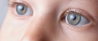
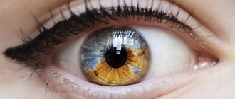
- the pupil becomes whitish.
Types of diabetic cataracts
Diabetic cataracts are divided into two types:
- True cataract;
- Senile or senile cataract.
True cataracts develop as a result of diabetes itself, due to disturbances in carbohydrate metabolism.
Senile cataracts develop due to old age.
Cataracts can develop over a long period of time until a person consults an ophthalmologist. According to the stages of development, experts distinguish 4 stages of cataracts:
- Initial stage;
- Immature stage;
- Mature stage;
- Overripe stage.
Depending on the stage of cataract development, treatment methods are selected.
Prices for treatment for diabetic cataracts
The cost of cataract removal with implantation of a rigid anterior chamber or posterior chamber lens (1 eye) at MGK is 70,000 rubles. The final price is determined after consultation with a specialist and depends on the model of the lens being installed and the condition of the patient’s eyes. You can find out the price of procedures, as well as various models of intraocular lenses, by calling 8 (800) 777-38-81 and 8 (499) 322-36-36 or online, using the appropriate form on the website.
Go to the “Prices” section>>>
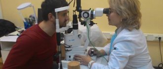
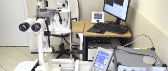
Detection of cataract development
If you suspect the development of cataracts, consultation with both an endocrinologist and an ophthalmologist is necessary.
The endocrinologist should check the blood sugar level and the level of glycated hemoglobin.
The ophthalmologist will conduct a number of examinations, including:
- Visual acuity testing;
- Examination of the eyeball - at this time, individual flakes of opacities, if any, may be noted;
- Fundus examination to rule out the development of diabetic retinopathy;
- An ultrasound examination of the eye is possible.
Stages of treatment
When diagnosing diabetic cataracts, the patient should be observed by an ophthalmologist and an endocrinologist. It is important to compensate for diabetes and stabilize the patient's condition. For these purposes it is necessary:
- normalize the metabolic process;
- choose the right diet;
- minimize blood glucose levels by prescribing adequate insulin therapy or selecting hypoglycemic drugs;
- add vigorous physical activity.
This will allow for more effective diabetes compensation. If the condition improves, you can begin directly to treat complications. The operation is recommended to be performed in the early stages of the pathology, before the progression of inflammatory eye diseases.
What contributes to the development of diabetic cataracts?
The development of cataracts is influenced by the presence of diabetes mellitus. Particularly important points are:
- Duration of diabetes – the longer a person lives with diabetes, the higher the risk that they will develop cataracts;
- Diabetes compensation - the worse the compensation, the higher and longer the periods of hyperglycemia, the higher the risk of developing cataracts;
- Diabetic acidosis – Acidosis activates enzymes that cause the lens to become cloudy.
With increased sugar, damage to the fibers of the lens occurs, since hyperglycemia causes disturbances in water metabolism and interferes with normal moisture circulation. This leads to the development of clouding of the lens.
The second way cataracts form is that the accumulation of glucose leads to its transition to the form of sorbitol, which accumulates in the anterior chamber of the lens. This also leads to the development of cataracts.
Causes
The leading etiological factor of diabetic cataract is an increase in blood glucose levels in type 1 and type 2 diabetes. In insulin-dependent diabetes, the clinical picture of the disease is revealed at a younger age, this is due to chronic hyperglycemia against the background of absolute or relative insulin deficiency. In non-insulin-dependent diabetes, the interaction of cells with the hormone is disrupted; such changes are more typical for patients in the middle age group.
The risk of developing cataracts directly depends on the diabetic “experience”. The longer a patient suffers from diabetes, the higher the likelihood of developing lens opacities. An abrupt transition from oral tablet forms of hypoglycemic drugs to insulin for subcutaneous administration can become a trigger that starts a chain of pathological changes. It should be noted that with timely and adequate compensation for carbohydrate metabolism dysfunction, such disorders can be avoided.
Treatment of cataracts in diabetes mellitus
Cataracts respond well to treatment. Of course, the earlier the diagnosis is made and treatment carried out, the higher the likelihood of a favorable outcome.
Surgical methods of treatment
Treatment for diabetic cataracts involves replacing the clouded lens with a new one.
Nowadays, phacoemulsification (lens replacement surgery) takes place with virtually no side effects and is easily tolerated by patients.
It is important to detect the development of cataracts in time in order to perform surgery in the initial stages. In this case, the results will be most effective. If the operation is performed in the later stages, then it will not be possible to completely achieve the original vision that was before the development of cataracts.
The operation requires certain preparation and compliance by the patient with a postoperative regimen.
Conservative treatment methods
Conservative treatment methods are effective only in the initial stages of cataract development.
Normalizing compensation can reverse the changes caused by early stage cataracts. The sooner normoglycemia is achieved, the higher the likelihood that the effects of cataracts will go away forever.
When establishing compensation, for a better result, eye drops are prescribed that help get rid of the effects of clouding.
Is surgery always necessary?
To treat cataracts, surgery is not always necessary, although, in fact, this is the only way to radically solve the problem. During surgery, the lens is replaced with its artificial counterpart, due to which the patient’s vision is restored. But in the early stages, with the help of medications and blood sugar control, you can try to stop the development of the disease. If the cataract does not progress, then the patient has every chance of maintaining normal vision for a long time without surgery.
Surgical intervention allows you to get rid of cataracts in advanced cases, but it is possible only in the absence of contraindications. For example, severe retinopathy, which affects a large part of the retina, can be a serious obstacle to surgery. Difficulties also arise when small blood vessels grow on the iris of the eye. In these conditions, the question of the advisability of surgical treatment should be decided by several ophthalmologists based on data from an objective examination and instrumental examinations.
Another contraindication to surgery is inflammatory eye diseases. First, it is necessary to eliminate the acute process with the help of drug treatment and local procedures, and only after that plan to replace the lens. Modern surgical techniques allow the intervention to be performed under local anesthesia and with a minimal incision area. For this purpose, laser equipment and artificial analogues of the lens made of reliable polymer materials are used.
Preventive measures
Unfortunately, diabetes mellitus totally, in almost all aspects, affects the quality of life. The patient has to remember and observe numerous restrictions, follow recommendations, monitor the composition of the blood, regularly visit the supervising endocrinologist - in order, among other things, not to miss the onset of the development of one of the possible complications of diabetes and to take timely measures to prevent such complications. Periodic examinations and consultations with an ophthalmologist in this regard are mandatory.
- Visual acuity testing, examination of the lens and fundus structures should be carried out at least twice a year. The fact is that intervention in the development of cataracts at its earliest stages makes the prognosis much more favorable, and therapeutic measures are truly effective. Even if indications for microsurgical operation are identified, it should be performed as early as possible, before more severe complications develop and become chronic.
- You should know and remember that there are a number of drugs specially designed for the prevention and protection of the visual organs in diabetes mellitus - for example, catalin, catachrome, taurine, quinax, etc. As a rule, the course of prevention takes 1 month and consists of daily eye drops. After a certain break, the course is repeated.
In some cases, periodic courses of cataract prevention must be taken for life; but this is much better than cataract itself with severe visual impairment and the risk of losing it completely.
It should also be taken into account that some of the drugs prescribed for diabetes mellitus have undesirable side effects. In particular, trental, which effectively stimulates blood circulation in the extremities, can negatively affect blood microcirculation in the ocular structures and even cause hemorrhage. Therefore, the observing ophthalmologist must be informed about which drugs and in what dosages are prescribed as part of the treatment of a general disease in order to take into account additional negative effects on the eyes and take adequate measures to neutralize these effects.
In particular, the drug “Anthocyan Forte” is distinguished by its high efficiency and complex action. Like many other ophthalmic preparations, it is borrowed from nature itself and contains natural extracts of blueberries, black currants, seeds of certain grape varieties, etc. A high concentration of vitamins, nutritional and protective microelements creates a powerful antioxidant effect (free radical oxides are one of the main immediate causes of lens clouding), strengthens the vascular system of the fundus of the eye, and helps maintain visual acuity in daylight and at dusk.
Features of the development and treatment of diabetic cataracts
Diabetes mellitus is a systemic disease, affecting not only the kidneys, blood vessels, reproductive and nervous systems, but also the eyes. Diabetic cataracts are a common complication of the disease.
A cataract is a clouding of the lens. The disease is natural for older people, but in young people it can be a consequence of injury or a complication of systemic diseases, including diabetes.
The only way to cure cataracts is surgery. Modern phacoemulsification allows to minimize complications and achieve good quality of vision even in patients with diabetes. If previously every fourth diabetic was denied implantation of an artificial lens, now the operation is performed even with serious disorders in the visual system.
Surgery for cataracts in a patient with diabetes requires careful preparation and planning, physician care, and longer postoperative follow-up. However, when phacoemulsification is performed correctly, complications are observed no more often than in patients without diabetes.
Cataracts in diabetes mellitus
A person with diabetes may develop true cataracts, caused by impaired carbohydrate metabolism, and senile (senile) cataracts.
Diabetic cataracts are divided into initial, immature, mature, and overripe. The degree of maturity will determine the choice of surgical technique and prognosis. It is believed that cataracts develop faster in diabetes.
Incidence of cataracts in diabetes
Research shows that 30% of patients who live with diabetes for more than 10 years develop cataracts. With a disease duration of 30 years or more, the frequency increases to 90%. It is noteworthy that women develop cataracts twice as often as men.
In patients over 40 years of age who suffer from diabetes, cataracts are diagnosed in 80% of cases. The risk of lens opacification in a diabetic increases with age, as well as with poor glucose control and concomitant diabetic retinopathy.
Mechanisms of development of diabetic cataracts
Cataracts in diabetes do not develop due to excess sugar in the lens masses, because this requires a lethal five percent concentration. However, there is a direct connection between the rate of lens opacification and the concentration of sugars in the aqueous humor of the anterior chamber of the eye.
A sharp increase in the level of sugar in the anterior chamber fluid in uncompensated diabetes leads to blocking of the glycolytic pathway of absorption and a transition to sorbitol. The conversion of glucose into sorbitol causes galactose cataracts, because biological membranes are impermeable to sorbitol. The accumulation of sorbitol in the lens leads to the development of true diabetic cataracts.
With endocrine disorders, direct damage to the lens fibers is also possible. Excess glucose causes a decrease in the permeability of the lens capsule, disruption of local metabolism and moisture circulation. As a result, metabolic processes and circulation in the lens are disrupted, which causes clouding. In diabetes mellitus, swelling and degeneration of the epithelium of the ciliary processes are also observed, which leads to deterioration in the nutrition of the lens.
Diabetic acidosis may also be a cause. With reduced acidity, proteolytic enzymes are activated, which can stimulate cloudiness. Diabetes also affects lens hydration as the osmotic pressure in tissue fluids decreases.
There is a photochemical theory of the development of cataracts in diabetes. It is based on the fact that excess sugar and acetone in the lens increases the sensitivity of proteins to light, which causes them to become cloudy. The exact pathogenesis of diabetic cataract is not fully understood, but each of the listed factors has its own influence.
Clinical picture of diabetic cataract
Dotted or flake-like white opacities appear in the surface layers. Subcapsular vacuoles can form both on the surface and deep in the cortex. In addition, water gaps form in the bark. Sometimes diabetic cataract has all the signs of a normal complicated one: color iridescence, vacuoles, opacities of the peripheral cortex in the center of the lens.
If carbohydrate metabolism is normalized in time, the initial diabetic cataract disappears within 2 weeks. Without treatment, deep gray opacities subsequently appear, and the lens becomes cloudy evenly.
Senile cataracts in diabetes develop at a young age, affect both eyes and mature faster. Brown nuclear cataracts and significant refractive changes toward myopia are often diagnosed, although cortical, diffuse, and posterior subcapsular opacities are also common.
Changes in the lens in diabetes are always combined with degeneration of the iris. Most patients also experience microcirculation disorders.
Conservative treatment
If you normalize sugar levels in a timely manner, you can not only delay the development of cataracts, but also achieve partial or complete resorption of the cloudiness. In the presence of gross opacities, clearing and delayed development of the disease are unlikely.
Therapy for rapidly developing diabetic cataracts with significant disturbances in carbohydrate metabolism consists of diet, oral medications or insulin injections. In patients with senile cataracts who suffer only from slight visual impairment and myopia, it is sufficient to compensate for diabetes and regularly use eye drops. A very popular mixture is riboflavin (0.002 g), ascorbic acid (0.02 g) and nicotinic acid (0.003 g) in 10 ml of distilled water.
Cataract drops:
- Vita-Iodurol. A preparation with vitamins and inorganic salts, which is prescribed for nuclear and cortical cataracts. It is based on calcium chloride dihydrate, magnesium chloride hexahydrate, nicotinic acid and adenosine. Chloride compounds improve nutrition of the lens, and acid and adenosine normalize metabolism.
- Oftan Katahrom. Drops with cytochrome C, adenosine and nicotinamide. Thanks to this composition, the drug has an antioxidant and nutritional effect. In addition to cataracts, Oftan Katahrom is effective for nonspecific and non-infectious inflammations in the anterior part of the eye.
- Quinax. The synthetic components of the drug prevent the oxidation of free radicals. The active ingredient is sodium azapentacene polysulfonate. It suppresses the negative effects on lens proteins and stimulates proteolytic enzymes in the intraocular fluid.
In the later stages of cataracts, conservative therapy is ineffective. In case of visual impairment, surgical treatment is recommended regardless of the degree of maturity of the clouding.
Surgery
Phacoemulsification with intraocular lens placement is the procedure of choice for diabetic cataracts. An artificial lens is called an intraocular lens. With its help, you can additionally correct refractive errors (myopia, farsightedness, astigmatism).
The best conditions for surgery are initial or immature cataracts, when fundus reflexes are preserved. Mature and overripe cases require increased ultrasound energy and, accordingly, greater load on the eye tissue. In diabetes, the eye tissues and blood vessels are very weak, so increasing the load is undesirable. Also, with mature cataracts, the lens capsule becomes thinner and the zonules of zonules weaken. This increases the risk of capsule rupture during surgery and complicates the implantation of an artificial lens.
Preoperative examination
Before surgery, the patient must obtain permission from a physician, dentist, and otolaryngologist. Preliminarily exclude the presence of HIV infection and hepatitis, check blood clotting and do an electrocardiogram. Before cataract removal, you must separately obtain permission from an endocrinologist.
The operation is not performed in severe renal failure, even if there is a risk of blindness. Contraindications to prosthetics would be lens subluxation and severe vitreoretinal proliferation in combination with iris neovascularization.
During biomicroscopy, the doctor should pay attention to the iris, since it reflects the state of the vascular system of the eyes. Neovascularization of the iris may be a sign of diabetic retinopathy.
Opacities may complicate ophthalmoscopy. Instead, an ultrasound B-scan is performed, which shows the morphological structure of the eye. Ultrasound scanning can detect hemophthalmos, retinal detachment, proliferation and vitreoretinal complications.
Preparing for surgery
For two days before surgery, it is recommended to instill Tobrex, Floxal or Oftaquix 4 times a day. Immediately before surgery, the antibiotic is instilled 5 times per hour.
On the day of surgery, the glycemic level should not exceed 9 mmol/l. In type I diabetes, the patient does not eat breakfast and does not take insulin. If insulin levels are not exceeded after surgery, it is not administered. At 13 and 16 hours, the glucose level is determined again, the patient is given food and transferred to the usual regimen.
In type II, pills are also discontinued. If the glucose level after surgery is below normal, the patient is immediately allowed to eat. When glucose levels are elevated, the first meal is postponed until the evening, and the usual diet and diabetes treatment are returned to the next day.
During surgery and for some time after, sugar levels may increase by 20-30%. Therefore, in severely ill patients, sugar levels are monitored every 4-6 hours for two days after the intervention.
Features of phacoemulsification in diabetes
The optimal treatment method for diabetic cataracts is ultrasound phacoemulsification with implantation of flexible intraocular lenses. It is necessary to take into account that diabetics have a smaller pupil diameter and it is more difficult to achieve mydriasis.
Since patients with diabetes often have defective vessels and vulnerable endothelium of the cornea, lens removal is performed through a puncture in the avascular part of the cornea. The puncture is only 2-3.2 mm and does not require sutures, which is also important for diabetes. Removing the suture injures the corneal epithelium, which, against the background of weakened immunity in diabetics, is fraught with viral and bacterial keratitis.
If subsequent laser treatment is recommended for the patient, lenses with a larger optical diameter must be used. The physician must use instruments carefully, as there is an increased risk of iris neovascularization and bleeding into the anterior chamber of the eye.
The phacoemulsification technique allows you to maintain the tone of the eyeball, which reduces the likelihood of hemorrhagic complications. With a combined intervention, phacoemulsification is first performed, and then vitrectomy with the introduction of silicone or gas. The intraocular lens will not interfere with the examination of the fundus during vitrectomy and photocoagulation.
Postoperative complications
Patients with diabetes require increased attention at all stages of treatment and even in the postoperative period. An inflammatory reaction is possible 4-7 days after surgery, requiring hospitalization of the patient. After cataract surgery, postoperative endophthalmitis may develop.
Macular edema after phacoemulsification is a very rare complication. However, some studies show that in people with diabetes, macular thickness may increase by 20 microns after surgery. As a rule, the swelling disappears by the end of the first week, and only in some cases the complication has an aggressive form and after 3 months develops into full-fledged macular edema.
Secondary diabetic cataract
Phacoemulsification and hydrophobic acrylic IOLs have reduced the incidence of secondary cataracts. The main reason for this complication is insufficient cleansing of the capsule from lens cells, which subsequently regenerate and become cloudy again. The design of the new IOLs prevents the ingrowth of cloudy cells into the optical zone.
It is noteworthy that in people with diabetes, the lens epithelium regenerates less, so secondary cataracts are observed half as often as in healthy people. However, in diabetic retinopathy, posterior capsule opacification is 5% more pronounced. On average, secondary cataracts develop in patients with diabetes in 2.5-5% of cases.
Cataracts are becoming more common in diabetes, but modern medicine can successfully treat them. Today, almost every diabetic can regain good vision without consequences.
Sources: https://beregizrenie.ru/katarakta/diabeticheskaya-katarakta/ https://sosudy.info/kavinton-i-davlenie
General information about the disease
Cataract is a disease of the visual system, during which cloudiness gradually accumulates in the lens. In the future, it causes a deterioration in the visibility of the eye, up to the onset of blindness. In most cases, cataracts are diagnosed in patients whose age exceeds 40-45 years.
The presence of diabetes mellitus aggravates the situation, so the disease can occur at an earlier age. Here we are talking directly about the development of diabetic cataracts. As the disease progresses, it goes through several stages of development:
- Stage 1 – absence of specific symptoms. As long as the patient's vision does not deteriorate, he does not feel any severe discomfort. Pathological changes can be seen by an ophthalmologist during a routine examination. Even if there are no complaints, it is necessary to be regularly examined by doctors.
- Stage 2 – visible damage to the central area in the lens. This type of cataract is called immature. Interference appears on the retina and vision deteriorates.
- Stage 3 – complete clouding of the lens. At the same time, its transparency is lost. Clear visibility is rapidly decreasing. It is difficult for the patient to distinguish between light sources. This type of cataract is called mature.
- Stage 4 – complete blindness. In this case, processes occur that cannot be stopped. Because of them, the tissue in the lens is destroyed as much as possible.
Preparing for cataract surgery
Before performing surgery, a patient with diabetes mellitus must undergo a complete examination. It includes a number of laboratory tests and consultation with an endocrinologist: it is important to achieve compensation for the disease to reduce the risk of complications in the postoperative period.
Modern methods of removing the lens for cataracts have a short rehabilitation period: no large incisions are made and no stitches are applied. To monitor the effectiveness of the operation, the doctor prescribes an examination at a certain time. Compliance with all medical recommendations allows you to achieve excellent results in the form of a significant improvement in vision.
The article is for informational purposes only. To make a diagnosis and choose a treatment method, consultation with a specialist is necessary.