Causes
The main cause of the disease is herpes infection. It is caused by the following strains of the pathogen:
- herpes simplex types 1 and 2;
- zoster;
- cytomegalovirus.
A predisposing factor may be the presence of the Epstein-Barr virus in the patient’s blood, which suppresses the activity of the immune system.
We recommend reading: What is keratitis
When pathogenic microorganisms first enter human blood, the disease does not manifest itself in an obvious form. The patient may develop rashes on the lips, which subsequently disappear without forming a connective tissue scar. Next, the virus penetrates the nervous tissue where it is localized. There he is dormant and does not show activity.
If a person is exposed to negative environmental factors, for example, extreme cold, frequent colds, swimming in polluted waters, the virus is activated and moves along the nerve fibers. From there it travels to the eyeballs, where it begins to develop.
The virus itself does not cause destruction of eye tissue in the form of ulcers, erosions, or foci of necrosis. This is facilitated by the activation of the immune system, that is, immune cells infiltrate the eye tissue, releasing inflammatory mediators. All this leads to characteristic clinical symptoms and the formation of lesions.
Treatment of the disease
As mentioned above, the treatment of herpes keratitis depends on its form.
For the tree-like form without stromal damage, the “gold standard” is a combination of topical application of Ganciclovir and oral administration of Acyclovir. Ganciclovir in the form of an ophthalmic gel is prescribed for use 5 times a day, Acyclovir in tablets of 200 mg - 5 times a day for 10 days. Their intake ensures rapid suppression of developing infection in the tissues of the eye.
It may also be advisable to prescribe Valacyclovir orally.
When the stroma is damaged, it may be necessary to take antihistamines. As a rule, in these cases, prednisolone drops are prescribed every two hours for 10-15 days. In parallel, systemic antiviral therapy is carried out with Acyclovir or Valacyclovir.
Diclofenac solution is also used as an anti-inflammatory agent. If necessary, the edges of ulcers can be shaded with brilliant green, and when the ulcers heal, Korneregel and Actovegin are prescribed to accelerate tissue regeneration.
For metaherpetic ulcers, diclofenac and regenerating drugs are also prescribed, and sometimes glucocorticoids for more gentle scarring.
Additional procedures may include washing the tear ducts and using B vitamins.
Finally, in some cases, after a severe course of the disease, surgical intervention may be required: keratoplasty, the use of autologous blood, the application of therapeutic contact lenses or cadaver corneas.
In any case, treatment of herpes keratitis is carried out exclusively in a hospital setting due to the complexity of all manipulations. Self-treatment at home can be dangerous not only because of the possible lower effectiveness, but also because of the risk of eye damage if the procedures are carried out inappropriately.
Classification
There are several forms of disease, each of which has specific clinical symptoms:
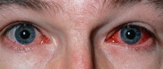
- Classic shape. The patient's cornea becomes cloudy, bubbles with transparent contents appear on it, inside which the herpes virus is located. The eyes become very red and inflamed.
- Tree or geographic form. The patient's cornea becomes covered with ulcers that appear as a result of a herpetic infection. These formations are located before the eyes in the shape of a tree or a branch from a river. The surface structure of the eyes is significantly destroyed. Gradually, the form changes from tree-like to geographical, that is, in the form of continents. This means that the eye tissue is severely damaged.
- Stromal or discoid form. It is not the superficial part of the cornea that is damaged, but its internal region, that is, the stroma.
- Necrotic form. This is the most dangerous type of disease. With it, extensive foci of necrosis are formed, which quickly spread throughout the eyeball. Without emergency treatment measures, this form of the disease leads to blindness.
All forms of herpetic eye damage are curable with timely identification of the disease and the cause of the pathogen.
How is the disease treated?
Depending on the cause and classification of herpetic keratitis, drug or surgical treatment is prescribed. To suppress the virus, the ophthalmologist prescribes:
- “Vidarabine.” Helps against particularly resistant infections.
- “Acyclovir.” Available in ointment form.
- "Florenal". Used as drops and ointment for superficial herpetic keratitis.
Along with antiviral drugs, agents are prescribed that reduce the sensitivity of cells to the virus. The drugs are called immunomodulators. Popular among these are “Poludan”, “Pyrogenal”, “Interferon”, “Polyacrylamide”.
When medications fail and complications develop, surgery is prescribed. The main indications for its implementation are considered to be rapid deterioration of vision and damage to the central part of the cornea. There are 2 types of operations for herpetic keratitis:
- Scraping. During the operation, the inflamed epithelium is removed using a special instrument. Afterwards, the cut areas are washed with an antiseptic.
- Keratoplasty. Prescribed if scraping does not help. Keratoplasty can be through or layer-by-layer. In the second case, the affected area is completely removed. Penetrating keratoplasty involves preliminary treatment of eye diseases. Eating is prohibited before surgery.
During surgical operations, painkillers and a microscope are used.
Symptoms
The herpetic form of keratitis is formed in the form of characteristic clinical symptoms, by which the doctor can assume the disease:
- increased production of tear fluid, which can be replaced by dryness of the membrane, which leads to additional damage to the corneal layer;
- the eyelids are in a state of spasm, in which the patient cannot open his eyes;
- photophobia, that is, strong sensitivity of the eyes to the action of light, especially bright light, which causes spasm of the eyelids and lacrimation;
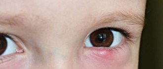
- severe redness of the superficial structures of the eyes;
- a feeling of a foreign body under the eyelids, which causes a person to rub their eyes, which worsens the condition;
- painful sensations in the eyes: itching, burning, pain;
- at the initial stage, bubbles filled with transparent contents appear on the cornea; if they burst, the herpes virus easily infects surrounding people.
A characteristic feature of herpetic rashes is that after the blisters burst, no scars form on the mucous membrane. The tissues heal quickly, but cause severe pain and redness. If the true cause of herpes is not eliminated, keratitis will become chronic.
Gradually, erosions and ulcers will appear in the patient’s eyes.
Chronic herpetic keratitis is associated with the fact that the pathogen persists in the nerve tissues. If a person is affected by negative environmental factors, it moves to the eyeball and infects it.
Causes and symptoms of the disease
Contact with the herpes virus on the mucous membrane of the eye is the main cause of herpetic keratitis. There are 2 types of herpes. In the first case, infection occurs by airborne droplets. The second type of disease is transmitted through unprotected sexual intercourse, during childbirth, from mother to child.
Most people are considered carriers of the virus. But it is almost always in the body in a state of sleep. Factors that activate the virus:
- previous infections;
- disruptions in the functioning of the endocrine system;
- weakened immune system;
- Severe ARVI, influenza epidemic;
- periodic stress, nervous breakdowns;
- hypothermia;
- overheating;
- excessive exposure to ultraviolet radiation on the body.
The first sign of the development of eye pathology is irritation and clouding of the cornea. The man avoids the light. The inflammatory process spreads to the sclera, the ciliary body, and the iris. Herpetic keratitis is characterized by a symptom such as corneal edema.
Diagnostics
To determine the cause of the disease, it is recommended to consult an ophthalmologist. He will conduct several diagnostic tests, based on which he will make a reliable diagnosis.
- Interviewing the patient or his caregivers. There are complaints of redness, pain in the eyes, and blisters often form. It is with the last symptom that the ophthalmologist can assume a herpetic form of the disease.
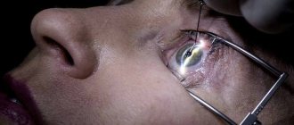
- General inspection. For this, the doctor uses a slit lamp. But the patient may develop photophobia and spasm of the eyelids, so in such cases, examination in daylight is avoided. Severe redness and inflammation of the eyes are detected, rashes, erosions, ulcers, and foci of necrosis may appear.
- General clinical analysis of blood and urine, blood biochemistry. An increase in leukocytes and ESR will be detected in the blood, which indicates an inflammatory focus in the body.
- Serological study. It is necessary to identify the viral pathogen. With its help, the doctor determines the true cause, for example, herpes virus type 1 or 2, zoster.
- Bacteriological culture. It often happens that a herpes infection is complicated by bacterial conjunctivitis. In this case, you will need to determine the pathogen and the antibiotics to which it is sensitive.
- Biomicroscopy. This is an intravital examination, with which you can determine the condition of the eye tissues without removing their area. Under a microscope, the doctor can see areas of necrosis, erosion, and other cell damage due to the action of the virus.
Based on the data obtained, the doctor can not only confirm the herpes infection, but also the exact pathogen that caused it.
Keratitis
Signs
The clinical picture is characterized by the so-called corneal syndrome (lacrimation, photophobia, blepharospasm), caused by irritation of the sensory nerves of the cornea. With keratitis, clouding of the cornea is observed, which develops as a result of its infiltration and is accompanied by a decrease in transparency and gloss, a violation of sphericity and sensitivity. The absence of specularity and shine against the background of unevenness of the anterior surface of the cornea in the area of infiltration indicates ulceration. Patients also experience pericorneal injection, which, with concomitant conjunctivitis, takes on a mixed character. The inflammatory process may spread to the iris, ciliary body and sclera with the development of iritis, iridocyclitis and scleritis. Prolonged or severe course of the disease can lead to complications such as corneal perforation, complicated cataracts, secondary glaucoma, optic neuritis, endophthalmitis.
Description
The most common are infectious keratitis, among which viral diseases predominate. The main causative agents of viral keratitis are herpes simplex viruses and adenoviruses; they also occur in viral diseases such as chicken pox and measles. Among infectious keratitis of non-viral origin, bacterial ones caused by coccal flora, Pseudomonas aeruginosa, pathogens of tuberculosis, and syphilis are especially common. There are also chlamydial, fungal, parasitic (with toxoplasmosis, onchocerciasis, leishmaniasis, amebiasis, etc.) keratitis. There are traumatic keratitis caused by the direct influence of damaging factors on the cornea (mechanical, thermal, chemical, radiation). The impact of these factors on the trigeminal nerve can lead to the development of so-called neuroparalytic keratitis. There are keratitis of immune origin (corrosive Moraine's corneal ulcer, sclerokeratitis in rheumatoid arthritis and necrotizing nodular polyarthritis, dry keratoconjunctivitis in Sjögren's syndrome, corneal pemphigus, etc.), allergic keratitis (atonic, in spring catarrh, hay fever, medicinal giant papillary keratitis toconjunctivitis), and also keratitis associated with metabolic disorders (rosacea keratitis, keratitis in diabetes mellitus, gout, psoriasis,
The state of general and local immunity is important in the development of the disease; it also largely determines the nature of the course of keratitis and the severity of the pathological process.
The main morphological sign of keratitis is swelling and infiltration of corneal tissue. Infiltrates consisting of lymphoid, plasma cells or polynuclear leukocytes have unclear boundaries, different shapes, sizes, and colors. The latter depends mainly on the cellular composition of the infiltrate (with a predominance of lymphoid cells, its color is whitish-grayish, with purulent infiltration it acquires a yellowish tint). The process can cover no more than 1/3 of the thickness of the cornea - the epithelium and upper layers of the stroma (superficial keratitis) or spread throughout the entire stroma (deep keratitis). In severe cases, corneal necrosis occurs, leading to the formation of abscesses and ulcerations.
A sign of compensatory and restorative processes in keratitis is the vascularization of the cornea - the ingrowth of newly formed vessels from the edges of the looped network into it. The nature of vascularization depends on the depth of the lesion; with superficial keratitis, the vessels, dichotomously branching, pass through the limbus from the conjunctiva to the cornea towards the infiltrate; with deep keratitis, they have a linear course and grow through the thickness of the cornea in the form of a brush.
Herpetic and bacterial keratitis are of greatest importance in clinical practice due to the frequent occurrence and severity of the consequences. Herpetic keratitis is caused by the herpes simplex virus. Its main forms are dendritic, metaherpetic and discoid keratitis. Arborescent keratitis is a typical superficial keratitis. An early sign of it, determined by biomicroscopy of the eye, is the rash of small bubbles in the corneal epithelium, which tend to open and leave behind an eroded surface in the form of characteristic figures, often in the form of tree branches, less often snowflakes or stars. Sometimes such a lesion is accompanied by the formation of small point infiltrates located in the epithelium and anterior layers of the stroma.
Metaherpetic and discoid keratitis are characterized by damage to the deep layers of the corneal stroma, accompanied in a significant proportion of patients by involvement of the anterior part of the choroid (keratoiridocyclitis). With metaherpetic keratitis, severe damage to the corneal stroma is observed in the form of ulcerations of various shapes and sizes. A stromal ulcer often resembles a river bed. In half of the patients, biomicroscopy of the eye reveals tree-like figures against the background of stromal infiltrate. Recurrence of the process is accompanied by vascularization of the cornea. The disease often occurs with severe pain, is persistent, and is often accompanied by perforation of the cornea. Discoid keratitis begins with swelling of the epithelium and stroma. Subsequently, more often in the central zone, a rounded lesion forms. The sensitivity of the cornea is significantly reduced, and the degree of vascularization may vary. If the infiltrate is replaced by intense clouding, visual acuity sharply decreases.
The second place in frequency of occurrence after herpetic is purulent keratitis (purulent, or creeping, corneal ulcer), the typical pathogens of which are coccal flora (pneumococcus, streptococcus, staphylococcus), as well as diplobacillus Morax-Axenfeld. Due to the widespread use of antibiotics, opportunistic gram-negative bacteria, mainly Pseudomonas aeruginosa, have become a frequent causative agent of purulent keratitis. Cases of purulent keratitis have become more frequent in patients with reductive herpetic keratitis treated with corticosteroids. The development of the disease is often preceded by microtrauma of the cornea or chronic dacryocystitis, blepharoconjunctivitis. Initially (usually in the center or paracentral zone of the cornea), a gray infiltrate is formed, which subsequently acquires a purulent character. An ulcer appears in its place. One edge of the ulcer is usually raised and undermined, and has a crescent shape. Melting of tissues begins in this zone. The process progresses quickly and within 3-5 days can cover the entire cornea. In the center, the cornea completely melts, and therefore in this area you can find a protruding dark-colored bubble of a stretched Descemet's membrane - a descemetocele. The process usually involves the iris and ciliary body. At the bottom of the anterior chamber of the eye, pus (hypopyon) accumulates, occupying more than half of its volume. The most malignant course is purulent keratitis caused by Pseudomonas aeruginosa, in which the process develops especially quickly and spreads not only deep into the cornea, but also to the surrounding sclera (sclerokeratitis). After spontaneous perforation, self-healing can occur with the formation of a cataract, or the purulent process spreads to the deep parts of the eye with the development of endophthalmitis and atrophy of the eyeball, the latter is especially characteristic of purulent sclerokeratitis.
Keratitis of a specific origin is characterized by focal or diffuse infiltration of the deep layers of the cornea. In children suffering from tuberculous bronchoadenitis, tuberculous-allergic lesions of the cornea and conjunctiva often occur with the formation of small nodules on them - conflicts.
Diagnostics
The diagnosis of keratitis and its etiological forms is established on the basis of the clinical picture and medical history, which is of greatest importance in identifying herpetic keratitis, which is characterized by significant polymorphism of changes. Common signs for various clinical forms of herpetic keratitis are the connection of keratitis with an infectious disease that occurs with fever, the presence of herpetic rashes on other parts of the face, the neurotrophic nature of the lesions (for example, decreased sensitivity of the cornea), pain along the branches of the trigeminal nerve, delayed regeneration of the process, failure antibiotic therapy, frequent (in approximately 50% of patients) tendency to relapse. The main research method for keratitis is eye biomicroscopy, which allows one to accurately determine the size and nature of the lesion, as well as detect signs of keratitis in the early stages of the disease. If a purulent lesion of the cornea is suspected, as evidenced by a yellowish tint of the infiltrate, an urgent bacterioscopic examination is carried out and the patency of the lacrimal ducts is determined. The presence of ulceration is confirmed by a fluorescein test. Also, for keratitis, laboratory methods are widely used - bacteriological and cytological examination of the epithelium of the conjunctiva and cornea, immunological research methods, allergic diagnostic tests with various antigens (antiherpetic vaccine, tuberculin, brucellin, etc.), the method of mirror microscopy of the posterior corneal epithelium.
Treatment
Treatment, especially for deep forms of keratitis, is carried out in a hospital. The nature of treatment is determined by the cause of the disease. Treatment of viral keratitis is based on the use of antiviral agents. Immunoglobulin is instilled into the conjunctival sac, and immunomodulators are used orally and parenterally. For ulcerations, antiviral therapy is recommended to be combined with microsurgical interventions (microdiathermo- and laser coagulation, cryoapplication). In case of herpetic keratitis occurring with ulceration, local use of corticosteroid drugs is strictly contraindicated due to the risk of developing severe complications - bacterial infection and corneal perforation. If there is no effect from conservative therapy within 1 month and a sharp decrease in visual acuity, patients undergo a cornea transplant. To prevent relapses of herpetic keratitis, the administration of an antiherpetic vaccine is indicated.
When treating bacterial keratitis, sulfonamides and broad-spectrum antibiotics are prescribed in the form of instillations, ointments and medicinal films, taking into account the sensitivity of the pathogen. For severe ulcerative lesions of the cornea, which are most often caused by staphylococcus and Pseudomonas aeruginosa, antibiotics are also administered subconjunctivally, intramuscularly or intravenously. For corneal ulcers, microdiathermocoagulation and other microsurgical interventions are indicated. In case of progressive purulent infection of the cornea, emergency care is provided in a clinic (diathermocoagulation of the ulcer, administration of antibiotics and sulfonamides), after which the patients are immediately sent to the hospital, where, if conservative measures are ineffective, they undergo a corneal transplant.
In the treatment of tuberculous keratitis, anti-tuberculosis chemotherapy drugs and hyposensitizing agents are used, while syphilitic keratitis is treated with antisyphilitic therapy.
Along with specific drugs for keratitis of various etiologies, mydriatics, novocaine blockades along the superficial temporal artery, antiseptic solutions, and agents that promote epithelization of ulcers are prescribed locally. When visual acuity decreases, electro- and phonophoresis with enzymes and biogenic stimulants are prescribed. According to indications (decreased visual acuity as a result of scar changes, secondary glaucoma, etc.), keratoplasty, antiglaucomatous and other operations are performed.
The prognosis is largely determined by the cause of keratitis, the localization and nature of the infiltrate, and associated complications. With timely and rational treatment, superficial infiltrates completely resolve or slight cloud-like cloudiness remains. Deep keratitis, occurring both with and without ulceration (with central and paracentral localization of the infiltrate), can lead to a significant decrease in visual acuity due to the development of corneal opacities of varying degrees of severity of irregular astigmatism. Keratitis complicated by endophthalmos leads to complete loss of vision.
Prevention
Prevention of keratitis consists of preventing injuries (including microtraumas) of the eye, timely detection and treatment of blepharitis, conjunctivitis, dacryocystitis, immunodeficiency conditions and various diseases that contribute to the development of keratitis.
© Medical encyclopedia of the Russian Academy of Medical Sciences
Treatment
To treat a herpes infection, the patient is prescribed complex therapy to not only eliminate the activity of the virus, but also normalize the condition of the eye tissue:
- Systemic antiviral drugs. For most viruses, the effect of such drugs has not been proven. But for the herpes virus there is a drug called Acyclovir, which only affects this pathogen.
- Local antiviral drops. They can be used over a long period of time; the pathogenic microorganism does not become addictive. They use Actipol, Poludan.
- Moisturizing drops. They are applicable if the patient develops dry eye syndrome. The advantage of such drugs is the possibility of frequent use, there are no side effects, and the body does not become addicted.
- Antibacterial drops. They are necessary if the patient has a complication in the form of a bacterial infection. They are used for no more than 7 days, after which the bacteria develop resistance. They use Vigamox, Tobrex, Levomycetin. Ophthalmologists recommend applying antibacterial ointment under the eyelids at night. For this, Tetracycline and Erythromycin ointment are used.
- Non-steroidal anti-inflammatory drops. They reduce swelling, inflammation, redness and pain.
- Vasoconstrictor drops can be used for no more than 4 days, since the body becomes addicted after this time. Vizin is suitable for such purposes. It effectively eliminates redness and eye irritation.
To prescribe individual therapy, it is recommended to consult an ophthalmologist. The patient may experience side effects if the drug was prescribed independently. This will worsen the patient’s health, his health will become worse.
Treatment of herpetic keratitis
Treatment of the disease must begin quickly, since damage to the cornea leads to clouding, which can lead to blindness.
In the treatment of the disease, antiviral drugs are used, which are prescribed in the form of drops and ointments locally. These are medical products - analogues of interferon, or substances that activate the production of its own interferon, which protects the body from viruses. In case of severe inflammation, systemic antiviral agents are also used.
An intense inflammatory process that continues for several weeks without a response to drug therapy requires surgical intervention - layer-by-layer or penetrating keratoplasty, in which a donor cornea is transplanted for therapeutic purposes.
If clouding has formed, it is also possible to perform a layer-by-layer or through-the-hole corneal transplant. In this case, it is performed to improve vision. The operation is prescribed after all manifestations of keratitis have subsided.
Complications
If therapy was not carried out on time or completely, and the patient was treated independently, this can lead to complications for the patient:
- the appearance of erosion and ulcers on the cornea, which causes clouding and deterioration of visual acuity;
- blindness in one or both eyes, if the disease has reached the terminal stage and no treatment has been carried out;
- in people with immunodeficiency, the herpes virus can spread throughout the body, causing other lesions, for example, on the skin, genitals, or mouth;
- spread of herpes infection to the brain;
- a bacterial infection that occurs as a result of a disruption in the composition of the normal microflora of the superficial structure of the eyes.
If the patient is a child under 3 years of age, herpes infection always causes a sharp increase in body temperature, more than 39 degrees. In the absence of antipyretic drugs, he may develop seizures, which will lead to damage to small or large parts of the brain.
What complications can there be?
Untimely or incorrect treatment of herpetic keratitis can not only increase the number of relapses, but also cause a number of complications that will cause irreparable harm to health.
Such complications include:
- glaucoma;
- cataract;
- corneal clouding;
- partial or complete loss of vision;
- thorn;
- retinal detachment.
If help was provided on time and the treatment was effective, then this eliminates the possibility of consequences.
Prevention
Once the herpes virus has entered the bloodstream, it is impossible to eliminate it from there. It will forever remain in the human nervous tissue. It is possible to prevent only its periodic activity, which occurs due to exposure to negative environmental factors. To do this, it is recommended to take the following measures:
- proper nutrition containing vitamins, microelements, minerals and other beneficial substances;
- in the autumn-winter period there are not enough nutrients in food, so it is recommended to take multivitamin preparations;
- frequent walks in the fresh air to allow early oxygen to enter the body;
- clothing according to the weather, that is, you cannot wrap yourself up or, conversely, wear clothes that contribute to hypothermia;
- It is not recommended to swim in public bodies of water with stagnant water in the summer, as the largest number of pathogenic microorganisms that infect all people are concentrated there.
In most cases, a herpetic infection is not dangerous for the patient. It is easily treated with medications. But in some cases, herpetic keratitis can form, which is dangerous by reducing the patient’s visual acuity. It is recommended to consult a doctor promptly and carry out proper comprehensive treatment. In this case, the patient's vision can be completely preserved.
FAQ
How does the virus spread?
The spread of herpetic keratitis usually occurs through direct contact between an active herpetic lesion in any other part of the body and the eyes . To prevent this, you must strictly maintain contact lens hygiene; if you suffer from herpetic keratitis in one eye, do not spread it to the other.
Other measures to prevent herpetic keratitis and its relapses are :
• Avoid touching your eyes if you have a cold or inflammation in another part of the body.
• Do not wear contact lenses if you have active ocular herpes.
• Do not use cortisone ophthalmic drops if you have an active herpes infection, as this may allow the virus to multiply and, in the case of a previous herpes infection, may contribute to a new outbreak.
Possible complications
Complications of keratitis:
• Inflammation and damage to the cornea, which can leave scars on the cornea
• Corneal ulcers with risk of bacterial infection
• Chronic or recurrent viral infections of the cornea
• Chronic inflammation of the cornea
• Temporary (due to inflammation during active infection) or permanent (due to residual scar) loss of vision.
How long does it take to treat the disease?
The duration of herpetic keratitis will depend on the size, depth and cause of it . Eye ointments or oral antiviral medications commonly used to treat herpetic keratitis are prescribed by an ophthalmologist.
Despite this, the infectious period lasts from 7 to 10 days . After this period, corneal damage and inflammation persist until tissue regeneration begins. It may be complete and without complications in cases of superficial epithelial infection or with consequences due to residual scar in cases of stromal lesions.
Are people who wear contact lenses more likely to develop this type of condition?
Yes, people who wear contact lenses are 6 times more likely to develop herpetic keratitis, especially if they wear them for many hours a day or at night, as this causes small epithelial wounds through which the herpes virus can enter. This is why it is very important to practice good hand hygiene (especially if you have an active herpes infection in another part of the body), use lens cleaning and preservative solutions correctly, do not overuse contact lenses, do not swim with them, and remove them if you notice any discomfort.
Can the infection come back?
Although precautions are taken, it is very common for herpetic keratitis to reactivate due to the following factors:
• Stress
• Ultraviolet radiation
• Injuries or bruises on the body
• Use of certain medications
• Menstruation
• Instilling some eye drops
Patients with immunodeficiency or atopy are more prone to recurrent or refractory herpetic keratitis. Relapse occurs due to the fact that the herpes viral infection affects a large part of the population (the first infection can appear in early childhood, even in the form of a cold). After the first infection, which never goes away, but our immune system controls it and blocks the virus in the lymph nodes of the body. In the previously mentioned situations, when a certain immunosuppression occurs, the virus takes the opportunity to leave the ganglion and affect some parts of the body, mainly the lips.
Forms of the disease according to external signs
Depending on the intensity of the signs and the nature of the damage to the cornea, the primary type of keratitis has two types:
- Superficial keratitis : herpetic rashes damage only the epithelium with the appearance of ulcerative formations and irritation of the nerve roots. Signs: feeling of a speck getting into the eye, pain in bright light, profuse lacrimation, red mucous membrane.
- Deep keratitis : spread of the inflammatory process to the connective tissue, formation of a purulent focus involving blood vessels. Signs: severe pain, decreased visual acuity, damage to the organ of vision by a cataract. This condition carries risks of complications such as the appearance of an extensive ulcer, perforation of an ulcer, which often leads to complete loss of vision.
Superficial, in turn, is divided into two forms:
- Vesicular : mild stage of the disease. Small blisters of herpetic rash appear on the epithelium, in the place of which, after their independent rupture, erosions form.
- Tree-like : The rash affects the cornea in a twig-like shape.
Deep for three:
- Metaherpetic : extensive damage to the corneal stroma with the formation of a deep ulcer. Characterized by frequent relapses.
- Discoid : a chronic condition that causes the cornea to thicken and become ingrown with blood vessels. The signs are general. A scar remains at the site of the inflammatory process.
- Diffuse appearance : complex and severe stage of disease development. It is characterized by total damage to the organ of vision, the development of corneal necrosis, cataracts and secondary glaucoma.
With deep herpetic keratitis of the diffuse type, the outcome of the disease is rarely positive; surgery is required; there is a high risk of loss of vision, or the entire eye.
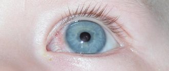
You can see what the various manifestations of herpetic keratitis look like in the photo below:
Reasons for appearance
Why does this disease make the world medical community nervous? The main causative agent is herpes simplex virus type I. It affects the patient’s body, bringing him a bunch of dermatological, neurotropic and mesodermotropic problems.
Attention! The first eye problems appear in people whose immunity suddenly weakens. The body is weakened and cannot resist viruses. Children under 2-5 years of age and adolescents are at risk.
When diagnosing, different types of keratitis are revealed, in particular epithelial in 70 percent of cases, stromal in 20 and metaherpetic in 10. The source of all problems - HSV-1 does not greatly harm the body: all lesions are superficial.
Much more dangerous is HSV-2, which has an even more severe course and often transforms into stromal keratitis. HSV-1 has a long course of illness. In patients, the mucous membranes, eyelids and other integuments are affected . Very rarely they develop an inflammatory process in the optic nerve.
Disease therapy
Treatment is complex and includes the following:
- Instillation of drugs with antiherpetic spectrum of action.
- Use of local spectrum steroids.
- Undergoing physiotherapeutic procedures.
- In case of total damage to the cornea - surgical intervention.
It is allowed to use traditional medicine recipes as an auxiliary therapy to relieve symptoms and alleviate the general condition.
Medicines
For superficial forms of the disease, the following drugs are used:
- Idoxyuridine solution 0.1% : for instillation. Number of procedures – up to 8 per day. Duration of use is no more than 14 days; if longer, toxic effects on the body are possible.
- Local spectrum drugs: Acyclovir , Zovirax, Virolex . Directions for use: Apply under the eyelids 3 times a day. The drugs have a depressing effect on the herpes virus, help to quickly relieve swelling and reduce the inflammatory process.
- Medicines for instillation: Interlock (dilute 10,000 IU per 0.1 ml of phosphate buffer). Dosage and duration of use are individual.
- Reaferon (5000 IU per 1 ml of distilled water). Directions for use: 2 drops 1 time per day. Duration of use – 6 days;
- Fron : 2 drops 6 times a day. Duration of use is 1 week.
Intramuscular administration of vitamins B1, B2, intake of ascorbic acid.
In case of deep form, the following is added to treatment:
- Ointment Zovirax, Virolex, Acyclovir (3%): apply the product to the skin of the eyelids up to 5 times a day.
- Reaferon for instillation.
Often, the doctor may prescribe additional intramuscular administration of immunomodulators and vitamin B2. In the case of a pronounced pain symptom, lidocaine injections are prescribed.
Symptomatic treatment includes taking antibacterial drugs and using antiseptics. In case of protracted course of the disease, non-steroidal anti-inflammatory drugs and steroids are prescribed.
Physiotherapeutic procedures
To quickly suppress the activity of the virus and relieve inflammation, electrophoresis is prescribed. How it is carried out: using medicinal ophthalmic agents with an antiherpetic spectrum of action, with which cotton swabs are impregnated and applied to the cornea. In case of frequent relapses of the disease, vaccination is recommended in the autumn.
Traditional medicine recipes
To relieve pain and reduce inflammation, you can also seek help from the following folk recipes:
- Sea buckthorn oil : 1 drop every 60 minutes for 2 days, then 1 drop every 2-4 hours. The product reduces the intensity of pain and removes symptoms such as photophobia.
- Celandine : used in the presence of a purulent focus. How to use: celandine juice (1 part), propolis solution (3 parts), mix. Use the resulting solution as eye drops before bed, dosage 2 drops. If irritation occurs, dilute celandine with propolis in a ratio of 1 to 5.
- Clay compress : place a 3 cm layer of thick clay on a napkin. Apply to the forehead and hold for 1.5 - 2 hours. Apply 2 times a day.
Before using folk recipes for eye drops, you should consult your doctor. There may be contraindications depending on the severity of the clinical case.
Surgical intervention
Indications for the operation:
- Lack of positive dynamics from drug therapy for 1-3 months.
- Decreased visual acuity.
- Presence of complications (damage to blood vessels, hardening of the cornea).
The type of surgical intervention depends on the severity of the clinical case. The part of the epithelium that was affected by the virus is scraped off to prevent further spread of the pathological process. In case of severe, extensive damage, keratoplasty is performed - resection of parts of the epithelium affected by the virus.