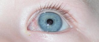
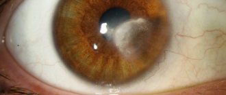
A cataract is a major disease accompanied by clouding of the cornea. The main reason for its development is cicatricial changes in the cornea (wide genesis). As a result of scarring, it becomes opaque and ceases to transmit light normally. In addition, the cornea acquires a characteristic shade - white, porcelain. Over time, a network of blood vessels grows into the thorn, the processes of fatty degeneration begin, and the leukoma becomes yellow. The main method of treating pathology is surgery.
Causes of herpetic keratitis
Herpetic keratitis is activated against the background of decreased immunity for a number of reasons:
- as a result of a cold, fever or hypothermia;
- due to prolonged stress;
- hypovitaminosis;
- microtrauma of the cornea, conjunctivitis or other eye diseases;
- viral, fungal and bacterial infections;
- a number of chronic diseases (for example, syphilis, tuberculosis, malaria)
Under any unfavorable conditions, once the herpetic keratitis virus has entered the body, it appears on the eyelids or cornea of the eye.
“Doctors are hiding the truth!”
Even “advanced” herpes can be cured at home. Just remember to drink once a day...
>
The herpes virus is contagious and has several routes of transmission:
- contact and household;
- sexual;
- airborne;
- transplacental.
Most often, infection of the cornea causes a microdefect in the surface of the eye itself, which in 85% of cases is associated with the use of contact lenses. The source of spread is the storage solution or their handling if the user does not comply with hygiene rules.
Causes of eyesore
The cornea is the first line of the eye on the path of infections and pollution, so most inflammatory processes occur on it. One of the reasons for the appearance of cataracts is eye injuries. If the damage is not penetrating and is localized at the periphery of the cornea, it will most likely not affect visual acuity. The situation is much more complicated with extensive penetrating wounds, since it is the localization of the cataract that determines the degree of decrease in visual acuity.
This group also includes chemical burns of the eyes, the scars after which are especially dense and rough. Surgical treatment in this case is practically useless, since chemicals quickly penetrate into the deep layers of the cornea, and its damage becomes inoperable.
A thorn may be a direct consequence of keratitis - inflammation of the cornea caused by infections (bacteria, fungi, microorganisms). Characteristic signs of keratitis are redness of the eyes, burning, pain, tearing and purulent discharge. Without proper treatment, once the inflammation occurs, the cornea will begin to scar and form a corneal cataract. The cause of keratitis can be bacteria introduced into the eye through contact with dirty hands, etc., as well as certain microorganisms. There are known cases when plumbing amoebas were found on the membranes of the eye, which were brought there by contact lenses washed with ordinary tap water. Such cases are quite rare, but the most unpleasant thing about them is that any infectious advanced inflammation can lead to complete blindness.
A thorn in the cornea of the eye can be a consequence of syphilis, chlamydia, HIV, tuberculosis and other serious diseases. As a rule, such manifestations can be observed in advanced cases, in which they are only secondary symptoms of the disease. Despite the fact that pathogens do not cause specific eye diseases, these infections tend to become chronic and affect all organs and systems.
Classification of herpes keratitis
| Primary | Herpetic blepharoconjunctivitis (follicular, membranous) | |
| Epithelial keratitis | ||
| Keratoconjunctivitis with ulcerations, vascularization of the cornea | ||
| Post-primary (recurrent) | Surface | Vesicular |
| Arborescent keratitis | ||
| Deep (stromal keratitis or keratouveitis without/with ulcerations) | Metaherpetic (amoeba-shaped), discoid | |
| Focal, discoid |
- Primary – typical for children aged 1.5 to 5 years, accompanied by clouding of the cornea, a rash on the skin in the form of transparent blisters and enlarged lymph nodes. In 6 out of 7 cases the disease is mild, but the child becomes a carrier of this virus.
- herpetic blepharoconjunctivitis occurs in a pronounced follicular form, primary lobar conjunctivitis occurs much less frequently; during the period of the disease, fibrin films are very easily removed;
- keratoconjunctivitis - the inflammatory process extends not only to the conjunctiva, but also to the cornea.
- epithelial - characterized by the appearance of whitish dots or gray foci of turbidity and the appearance of bubbles that deform the epithelium.
- Secondary (recurrent) keratitis has the following types:
- tree-like - characterized by compactions arranged in the form of a tree in accordance with the location of the vessels, proceeds sluggishly, gradually affecting the iris and ciliary body;
- metaherpetic - the virus penetrates into the deep layers of the eyes, causing clouding of the cornea and the formation of erosion on it. Scars form, causing vision loss. Inflammation affects the vessels in the anterior part of the eye;
- discoid - manifested by compactions in the form of discs, which are accompanied by swelling and damage to soft tissues. Infiltrates, bypassing the stage of ulcers, form scars. Complications such as creeping ulcers or glaucoma are possible;
- vesicular / vesicular keratitis (epithelial and subepithelial) - characteristic symptoms are blisters with a grayish liquid inside, which then transform into ulcers and subsequent scars, leaving opacities on the cornea.
Secondary keratitis can cause fragility of the corneal epithelium, decreased sensitivity of the cornea and gradual loss of vision. Superficial forms are mainly characterized by external manifestations, while stromal forms are characterized by internal changes.
The first signs and symptoms of the disease in humans
Provoking factors activate the pathogen, which spreads from nerve cells along their processes to the skin. In the initial stage of the disease, a person experiences characteristic symptoms:
- in the area controlled by the affected nerve, pink irritations of about 0.5 cm appear, disappear after 1-2 days, forming bubbles;
- increased fatigue;
- temperature 37 - 37.5 degrees;
- cold symptoms;
- gastrointestinal upset, possible nausea;
- itching and pain at the site of future rashes.
After a few days, your health worsens:
- temperature increases to 39-40°C;
- severe weakness, constantly falling asleep;
- itching and pain with the appearance of blisters, which subsequently dry out and form crusts, intensify.
Neurological symptoms of herpes zoster:
- severe pain that worsens at night;
- a diseased nerve contributes to loss of control over muscle function;
- the occurrence of pathological sensitivity or its loss in certain areas of the skin.
This feeling continues until crusts form at the site of the blisters, and itching and pain may persist even after other symptoms disappear.
Symptoms of eye damage with herpesvirus keratitis
Common symptoms for all forms of viral keratitis:
- spontaneous contraction of the orbicularis muscle of the eye area, provoking spasmodic closure of the eyelids;
- fear of light;
- increased tearfulness;
- often the appearance of vesicles on the cornea, sometimes turning into ulcers;
- sensation of a foreign body in the eye;
- decreased sensitivity of the cornea;
- the entire anterior part of the eye may be affected.
In the discoid type of the disease, the infection is localized in the center of the cornea, causing it to thicken more than 2 times. This pathology can manifest itself against the background of other diseases, for example, tuberculosis or syphilis, which complicates the diagnosis of the disease. External manifestations of the disease can be seen in the photo.
Differential diagnosis
Table 1 Differential diagnosis of keratitis
| Anamnesis | Exogenous keratitis | Endogenous keratitis | Neurotrophic keratitis | |||
| Fungal | Acanthamoeba | Herpetic | Bacterial | |||
| getting wood or earth into the eye; the patient's stay in the bathhouse. Hormonal disorders in women. | wearing contact lenses | previous acute respiratory viral infection, hypothermia, stress, climate change, general decrease in immunity | infection due to poor hygiene, trauma, entry/removal of a foreign body | past or existing infectious diseases, general somatic diseases | previous strokes, neuritis, lesions of the facial, trigeminal nerve | |
| Severity of corneal syndrome | moderately expressed | sharply expressed | may be missing | sharply expressed | moderately expressed | may be missing |
| Characteristics of infiltrate | white, with a diffuse edge, unclear boundaries | annular | discoid tree-like landscape-shaped | — | deep | — |
| Corneal sensitivity | saved | may be reduced | absent | saved | saved | absent |
| Laboratory research | positive tank culture on Sabouraud medium | — | positive ELISA tests for HSV | positive tank culture on standard media | positive blood tests for special infections | — |
Which doctor should I contact?
If you suspect herpesvirus keratitis, you should not self-medicate, you should immediately contact:
- An ophthalmologist is a doctor whose field of activity is limited to eye diseases.
- A virologist is a doctor who knows more about viruses than other specialists.
- An infectious disease specialist is a professional in the treatment of infectious diseases of various origins.
Any of the above specialists will conduct a competent examination and make the correct diagnosis based on the results of laboratory tests.
An eyesore for cats and dogs
The causes of eyesores in cats or dogs are generally the same as in humans. The problem is that in the initial stages the thorn may be visually invisible, and the animal already experiences discomfort and does not distinguish objects when the owner does not even suspect it. For cats and dogs walking on the street without the supervision of their owners, visual impairment can be fatal, because The street is full of dangers, ranging from other animals to passing cars.
Please note when:
- You notice purulent discharge in the corners of your pet’s eyes;
- The animal behaves restlessly, disoriented, hits the corners of furniture, does not respond to your gestures;
- A cat or dog tries to scratch or rub its eyes;
- An outwardly active pet sits out in shady corners and avoids bright light.
To treat an eyesore in a cat or dog, a surgical method is used. The owner can maintain the animal's condition with drug therapy. Every day, with a cotton swab dipped in hydrogen peroxide or boric acid solution, you need to remove pus and discharge from the eyes and put tetracycline or chloramphenicol ointment into the conjunctival sac. If possible, it is better to immediately contact a veterinarian who will prescribe the appropriate treatment.
Diagnostic methods
To diagnose herpetic keratitis in humans, the following is used:
- An enzyme-linked immunosorbent test capable of detecting markers of herpes infection.
- A polymerase chain reaction method aimed at identifying antibodies that arise in the body when infected with the herpes virus.
In order for the analysis result to be accurate, the biomaterial required for the study is taken on an empty stomach.
Treatment of herpetic keratitis
Depending on the severity of the disease, complex treatment or one of the following types of therapy is used:
- medicinal;
- physiotherapy;
- surgical treatment.
Drug treatment
Drug therapy is carried out comprehensively using:
- antiviral drugs (Acilovir, Zovirax, Virolex, Nucleovir, Interferon, Poludan, Pirogeshal) suppress viral activity in the body;
- topical agents (ointments Kerecid, Tebrofen, Florenal) act specifically on the lesion;
- immunostimulants (Levamisole) increase the body’s natural ability to fight infection;
- drugs aimed at relieving symptomatic manifestations (Dexamethasone, Succinic acid).
Drug treatment of keratitis is best carried out in a hospital setting under the constant supervision of specialists.
Electrophoresis
Electrophoresis has proven its effectiveness in treating the disease:
- The procedure with Novocaine, vitamin B6, or aloe improves trophic processes.
- Medicinal mixtures are used in physiotherapy to relieve inflammation of the iris.
- For dendritic keratitis, Heparin electrophoresis has a positive effect.
This type of physiotherapy acts locally, without placing additional stress on the liver and gastrointestinal tract.
Surgical intervention
If the use of physiotherapy and drug treatment does not help, the only possible effective method remains - surgical treatment:
- keratoplasty is a surgical method of transplanting a donor cornea;
- laser coagulation, which allows timely prevention of degeneration and dystrophy of the retina.
Possible consequences and complications
If the disease is not treated, complications may arise:
- corneal clouding;
- decreased transparency and shine of the eye;
- loss of sensitivity in the eyeball;
- cataract develops.
In addition, cases of optic neuritis and glaucoma in the absence of necessary therapy have been recorded in ophthalmology.
Symptoms of corneal thorn
- The appearance of a white or pale yellow spot of any shape on the eye;
- A veil before the eyes, distortion of the shape and images of objects;
- General decrease in the quality of vision.
To accurately diagnose corneal clouding, the doctor uses an ophthalmic lamp. Sometimes leukomas are small and cannot be seen with the naked eye in the mirror, but cause significant discomfort if they are in the visual area. The thorn can grow and increase in size if measures to treat the concomitant disease are not taken in time.
An eyesore does not always have a marble-white color; sometimes fats begin to be partially deposited in it (the so-called hyaline degeneration of leukoma), giving it a pale yellow tint.
Prevention
In order to prevent the occurrence of herpetic keratitis, you need to follow simple rules:
- avoid contact with people who have signs of herpes;
- Visit a doctor at the slightest sign of illness.
Compliance with basic rules of personal hygiene will help avoid not only herpes infection, but a number of other viral diseases.
We recommend! No more rashes, itching, burning and other symptoms of HERPES! Our readers have been using this method to treat herpes for a long time. Continue reading >>>


