Under the influence of viruses and bacteria that penetrate the mucous membrane of the eye, keratitis develops in children. This is the most common ophthalmological disease, characterized by inflammation of the cornea. This causes discomfort and pain in the eyes. As the disease progresses, the symptoms increase, and if treatment is not started in time, complications in the form of vision problems are possible. The younger age category is at risk; the disease can develop in patients of different years.
Keratitis is a contagious disease, easily transmitted through household contact. Therefore, the first thing to do if suspicion arises is to isolate the child from other children.
Essence of the disease
When a child is diagnosed with this, many parents do not even know what it is, although the disease is quite common in childhood. According to the medical reference book, keratitis is an inflammatory process in the cornea of the eye (the front, most convex transparent part of the eyeball). It has a different nature of origin, depending on which there are several types and forms of the disease.
Origin of name. The term "keratitis" comes from the Greek word "keratos", which translates as horny substance + postfix -itis.
Treatment of keratitis
Treatment of keratitis depends on the cause of its occurrence, i.e. pathogen. Antibiotics (usually broad-spectrum), sulfonamides, antiviral or antifungal agents are prescribed. These medications are administered in different ways: tablets, eye drops, ointments. Increases the effectiveness of treatment of keratitis by testing the sensitivity of the pathogen to drugs. This is a simple analysis in which the causative agent of the inflammatory process is isolated and it is determined which medications can best eliminate it. When treating tuberculous keratitis, anti-tuberculosis drugs are used, for syphilitic keratitis - antisyphilitic drugs, etc.
Treatment of keratitis includes immunomodulators - means to increase local and general immunity. It is also necessary to improve nutrition and regeneration of the cornea. In connection with taking a complex of drugs, antiallergic drugs must be prescribed. For severe corneal lesions with the formation of ulcers, antibiotics are administered subconjunctivally, intravenously or intramuscularly. For corneal ulcers, microdiathermocoagulation and other microsurgical interventions (for example, laser coagulation, cryoapplication) are used. Their goal is to preserve vision and prevent retinal detachment.
Causes
To avoid this disease, parents should know the causes of keratitis in children. Then they will be able to protect the child from dangerous factors that provoke the development of inflammation of the eye cornea. They can originate inside the child’s body (endogenous) and attack from the outside (exogenous).
Endogenous factors:
- diseases of the eyelids and meibomian glands;
- corneal erosion;
- bacterial infections;
- fungus;
- allergy;
- neuroparalytic diseases and use of psychotropic substances;
- weakened immune system;
- avitaminosis;
- some autoimmune diseases: diabetes, psoriasis;
- acute respiratory infections, ARVI;
- helminthic infestations.
Exogenous factors:
- physical, mechanical, chemical, radiation injury to the eye (burns, scratches);
- failure to comply with personal hygiene rules, contamination of the eye mucosa;
- hypothermia of the body;
- improper wearing of contact lenses.
Depending on the causes, keratitis in children can be of different types. In ophthalmology there is a whole classification of this disease. Each form has its own characteristics, may manifest itself differently and requires separate, individual treatment.
Helpful advice. To avoid keratitis, parents need to closely monitor the condition of their child’s eyes. They must always be clean.
Treatment methods for eye keratitis
As a rule, the patient remains in the hospital throughout the entire course of treatment for keratitis. Drugs and treatment methods are determined by the nature and cause of the disease.
Traditional methods
Keratitis caused by a virus is eliminated with the help of antiviral drugs. In particular, immunoglobulin is used, which is instilled into the eye, and immunomodulators are used parenterally.
In case of keratitis caused by the herpes virus, the use of corticosteroid drugs is strictly prohibited. Severe complications are possible.
In the most severe manifestations of the disease, patients undergo a cornea transplant. In order to avoid relapses, it is necessary to administer an antiherpetic vaccine.
In the case of bacterial keratitis, treatment is based on taking broad-spectrum antibiotics and special ointments. Surgical treatment methods may be used.
In addition to the listed treatment methods, the following are used:
- novocaine blockades;
- antiseptic solutions;
- agents that activate the epithelization of ulcerative formations;
- electrophoresis;
- biogenic stimulants;
- keratoplasty.
It should be noted that the prognosis regarding recovery and possible relapses is determined by several factors:
- zone of infiltrate formation;
- the nature of the infiltrate;
- additional complications.
If the help of an ophthalmologist was provided in a timely and correct manner, then superficial infiltrates, as a rule, completely disappear.
Keratitis, which develops deep in the tissues of the cornea, can cause decreased visual acuity and even complete blindness.
ethnoscience
In addition to traditional methods of treatment, methods proposed by traditional medicine can also be effectively used. Moreover, the combination of traditional medicines with folk ones leads to a positive result.
However, you should start treatment with traditional methods only after consultation with an ophthalmologist and after determining the diagnosis.
Sea buckthorn oil
As a result of the effect of the drug on the body, the signs and symptoms of keratitis, in particular, pain and photophobia, completely disappear.
As a treatment, it is necessary to instill sea buckthorn oil into the eyes every hour, one drop.
After two to three days, the frequency of the procedure can be reduced to once every three hours. The drug is excellent for treating corneal burns and can improve visual acuity.
Celandine and propolis
To prepare the drug, you need to squeeze out the juice of greater celandine and add an aqueous extract of propolis. Ratio: 1 part celandine juice and 3 parts propolis extract. Apply 2 drops of the resulting solution to your eyes at night.
If a burning sensation is felt during the procedure, aqueous propolis extract must be added to the solution. The drug helps well with pus discharge and the formation of a cataract.
Aloe and mumiyo
In medicine, as a rule, a plant that is more than three years old is used.
To prepare the drug, you need two or three large aloe leaves, which should be wrapped in paper and left in the refrigerator for a week. Then squeeze the juice out of them, filter and add a little mumiyo (a particle the size of a grain of wheat). Mix all ingredients and pour into a glass jar.
Place one drop in your eyes once a day. After a month, the procedure can be carried out with pure aloe juice without mumiyo.
Classification
In ophthalmology, there is the following classification of keratitis in children.
Depending on the reasons
- Allergic keratitis
It represents the child’s body’s reaction to some allergen. This can be plant pollen, animal hair, eaten citrus fruits, household dust and much more. Includes vernal keratoconjunctivitis and onchocerciasis keratitis.
- Autoimmune
The causes are diseases such as psoriasis or diabetes.
- Bacterial
Causes: trauma, wearing contact lenses. Active bacteria - Staphylococcus aureus, Pseudomonas aeruginosa, coccal flora (staphylococcus, pneumococcus, streptococcus). A variety is amoebic keratitis due to early or improper wearing of contact lenses by a child. The main symptom is the formation of a gray infiltrate (blood clot) in the center of the eye, followed by yellowing (pus accumulates). Varieties of this form of keratitis in children:
- staphylo-pneumo-diplo-streptococcal;
- tuberculosis;
- syphilitic;
- malarial;
- brucellosis.
In the absence of the necessary treatment, this form of the disease can cause blindness in the future.
- Vernal keratoconjunctivitis
Symptoms are severe inflammation (redness) and ulceration of the cornea of the eye. The reason is allergies.
- Viral
The cause is viruses, 70% of them are herpes. Varieties:
- adenoviral;
- herpetic;
- measles;
- smallpox
The consequences are decreased vision.
- Herpetic
The cause is the herpes simplex or herpes zoster virus. Within this group, there are several more forms of keratitis:
- Primary is diagnosed in children under 5 years of age. The reason is weak immunity. It is difficult and manifests itself as herpetic blisters.
- Post-primary. The reason is weak immunity. Symptoms: pain and pain in the eyes, convulsive contractions of the eyelids.
- Discoid. Symptoms: inflammation in the center of the cornea in the shape of a disc, redness of the eye, swelling of the eyelids, conjunctiva, increased intraocular pressure.
- Post-traumatic. Causes: careless scratch, foreign body, ultraviolet, thermal or chemical burns. Symptoms: ingrowth of blood vessels into the cornea, convulsive spasm of the eyelids, yellow purulent discharge from the eyes.
The consequences are “dendritic ulcers”, constant relapses.
- Fungal
The cause is parasitic fungi. Symptoms: corneal syndrome, pain, mixed eye hyperemia, ulceration of the superficial and deep layers of the cornea. The choroid is affected. The prognosis is the appearance of a cataract and a significant decrease in vision. Diagnosis of this form of keratitis in children is often difficult, which can lead to errors in treatment.
- Infectious
The reason is infection of the cornea with viruses, adenoviruses, fungi, amoebas, and chlamydia. Includes viral, herpetic and fungal keratitis.
- Regional superficial
The cause is infectious eye diseases: blepharitis or conjunctivitis. The cornea is affected only from the edge. Gray small inclusions are formed, which over time either resolve or form an ulcer.
- Non-ulcerative
A distinctive symptom is swelling of the epithelium without the formation of ulcers on the cornea. The reason is the entry of gram-negative bacteria into the eye.
- Exchange
The reason is metabolic disorders, vitamin deficiency. Most often it begins as a result of a lack of vitamin A (retinol). Forms of this keratitis:
- amino acid (protein);
- avitaminous.
The consequence is weakened immunity.
- Onchocerciasis
The reason is allergic reactions. The anterior and posterior parts of the eyes are affected. Prognosis: sclerosis of the eye membranes, decreased vision, blindness. An early sign is conjunctival-corneal syndrome: lacrimation, itching, photophobia, blepharospasm. Hyperemia, swelling of the conjunctiva, and the formation of a ridge around the limbus are also diagnosed.
- Creeping corneal ulcer
The cause is superficial trauma by small foreign bodies, dacryocystitis (purulent inflammation of the lacrimal sac, which is not uncommon in children). Usually the course of the disease is severe. Dangerous complications include corneal perforation.
- Toxic-allergic
The reason is hypothermia, acute respiratory infections, flu, helminthic infestations. The main symptoms are swelling and redness of the cornea with the appearance of inflamed tubercles with blood vessels crossing the mucous membrane, clouding. Varieties:
- phlyctenulous (scrofulous);
- allergic.
This form of keratitis is very difficult for children and adolescents to tolerate.
- Traumatic keratitis
The reason is various types of eye injuries.
- Photokeratitis
The reason is a burn to the cornea after a child has been in the sun for a long time or from a welding machine.
Depending on location
There is a classification of keratitis in children according to localization (location of the inflammatory process):
- central;
- paracentral;
- limited;
- peripheral;
- diffuse.
This form of keratitis is detected only in laboratory conditions.
Depending on the shape
Another classification of childhood keratitis is based on the form of inflammatory infiltrates on the cornea of the eye:
- landscape-shaped;
- point;
- in the form of branches;
- coin-shaped.
All these forms of keratitis in children differ in their symptoms and causes. Classification is needed in order to choose the right course of therapy. If the traumatic form is treated with conventional anti-inflammatory drugs, then for viral and bacterial varieties more powerful agents will be required aimed at destroying the harmful microflora that has entered the cornea of the eye. Parents do not need to understand all of these groups: they need to promptly notice the first signs of keratitis in their child and consult a doctor.
Keep in mind. One of the most dangerous consequences of herpetic keratitis in children is “dendritic ulcers,” which are ruptured nerve cells.
Keratitis
Keratitis is considered one of the most serious forms of corneal infection. Fortunately, it does not occur very often: this diagnosis is made in only 4.2% of cases of the total number of inflammatory diseases of the eye membranes.
However, inflammation of the cornea must be taken seriously.
If you suspect you have this disease, you should immediately consult a specialist, and then carefully follow the prescribed treatment.
The consequences of trying to cope with the disease on your own can be its transition to a chronic form, the appearance of complications (from the appearance of ulcers, scars, scars up to a cataract and loss of vision.
So, keratitis is an inflammatory disease of the cornea of infectious or traumatic origin, which can lead to partial or complete loss of vision. Let's take a closer look at its symptoms and manifestations, the reasons why it occurs, as well as methods of treatment and prevention.
Causes of corneal keratitis
Reasons that can lead to inflammation of the cornea include:
- damage to the eyes by viruses (for example, of the herpetic group - in more than a third of all cases, adenoviruses. Viral herpetic keratitis of the eye is also called dendritic);
- bacterial flora (often the appearance of keratitis is caused by streptococci and staphylococci, pale spirochete, Haemophilus influenzae and Pseudomonas aeruginosa, etc.);
- mycotic infection by candida and other fungi (most often develops with long-term use of penicillin antibiotics);
- damage by amoebas, more common in contact lens wearers;
- getting particles of dirt, dust and other foreign bodies into the eye;
- development of complications in dry eye syndrome of various etiologies (neurological, hormonal nature, vitamin deficiency, physiological features of the structure of the eye);
- long-term use of certain medications: for example, ointments with corticosteroids, etc.;
- allergies, the manifestations of which affect the organ of vision;
- traumatic effects of bright light: direct sunlight, welding, etc.;
- improper use of contact lenses.
It happens that even a specialist cannot determine the cause of the disease.
In addition, there are factors that may contribute to the development of the disease. This:
- decreased immunity, fatigue;
- endocrinological pathologies;
- autoimmune diseases;
- vitamin deficiency (especially B vitamins, vitamins A and C).
- uncontrolled use of certain eye drops, for example, hormonal ones.
Symptoms of eye keratitis
You can suspect that you have this terrible disease if you have a combination of symptoms such as:
- discomfort turning into pain;
- headache;
- photophobia;
- hyperemia of the sclera;
- blepharospasm (the eyelids close spastically, and it is difficult for the patient to open his eyes);
- increased secretion of tear fluid;
- purulent discharge;
- formation of infiltrate;
- fog before the eyes;
- change in corneal transparency. Its deformation, loss of sensitivity and shine are also possible;
- the appearance of ulcers, erosions;
- decreased visual acuity.
Different forms of keratitis have similar symptoms. In order to determine what exactly caused the disease, you need to consult an ophthalmologist.
Classification of keratitis
Researchers have based the classification of keratitis on various factors: origin, location, causes of development, forms of progression, degree of damage.
So, according to where the disease “came” from - from within the body or from the external environment, keratitis is divided into:
- endogenous. When the disease was caused by the body’s own specific bacteria, lack of vitamins, general diseases and allergies;
- exogenous. Developed as a result of injuries, foreign bodies, burns, etc.;
- unknown etiology.
Depending on the depth of the lesion, the disease can be:
- superficial. The lesion extends no more than one-third of the thickness of the corneal layer;
- deep. When the inner, deeper layers of the cornea are affected.
The division of keratitis into:
- peripheral. Do not enter the iris and pupil;
- paracentral. Located in the area of projection of the iris;
- central. Located on the projection of the pupil zone.
Diagnosis of keratitis
The diagnosis is made based on a general examination, medical history and a special fluorescein test that uses dyes.
In addition, the specialist will examine the eye using a slit lamp, assess the nature and extent of the lesion, check visual acuity and collect material for research using the PCR method - polymerase chain reaction.
This way he will determine which pathogen caused the disease. The following may also be prescribed:
- biopsy of eye tissue;
- allergy tests;
- tests for tuberculosis;
- bacterial sowing, etc.
Treatment of keratitis in adult patients
Treatment of the disease can be either conservative or surgical. The doctor determines how to treat keratitis.
He may prescribe antibiotics, antihistamines, and antiviral drugs for oral administration.
Locally - antibacterial, antiseptic, moisturizing drops, anti-inflammatory ointments and gels, agents that defeat the herpes virus. In addition, the specialist will select medications that promote regeneration.
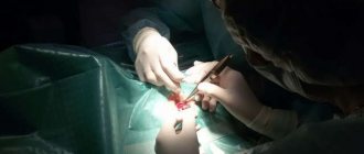
If the process has gone too far, surgery may be indicated, which may involve excision of the affected area of the cornea, replacing it with a graft, and removing superficial scars and scars.
Prevention of keratitis of any etiology consists of maintaining immunity, timely treatment of general and eye diseases, eye hygiene and timely examinations by an ophthalmologist.
Source: https://ultralinzi.ru/articles/keratit/
Symptoms
Having studied the classification, you can see how diverse the symptoms of keratitis in children are, and each of them may indicate a specific form of the disease. Despite these nuances, there are typical signs by which one can recognize the inflammatory process of the cornea at the initial stage of its development. These include:
- photophobia;
- hyperemia (redness) of the conjunctiva;
- lacrimation;
- cutting pain;
- complaints of a foreign body in the eye;
- decreased visual acuity: feeling of a veil, fog before the eyes;
- the vessels on the surface of the eye become bright red and look like a branched tree;
- blepharospasm: involuntary contraction of the eye muscle, which leads to spasmodic closure of the eyelids;
- itching;
- swelling, dryness, clouding of the cornea;
- formation of infiltrate (turbidity), in place of which an ulcer forms over time;
- filamentous mucus in the conjunctival sac.
Sometimes parents worry whether keratitis is contagious or not, since there may be other children in the family, and whether they need to be isolated from the sick baby. Yes, it is transmitted by contact. Many symptoms of keratitis are similar to those of allergic conjunctivitis. Even doctors require multi-stage and thorough diagnostics in order to separate these two diseases and ultimately prescribe the correct treatment. Therefore, modern diagnostics plays an important role here.
Helpful advice. There is a category of parents who, having seen redness or clouding of the mucous membrane of the child’s eye, do not attach much importance to it. They rinse the eye, use the first drops found in the medicine cabinet, thinking that it is due to overwork or insomnia. Any eye problems in children should force parents to immediately consult an ophthalmologist.
Symptoms
To identify keratitis, you need to know its symptoms. The disease is accompanied by narrowing of the palpebral fissure (blepharospasm), lacrimation, photophobia, cutting pain in the eye or foreign body sensation, itching. The reason for these phenomena is irritation of the endings of the trigeminal nerve. The brighter the manifestations, the closer to the surface of the cornea the inflammation is located. Quite often, swelling of the upper edge of the eyelids is observed.
Objectively, the disease is accompanied by a loss or decrease in tactile sensitivity, impaired specularity and shine, smoothness, transparency of the cornea, mucous, purulent or mucopurulent discharge from the conjunctival sac. Herpes and neuroparalytic keratitis are not accompanied by such discharge.
Also, at the end of the second or at the beginning of the third week, with some types of illness, newly formed vessels begin to sprout in the cornea, and a corneal ulcer may form - rejection of the surface layers.
Complications of the disease occur when the infection penetrates deep into the eye, resulting in ulcerative damage to the cornea.
In especially severe cases, the purulent process can invade other membranes of the eye, melt the cornea, which will lead to the death of the visual organ.
Diagnostics
To avoid making a mistake in diagnosis, the doctor carries out a number of diagnostic measures. Among them:
- taking anamnesis (questioning parents);
- scraping of the infiltrate and its microscopic examination;
- laboratory and general tests to identify other internal diseases in the child that could cause the development of an inflammatory process in the cornea of the eye;
- eye biomicroscopy using a slit lamp;
- conjunctival smear;
- immunological research methods;
- diagnostic tests with various antigens;
- specular microscopy of the posterior corneal epithelium.
After all the data during the diagnosis is received and analyzed, the doctor will be able to make an accurate diagnosis and determine the type of keratitis in the child. The methods and duration of treatment will depend on this.
Educational program for parents. One of the devices for diagnosing keratitis in children is the slit lamp. It is a device for conducting microscopic analysis of parts of the eye - the conjunctiva, eyelids, sclera, iris, cornea, lens.
Herpetic keratitis treatment with folk remedies
The most common type of keratitis is herpetic keratitis (ophthalmoherpes), which manifests itself in people of all ages. This type can cause significant complications, change the color of the lens and impair the vision of the eye itself.
A significant reason is infection with the herpes virus, which is unfavorable for any person. Mostly people who suffer are those who have significantly weakened and neglected their own immunity, children and even many teenagers.
Herpetic keratitis has different and difficult symptoms. Herpes can quickly affect the nerves of the eye, which results in quite severe pain. The eyes become red (much like with conjunctivitis), the eyelids become inflamed, the cornea becomes cloudy and noticeably swollen, an extraneous pain is felt inside the eye, photophobia sets in, and many experience lacrimation and annoying itching.
Treatment of keratitis with folk remedies.
Unfortunately, it is almost impossible to completely get rid of herpetic keratitis using folk remedies. But, they can significantly alleviate and reduce pain syndromes and other symptoms. The main condition is that it is strictly necessary to obtain permission from a competent and trusted doctor.
Also, natural treatment can last a long time; it will be necessary to strictly adhere to the dosage, norms and timing of treatment. But still, they quite favorably relieve a person from the pain that has arisen and help reduce other unfavorable symptoms.
Sea buckthorn oil.
The initial period of treatment is 2 drops, always in each eye. Gradually change the frequency of instillation to 2 pipette drops, but exactly every next 3 hours. A great way to effectively reduce pain and save your eyes.
Plantain.
This natural plant has been renowned for its natural strength for centuries. Anyone can prepare the right decoction from dry medicinal leaves. You will need 300 ml of clean boiling water, pour only 1 tbsp. l plantain powder, then do not touch for up to 5 hours. Strain carefully and efficiently and you can rinse the affected eyes.
Kalanchoe.
This medicinal plant can be found in almost any housewife. Dilute the healing Kalanchoe juice in equal quantities with water and soak a napkin. Cover the affected eyes with a tissue for 10 minutes. It is allowed to use these medicinal lotions up to 5 times a day.
Treatment of keratitis with folk remedies reviews.
Maria is 24 years old.
Personally, sea buckthorn oil helped me a lot. I was in a lot of pain. This miraculous oil was really able to extinguish all the existing pain. But electrophoresis helped me to recover completely. The treatment was carried out in a hospital (but it is also possible on an outpatient basis).
Denis is 48 years old.
Aloe brought me real relief. I read advice about him on LiveJournal. Combining it with propolis doubles the power of its benefits and reduces pain to a minimum. Tearing almost completely stops.
Treatment
How much keratitis is treated will depend on the advanced stage of the disease and the timeliness of contacting an ophthalmologist. Parents must strictly follow all his recommendations after confirming the diagnosis. These can be both medications and folk remedies.
Drug treatment
Drug treatment of keratitis in children involves the use of the following drugs:
- ointments (once a day, at night): erythromycin, tetracycline, ciprofloxacin, colistimethate, roletetracycline, acyclovir, Zovirax;
- eye drops (up to 4 times a day): miramistin, lomefloxacin, ciprofloxacin, colistimethate, roletetracycline, diclofenac, cyclopentolate, atropine, tropicamide, ophthalmoferon, poludan, actipol;
- intramuscular injections of penicillin, non-steroidal anti-inflammatory drugs (diclofenac sodium), B vitamins, ascorbic acid;
- NSAIDs can be administered rectally (Voltaren suppositories) or orally (Indomethacin tablets);
- intravenous injections of solutions of glucose, hemodez, ascorbic acid;
- drugs to strengthen the immune system;
- metronidazole;
- at the same time, treatment of diseases that served as an impetus for the development of keratitis is carried out;
- mechanical extinguishing of the ulcer with an alcohol solution of brilliant green, an alcohol solution of iodine, cryo-, thermo, diathermocoagulation;
- laser stimulation, magnetic therapy using 20% Actovegin or solcoseryl gels;
- electrophoresis with lidase and collalysine;
- rinsing with antimicrobial agents;
- therapeutic keratoplasty.
Self-medication of keratitis in a child is excluded, otherwise it can lead to irreversible and dangerous consequences.
Folk remedies
Treatment of keratitis with folk remedies is considered very effective, but they are effective only if they are approved by an ophthalmologist and are used as ancillary to the main course of therapy.
- instillation of sea buckthorn oil, 1% aqueous extract of propolis, woodlice infusion;
- contrast compresses: applying napkins soaked in either cold or hot water to the eyes;
- lotions with infusions of sweet clover flowers, eyebright, calendula, chamomile.
Answers to the question of how long it takes to treat keratitis in children may vary, since the duration of the course of therapy depends on many factors. This includes the general health of the baby, the advanced stage of the disease, and its type. On average, this is 2-4 weeks in a hospital setting. If you don’t catch it in time and consult a doctor, the consequences can be very dire.
Special attention! Very often, recipes for folk remedies against keratitis suggest using garlic or celandine juice. Be aware that they are very irritating to the mucous membrane of the eye and can cause a burn.
Eye keratitis - treatment and symptoms. Blueberries and eyebright will help prevent illness
The human eye is similar to a complex optical device, but, nevertheless, it is an independent organ consisting of tissues, membranes, and muscles. And, like any other organ, the eyes are susceptible to diseases and get sick both completely and in “parts”. One of these “partial” diseases will be discussed further.
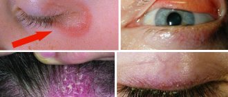
The main symptom of keratitis is clouding of the cornea
Keratitis and its types
On the front of the eyeball there is a transparent membrane - the cornea. Inflammation of this membrane is called eye keratitis.
Keratitis is an independent eye disease that requires specialized treatment and diagnosis, but since keratitis visually looks like the more common conjunctivitis, but is not, it is necessary to know and understand the differences between these diseases. The main symptom of keratitis is clouding of the cornea, which in normal condition should be completely transparent.
There are only two types of keratitis: superficial, affecting the outermost layer (epithelium) of the cornea, and deep (internal), affecting the remaining layers of the cornea, up to the stroma (the last retaining layer).
Superficial keratitis in most cases is a consequence of conjunctivitis, less often - a consequence of inflammation of the lacrimal sinus, inflammation of the eyelids.
Deep keratitis can be the result of neglected and/or untreated inflammatory processes, for example, conjunctivitis.
Types of keratitis: 1. Viral (general). Etiological factors are decreased immunity, the presence of adenoviruses in the body, for example, chickenpox, measles, ARVs.
2. Bacterial keratitis is mainly caused by the presence in the body of bacteria Staphylococcus aureus, Pseudomonas aeruginosa, Koch's bacillus, and spirochete pallidum. By the way, the pale spirochete, the causative agent of syphilis, is extremely dangerous as a cause of keratitis, especially for children. “Second place” in the list of reasons belongs to injuries and poor hygiene when using contact lenses.
3. Viral herpetic (tree-like). Almost 100% of the reason is the herpes virus, which is present in the body of any person, and when activated cannot be treated 100%. The tree-like variety of the herpes virus makes up half of the total percentage of cases.
It got its name because of the visualization in the form of a tree crown during diagnosis. It affects almost all layers of the cornea, including the stroma. Treatment is long, and relapses are frequent. Recurrent herpetic keratitis is observed in 90% of herpetic eye lesions.
It occurs more often in adults and in children over 5 years of age.
There are only two types of keratitis - superficial and deep
4. Fungal keratitis, in most cases, is the result of long-term use of drugs of the penicillin group. Symptoms of this form of eye keratitis: severe sharp pain, severe redness of the eye. Fungal keratitis comes in both types with the same percentage. As a result of fungal keratitis, a cataract may form and/or a sharp decrease in vision may occur.
5. Superficial marginal keratitis - an inflammatory disease of the cornea usually occurs as a complication of inflammatory processes of the eyelids (blepharitis), the mucous membrane of the eyes (conjunctivitis), and meibomites. Very difficult to treat.
In the medical literature, the following are identified as independent types: traumatic (light, chemical, mechanical); allergic (seasonal, allergies to pets, household dust); associated with metabolic disorders (diabetes, psoriasis).
Diagnosis and treatment
Diagnosing keratitis without fail, in addition to collecting an anamnesis, includes examining the patient using a slit lamp apparatus. Keratitis in its symptoms is very similar to other inflammatory diseases of the eye, for example, conjunctivitis, but an error in diagnosis, and therefore in treatment, will lead to irreversible consequences.
The course of treatment for keratitis depends on the diagnosis of one type or another. Viral keratitis - interferon and/or drugs containing it (poludan, actipol) are prescribed.
Bacterial keratitis - drugs of the antibacterial group (levomycin). Allergic keratitis - drugs of the antihistamine group; in some cases, hormonal ointments (dexamethasone) are introduced into treatment.
Be sure to avoid contact with the allergen.
Making a diagnosis of keratitis, in addition to collecting an anamnesis, includes examining the patient using a slit lamp apparatus.
When treating any type of keratitis, wearing contact lenses is prohibited.
Also, modern ophthalmologists have refused to introduce steroid tablets into the course of treatment, since statistics have shown that these drugs do more harm to the tissues of the eye and the cornea in particular than they help.
Considering that almost all types of keratitis have frequent relapses, modern ophthalmology offers a new type of treatment - ultrasonic sterilization of the cornea. In medicine, this procedure is called cross-linking.
Prevention of keratitis
Prevention of corneal inflammation involves: • Strict adherence to personal hygiene: if there is a suspicion of eye inflammation, it is advisable to use all household items individually (towels, dishes, office supplies, etc.). • If necessary, absolutely avoid contact with the allergen; if necessary, limit yourself to visiting places with large crowds of people (work, school, kindergarten).
• Take medications that support the immune system. Take medications to prevent vitamin deficiency (vitamins A, B, PP, etc.).
Source: https://simptom.org/keratit-glaza-lechenie-i-simptomy/
Consequences
Dangerous complications of keratitis in children can be irreversible if treatment was started too late or parents self-medicated the disease without consulting an ophthalmologist. These include:
- scars on the cornea;
- thorn;
- decreased vision;
- "dendritic ulcers";
- sclerosis of the eye shell;
- constant relapses;
- perforation (formation of a hole) of the cornea;
- blindness.
To ensure that the consequences of keratitis in children are not so dangerous, preventive measures must be taken in time. They will reduce the risk of illness, and if inflammation does begin, its form will not be so severe.
For consolation. Despite the fact that the consequences of keratitis are very dangerous for the health of children, they are diagnosed only in 4-5% of cases. Modern medicine and parental care prevent the development of such terrible complications.
Treatment of the disease
The patient must be treated in a hospital if the disease has reached an advanced stage.
Therapy primarily depends on the etiology and stage of the disease. Doctors also pay attention to the patient’s age and concomitant diseases. Treatment begins with isolation of the sick child; in advanced stages, hospitalization is required. Then medications are prescribed in the form of drops, tablets and injections; local remedies are considered the most effective. Surgeries are used if complications arise and scars form on the cornea. The choice of drug group is made by the doctor.
- Antibacterial, antiviral and antifungal agents. Prescribed for infectious nature of keratitis.
- Antihistamine drops. Necessary if you have been diagnosed with an allergy.
- Vitamins in the form of tablets for internal use. Used as maintenance therapy.
- Corticosteroid drugs. Prescribed after cleansing the eyes, as they relieve inflammation.
Full therapy lasts up to 4 weeks. As a maintenance treatment, ophthalmologists recommend the use of traditional medicine. They use lotions and herbal compresses that have a strong antiseptic effect. These include chamomile, calendula, sage, propolis, and chickweed. But it is impossible to cure keratitis using alternative medicine alone.
Prevention
Prevention of keratitis in children consists of the following measures that parents should take from the very birth of the child. Their main goal is to protect the cornea from infections, bacteria, and viruses. This is what ophthalmologists recommend for this.
- Isolation from carriers of keratitis.
- Compliance with personal hygiene rules.
- Prevent eye injury.
- Timely treatment of any diseases.
- Eliminating allergens from a child’s daily routine.
Keratitis is not so rarely diagnosed in children, although conjunctivitis is still more common. However, its consequences can be so serious that they can change the life of a child. Parents should always remember this, be vigilant, take all possible preventive measures, and at the first suspicion of inflammation of the cornea of the eye, immediately seek help from doctors.
source: www.vse-pro-detey.ru
Keratitis in children is an inflammatory disease that affects the transparent membrane of the eye (cornea). Occurs under the influence of bacterial, viral infections, mechanical injuries, vitamin deficiency. If left untreated, keratitis can cause vision deterioration, even loss .
Keratitis: what is it?
The cornea is a very strong, transparent shell of the eyeball that protects the iris, pupil and lens. It consists of five layers, depending on the damage to a particular layer, one can judge the consequences of scarring, as well as the transparency of the resulting stain.
Inflammation of the cornea of the eye occurs with the formation and accumulation of an infiltrate, in which cells of the immune system fight the source of inflammation. Leukocytes, antibodies, and lymphocytes are released from the bloodstream, which are opaque; they determine the color of the infiltrate.
As a result of the pathological process, corneal ulcerative formations appear, which are manifested by acute pain, since the membrane is saturated with nerve endings. If the inflammation is located within the first, outermost epithelial membrane and treatment occurs on time, the transparency of the cornea of the eye is not impaired. This layer is prone to rapid regeneration.
The more advanced the process, the deeper and more dangerous it becomes. The ulcers begin to scar, forming scars, the infiltrate thickens, smoothly flowing into a spot, which has three outcomes:
- A cloud is a transparent, slightly foggy formation.
- The spot is a translucent formation.
- A thorn is an aggressive, opaque formation.
Inflammation can spread to the internal structures of the eye, to the iris or lens, depending on what is the causative agent. Keratitis in children occurs in the same way, only all the symptoms are more pronounced. There is significant redness of the eye, the entire vascular network is clearly visible, this is explained by the need to increase blood supply at the site of inflammation to transport protective cells.
Kinds
The main types of keratitis that occur in childhood are: bacterial, herpetic, allergic, metabolic (vitamin deficiency) and post-traumatic.
Bacterial
Bacterial keratitis is characterized by the appearance of ulcers in a child's eye . It is rare in childhood. The causative agent is usually a coccal infection, but sometimes the disease occurs due to an injury to the eye. The disease is accompanied by eye pain, burning, and after eliminating inflammation, a thorn appears on the white part of the eyeball.
Toxic-allergic
Toxic-allergic keratitis has a complex course. Typically occurs in children from birth to 14 years of age and adolescents. The causes of the toxic-allergic form are previously untreated diseases (acute respiratory infections, acute respiratory viral infections), hypothermia, and helminthic infestations.
Toxic-allergic keratitis can be distinguished by characteristic tubercles with vessels crossing the eyeball. Vasodilation causes clouding of the transparent membrane of the eye (cornea) and permanent deterioration of vision .
Metabolic (vitamin-deficient)
Avitaminous keratitis occurs due to a deficiency of vitamins in the child’s body:
- vitamin A. The initial stage of the disease is accompanied by dry eyes, a grayish cloudiness forms on the cornea, and white plaques affect the conjunctiva;
- vitamin B deficiency and gastrointestinal dysfunction. A pinpoint clouding of the cornea forms, spreading in different areas of the eye. Over time, the cloudiness turns into ulcers that seem to tear the eyeball;
- Deficiency of vitamins PP and E causes eye inflammation.
Herpetic
Herpetic keratitis is the most common disease among young children. Occurs due to the activity of the herpes virus in the body. The causative agent of the disease is most often activated against the background of: hypothermia, psychological disorders (stress), eye injury, febrile manifestations (high temperature, chills).
The course of the disease is possible in mild and severe forms, with the manifestation of complications: the development of secondary glaucoma, cataracts, the appearance of lens transparency .
Primary
Primary herpetic keratitis occurs among children under 5 years of age. The main reason is that children’s immunity is not fully formed. Herpetic keratitis is characterized by a complex course, accompanied by herpetic blisters on the eyeball and throughout the body (arms, legs, torso, face).
Post-primary
Post-primary herpetic keratitis occurs, like primary, against a background of weakened immunity. Manifested by the following symptoms: pain and pain in the eyes, convulsive spasmodic contractions of the eyelids, no rash on the body.
Discoid
Disc herpetic keratitis involves deep areas of the cornea , in the center of which inflammation occurs in the form of a disc. The eyes turn red, swelling of the cornea, eyelids, and conjunctiva appears, and increased intraocular pressure occurs.
- You may have been looking for: how to cure red eyes in a child
Post-traumatic
Post-traumatic keratitis in children develops due to a careless scratch, entry and removal of a foreign body from the cornea, ultraviolet, thermal or chemical burns. The disease is characterized by direct ingrowth of vascular branches into the cornea , convulsive spasm of the eyelids, and yellow purulent discharge from the eyes.
Keratitis in children: what it is, symptoms, treatment, prevention
Keratitis is an inflammatory process that affects the cornea of the eye, its transparency, shine and specularity are impaired. A corolla of dilated vessels forms around it. In order to diagnose the disease and determine treatment tactics, a scraping is made for microbiological examination.
The disease has many varieties, depending on the reason why it occurs.
The cornea is affected mainly in the lower half, with pinpoint opacities, infiltrates and erosions occurring, but vision is not affected. The process simultaneously affects the membranes of the respiratory tract and the salivary glands.
If left untreated, blood vessels grow into the cornea, which causes it to become cloudy and, over time, lead to sudden loss of vision.
data-ad-format="auto">
Bacterial keratitis
- Injury.
- Surgical intervention.
- Immunodeficiency.
- Long-term exposure to adverse factors on the cornea;
- Dry eye syndrome.
- Wearing contact lenses.
- Severe general diseases.
- Trichiasis.
- Ionizing radiation - dry eye syndrome.
- Long-term instillation of steroid drugs.
- Use of keratotoxic drugs.
Certain clinical manifestations may indicate the causative agent of the disease.
- Pseudomonas causes rapidly progressing corneal ulcers with symptoms of leukomalacia. The process especially often affects young children and patients who use contact lenses.
- Moraxella causes conjunctivitis of the outer canthus.
- Staphylococcus spp.
- trauma, surgery or prolonged exposure to adverse factors;
- Staph, aureus can provoke the occurrence of a corneal ulcer with accompanying hypopyon.
- Streptococcus:
- use of contact lenses;
- local damage to corneal tissue;
- chronic dacryocystitis;
- rapidly progressing corneal ulcers with an undermined edge.
- Gonococcus.
- Gram-negative flora:
- E. coli;
- Aerobacter,
- Proteus spp.;
- Klebsiella spp.
They have an affinity for the cornea, especially in the presence of underlying diseases.
Keratitis caused by Pseudomonas in a newborn. Predisposing factors have not been identified
[16], [17], [18], [19], [20], [21], [22], [23], [24], [25]
Prevention of corneal inflammation
It is known that the best way to treat any disease is prevention. To protect yourself from keratitis, it is important:
- maintain hygiene ;
- store contact lenses and clean them with special solutions;
- protect eyes from injury and exposure to sunlight ;
- treat diseases of a bacterial and viral nature in a timely manner.
Eyeball injection in early childhood
- Conjunctivitis:
- discharge, conjunctival injection;
- lacrimation, visual acuity is not reduced.
- Keratitis:
- conjunctival injection, discomfort, lacrimation;
- discharge, photophobia.
- Endophthalmitis:
- pain, low vision, mixed injection;
- lacrimation, discharge.
- Uveitis:
- pain, photophobia, blurred vision;
- mixed injection, lacrimation.
- Chorioretinitis:
- low vision, floaters in front of the eye, injection of the eyeball;
- hemorrhages under the conjunctiva, injection of the eyeball.
- Glaucoma:
- pain, mixed injection;
- photophobia, low vision.
- Infiltration of the conjunctiva in leukemia:
- local infiltration;
- conjunctival injection.
- Malformations of the vascular system:
- Sturge-Weber syndrome;
- impaired development of orbital vessels.
- Sclerites:
- pain, deep injection;
- pain when moving.
- Episcleritis:
- local conjunctival and subconjunctival injection;
- lacrimation, mild discomfort, feeling of “dryness” in the eye, injection, scanty discharge.
- Foreign body:
- local injection, feeling of “sand” in the eye;
- foreign body sensation.
- Injury:
- direct trauma;
- closed head injury, causing the development of a carotid-cavernous fistula.
Viral keratitis
The main manifestation of viral keratitis caused by the herpes simplex virus is pinpoint opacities of the cornea. Sometimes, during acute primary infection, opacities transform into dendritic keratitis, usually combined with skin lesions. Antiviral drugs such as idoxuridine, triflurothymidine or acyclovir are prescribed.
There are keratitis characterized by the formation of deep infiltrates without signs of purulent inflammation (for example, discoid). In these cases, treatment is carried out with antiviral agents in combination with steroid drugs.
Other viral keratitis that is not prone to purulent inflammation and ulceration includes adenoviral keratitis, keratitis with molluscum contagiosum, papillomatous and warty forms of the disease, and Epstein-Barr virus.
In how many days can acute conjunctivitis be cured?
Conjunctivitis, treatment of which was started in a timely manner, can clear up in 5-7 days. How long does it usually take to treat a disease if adequate measures were not taken immediately and the symptoms managed to intensify? In this case, treatment may take about two weeks.
If you ignore doctor's instructions, insufficient treatment and improper care of the surface of the eyelids, acute conjunctivitis can become chronic and then it will be more difficult to fight the disease.
For example, patients often do not complete the course of antibiotics, having felt some improvements, which leads to new exacerbations, and then to serious complications of the disease.
How long does conjunctivitis last?
- Viral - this type of conjunctivitis is treated with antiviral drugs; in the absence of adequate and timely treatment, it lasts a long time, more than two weeks, but if antiviral drugs are used in the first days of the disease, the disease will subside in one to two weeks.
- How long does it take to treat bacterial conjunctivitis? Acute conjunctivitis, which is difficult to treat, can go away in 10-14 days if treated from the first day, and if complications arise (chronic), recovery lasts a long time, usually more than one to two months.
- Fungal - it is difficult to cure fungal conjunctivitis, because it is an acute, dangerous inflammation of the mucous membrane, it can be combined with keratitis, fungal blepharitis, and if complicated, it leads to entropion of the eyelid, perforation of the cornea, so treatment of the disease is long-term, more than 12-14 days.
- Allergic - how long does it take for allergic conjunctivitis to go away? Timely intake of antihistamines, decongestants, moisturizing drops, as well as avoiding contact with the allergen, will eliminate the symptoms of conjunctivitis in 5-7 days.
Conjunctivitis must be treated, otherwise recovery will have to wait a long time, since it will be difficult for the body to cope with bacteria without medications.
Can bacterial or viral conjunctivitis be cured with folk remedies? Popular folk recipes for herbal decoctions that are used to wash the eyes can only slightly ease the symptoms, but they are not able to stop the inflammatory process, and moreover, they often complicate the course of the disease, causing an allergic reaction.
Keratitis of fungal etiology
Keratitis caused by fungal flora occurs in weakened children or in the presence of concomitant diseases of the organ of vision. Examples include immunologically weakened children receiving general steroid therapy, patients with long-term non-healing wounds, as well as those who have suffered eye trauma or suffer from dry eye syndrome.
Bilateral Candida keratitis in a child with severe immunodeficiency
Source: https://medhouse-clinic.ru/bolezni/keratit-glaza-lechenie-narodnymi-sredstvami.html
Treatment
Immediate treatment under the supervision of a specialist is necessary for any type of disease. Keratitis is a contagious disease; treatment of sick children is carried out in hospitalization.
To identify the causative agent of the disease, the ophthalmologist examines the patient, prescribing a series of tests. Assistants wash the eyes, take a smear of the infiltrate, and collect a general blood test. After the result is obtained, complex treatment is prescribed.
Ointments and antimicrobial agents
Ointments for keratitis are used once a day at night; it is recommended to drop antimicrobial agents into the eyes at least 4 times.
- 1% erythromycin ointment;
- 1% tetracycline ointment;
- 0.3% ciprofloxacin ointment or solution;
- colistimethate ointment or solution, rolitetracycline;
- 0.01% solution of Miramistin;
- 0.3 solution of lomefloxacin.
Anti-inflammatory
Anti-inflammatory drugs are used to reduce inflammatory processes - 0.1% diclofenac solution, 1 drop 4 times a day. To reduce the risk of internal synechiae, solutions are used: 0.5 cyclopentolate, 1% atropine, 0.5% tropicamide.
Injections
Treatment of keratitis is carried out in conjunction with injections of antibiotics. Treatment is carried out with intramuscular and intravenous injections.
Intramuscularly
The most common and effective treatment is using penicillins . Injections are given at least 5 times a day, sometimes 10 injections can be prescribed.
Intravenously
Intravenous detoxification treatment is carried out with a 5% glucose solution, 200-400 ml of hemodez solution, 2 g of 200-400 ml of ascorbic acid. If healing takes a long time, drugs that stimulate the immune system are prescribed. Treatment is with metronidazole 500 mg per day or every other day.
How to treat keratitis: how long does treatment last, methods and medications
Keratitis can be treated in 1.5-2 months.
If injured eyes often water and turn red, there is a fear of light and cutting pain - these are signs of keratitis. Due to the formation of adhesions and corneal opacities, the pathology can lead to decreased vision. If you suspect such symptoms, an examination by an ophthalmologist is inevitable. In this article we will look at how to treat keratitis, how long treatment lasts, and we will analyze methods and medications.
How to treat keratitis?
Inflammation of the cornea of the eye can be quickly treated if you seek help in a timely manner and follow the instructions of a specialist. After conducting a series of medical examinations, the ophthalmologist makes the final decision on how to treat keratitis.
Depending on the etiology of the disease, the human body is treated comprehensively:
- drops containing antibiotics;
- vitamins and immunomodulators;
- general methods of combating viruses and bacteria.
The human body is susceptible to various types of viruses and bacteria.
Simple form
Patients with the disease without complications are treated on an outpatient basis. If the process worsens and is accompanied by the release of pus, there is a need to go to the hospital.
Important! Please note that keratitis takes a long time to heal.
First of all, the attending physician eliminates the causes of the disease, having thoroughly studied the etiology of the disease. Keratoconjunctivitis located internally indicates the presence of a disease inside, which has become the source of infection. Therefore, consultation with a phthisiatrician, allergist, venereologist or rheumatologist is required.
To determine the source of the disease, it is necessary to undergo diagnosis from several doctors
Acute form
The lesion of acute keratitis is blocked with antibacterial and sulfonamide drugs :
- penicillin,
- norsulfazole,
- chloramphenicol,
- gentamicin,
- tetracycline,
- erythromycin,
- furatsilin.
The antibiotic is selected taking into account the sensitivity of pathogenic bacteria to it. Severe corneal ulceration is treated with antibacterial agents: administered under the conjunctiva and orally.
Important! In order to prevent the development of intraocular adhesions, the doctor prescribes medications that dilate the pupil.
Dilated pupils help monitor the healing process
Based on the painful manifestations, glucocorticosteroid medications are prescribed - dexamethasone or betamethasone.
In addition, keratoprotective and epithelializing agents are used. In difficult cases, antibiotic injections are given under the mucous membrane of the eye.
Antibiotics or antiviral drugs are prescribed systemically.
How long it takes to treat acute keratitis of the eye is determined by the advanced stage of the disease and the individuality of the patient. Proper drug therapy will result in a quick recovery. Untreated pathology is accompanied by a protracted recovery.
Neurogenic keratitis
Having discovered neurogenic keratitis, the doctor prescribes a local treatment course:
- vitamin therapy;
- taurine;
- sea buckthorn oil;
- 20% Solcoseryl gel;
- triphosadenine;
- retinol;
- thiamine;
- pyridoxine;
- ascorbic acid.
When treating neurogenic keratitis, electrophoresis with a 1% diphenhydramine solution is prescribed, alternating with 1% riboflavin mononucleotide. The duration of the therapeutic event is 1-1.5 months. After this, electrophoresis is prescribed with a 0.1% solution of hydrocortisone and 0.1–0.25% ascorbic acid (two weeks).
Electrophoresis with a solution of hydrocortisone and ascorbic acid helps recovery
Scraping of inflammation and cauterization of damaged vessels is also done. You can speed up healing with biogenic stimulants:
- retinol;
- riboflavin;
- pyridoxine.
The specialist selects therapeutic procedures individually, prescribing local and systemic desensitizing treatment.
The best effect comes from complex treatment
Ways to destroy the disease:
- antiviral control;
- impact on the cornea;
- preventing the growth of ulcerations;
- stimulation of epithelial growth on damage;
- relief of iritis and cyclitis;
- reduction of scars and resorption of opacities.
Treatment method
When treating ocular keratitis in adults, the ophthalmologist uses physical methods:
- Bactericidal . It involves the use of electrophoresis with antibiotics: on closed eyelids;
- through an electrode-bath on an open eye;
- endonasal.
Ultraphonophoresis of the eye is one of the methods for treating keratitis
- Reparative - local darsonvalization, low-intensity microwave therapy. Involves low-intensity photosensitive therapy that causes polarization of free fluid molecules. Low-intensity waves stimulate the endocrine glands, inhibiting the activity of the affected cells.
- Defibrosing . It is noted by electrophoresis of defibrosating drugs - Lidaza 32 units, Papain 17.5 and others. Used in the treatment of regressive forms.
Ophthalmologists also prescribe penicillin, streptomycin, gentamicin, syntomycin.
All therapeutic measures carried out on time guarantee a positive result. The treatment course ends within 2 weeks .
At home
If the eye inflammation is minor, you can treat keratitis at home using available remedies.
The most common natural protectors against infection:
- sea buckthorn oil;
- aloe juice drops.
Sea buckthorn oil
Sea buckthorn oil has a positive effect on the body, subsequently causing the causes of the disease to disappear. Drops of sea buckthorn oil are applied to the eyes every hour. The oil provides effective relief for corneal burns and restores visual acuity.
Sea buckthorn oil is one of the most effective restorative agents
Aloe juice
Aloe juice contains a whole storehouse of nutritional vitamins. If you drip drops of aloe, the swelling and inflammation of the cornea will go away, and vision will be restored.
Aloe juice helps eliminate the virus and restore the affected areas
When preparing, maintain absolute sterility:
- Pour boiling water over the three-year-old leaves of the plant and place them in the refrigerator for 14 days.
- Next, we take the blackened leaves out of the refrigerator, cut off the spines, and remove the skin.
- Squeeze the pulp into a glass container and dilute it with injection water.
- Apply the prepared solution to the eyes 2 times a day, drop by drop.
In what cases is surgery required?
If the therapeutic course does not lead to recovery, the patient is prescribed surgery (keratoplasty, optical iridectomy). Modern medicine is famous for its wide selection of surgical techniques that bring positive results to patients.
Watch a video about the treatment of keratitis:
A timely visit to an ophthalmologist will help get rid of the infection and save your vision.
Source: https://bolezniglaznet.ru/kak-lechit-keratit/
Keratitis: symptoms and treatment of inflammation
Skip to content
IT IS IMPORTANT TO KNOW! An effective remedy for restoring vision without surgery or doctors, recommended by our readers! Read more…
Inflammation of the cornea of the eye is called keratitis. It occurs as a result of a person being injured, developing an infectious process, or exacerbating an allergic reaction. Keratitis manifests itself in the form of clouding or a sharp decrease in a person’s vision.
The main reasons for the development of keratitis
Doctors note that the disease can form as a result of exposure to the following factors:
- development of an infectious process in the patient’s body;
- due to prolonged wearing of contact lenses;
- the patient receives a corneal injury;
- exacerbation of an allergic reaction;
- development of photokeratitis, characteristic of people working with welding;
- with hypo- and vitamin deficiency;
- in cases of innervation disorders that occur as a result of damage to 1 branch of the trigeminal nerve.
Symptoms of keratitis
Keratitis can begin in such manifestations as the development of corneal syndrome. It is characterized by an increased flow of tear fluid from the eyes, the development of fear of light, and the formation of blepharospasms. Additional signs of keratitis:
- The appearance of pain in the eye and the presence of a foreign body in it: the patient is unable to open the eye on his own. These symptoms develop based on the innervation of the cornea.
- Development of pericorneal or mixed infection. A pericorneal infection usually affects the tissue near the cornea of the eye.
- Pus, hypopyons and precipitates localized in the back of the eye. Precipitates usually consist of lymphocytes, macrophages, plasma cells, and pigment dust, which is located in the chamber moisture.
- Decreased vision and formation of cloudiness in the eye.
Types of keratitis
According to its form, the disease is divided into the following types:
- spicy;
- chronic. This form is characterized by the formation of abscesses, necrosis, ulcerations, and damage in the cornea of the eye.
Also, doctors divide keratitis into the following types:
- By depth of damage: superficial and deep. With superficial keratitis, only the upper stromal layers of the skin are damaged. With the development of deep keratitis, damage spreads to all stromal tissues.
- According to the zone of localization of damage: central, paracentral, peripheral. Central keratitis is localized in the pupil of the eye. Thus, the larger the area of damage to the pupil of the eye, the worse the person’s vision. Paracentral keratitis is usually localized in the iris of the eye. Peripheral keratitis is localized in the limbus, the ciliary belt of the iris.
- Based on etiological characteristics, keratitis is divided into: exogenous, endogenous, allergic, neurogenic, infectious, herpetic, bacterial, fungal, avitaminous, viral and hypovitaminous. It is noteworthy that exogenous keratitis is characterized by erosion of the cornea of the eye, traumatic, bacterial, viral, fungal damage to eye cells, as well as damage to the eyelids, meibomian glands and conjunctiva of the eye. Endogenous keratitis develops as a result of the appearance in humans of diseases such as tuberculosis, malaria, syphilis, and brucellosis.
- Also, according to the degree of damage to the eye, keratitis is divided into such types as meibomian, filamentous and rosacea.
It is important to note that keratitis usually appears as a result of the development of keratoconjunctivitis, keratoscleritis, keratouveitis, iridocyclitis, and iritis. If a person has purulent inflammation of the three membranes of the eye, this can lead to premature death.
Main symptoms of keratitis
Doctors note that the development of keratitis in a patient is characterized by the appearance of the following symptoms:
- the presence of pain in the eye area;
- increased secretion of tear fluid;
- the appearance of a feeling of a foreign body in the eye;
- having problems opening the eye;
- the appearance of redness in the eyeball area.
It is important to remember that the appearance of one of the above signs indicates that a person needs to visit a doctor. The patient should not independently diagnose or self-treat the disease.
Diagnosis of keratitis
To make an accurate diagnosis, the following diagnostic methods are used:
- examination of the patient;
- performing a visual acuity test;
- Eversion of the eyelids by a doctor. This is necessary to exclude the presence of a foreign object in them;
- performing biomicroscopy. This method is based on studying the eyeballs using a microscope;
- staining the cornea with fluorescein. The substance helps improve the visual picture of the degree of damage to the cornea of the eye;
- performing analgimetry. This method helps to study human pain sensitivity.
We recommend!
To treat eyes without surgery, our readers successfully use a proven method. Having carefully studied it, we decided to offer it to your attention. Read more…
Additionally, the patient may be prescribed an eye microscopy and a bacterial culture from the cornea of the eye. These methods are necessary to identify sensitivity to the presence of hepatitis virus antibodies in a patient. He will also need to donate blood for an RW test and a rheumatic test. Additionally, he is prescribed fluorography and intradermal allergy tests.
Also, the patient must undergo an examination of the lacrimal ducts and have them sanitized before treatment. It is important to remember that this procedure is prescribed for severe stages of the disease.
In order to exclude the presence of foci of focal infection, the patient should visit an otolaryngologist and dentist.
If the disease appears as a result of endogenous causes, then the person should visit a general practitioner, rheumatologist, gynecologist, urologist, allergist, venereologist and phthisiatrician.
It is important for all patients to remember that only an experienced specialist can answer the question: “How to treat keratitis?” and you should not try to get rid of this disease on your own. This can lead to a worsening of the disease and worsening symptoms, as well as a deterioration in overall health.
How is keratitis treated?
Treatment of keratitis includes general and local procedures. Keratitis can usually be treated in an inpatient facility. In some cases, doctors allow the patient to be treated on an outpatient basis, but this is carried out under the supervision of the attending physician and with mandatory compliance with all his recommendations.
Outpatient treatment is possible if the disease is superficial.
Typically, treatment of keratitis in adults includes the following methods:
- Use of detoxification therapy. It includes the administration of a drug such as Reosorbilact 200.0 mg intravenously.
- Antibiotic, antiviral and antifungal agents are also used for internal use. They are selected depending on the type and sensitivity of the pathogen.
- Prescription of desensitizing therapy. It involves administering 10% sodium gluconate through an IV.
- The patient is also prescribed to take 1 tablet of multivitamins 1 to 2 times a day.
- Treatment with folk remedies includes: instillation of disinfectants and antibacterial drugs.
- Disinfectants include 20% sodium sulfacyl, miramistin.
- Antibacterial drugs include Floxal, Tobrex and Oftaquix.
- When the initial signs of the disease appear, all drugs are instilled every two hours, and after that every 4 hours. If there is no damage to the epithelium, the patient can use hormonal drugs. These include Oftan-Dexamethasone and Maxidex. They should be instilled 2 times a day.
- When viral keratitis develops, drops such as Okoferon and Oftan-ilu are used. Okoferon should be instilled at least 5 times a day. Oftan-ida should be used every hour, and then the dosage should be reduced to 3 times a day.
- Anti-inflammatory drugs used for keratitis include Naklof and Indocollir. They should be used within 4 to 6 hours.
- Mydriatic drugs that are also used for this disease include tropicamide, mesatone, and atropine. They help dilate the pupil of the eye and reduce the likelihood of adhesions appearing in it.
- To speed up the process of epithelization of the cornea, a product such as Korneregel is used. It should be used 2 to 3 times a day.
- When herpetic keratitis develops, Zovirax ointment and Acyclovir tablets are used. The last drug should be taken orally at a dose of 200 mg and this should be done at least 5 times a day. The course duration is from 7 to 10 days.
- If the lacrimal ducts become infected, they should be washed every day with a 0.25% solution of chloramphenicol. You can also use a 0.01% solution of miramistin or a diluted 1:5000 solution of furatsilin.
- If the innervation is impaired during the development of keratitis, then the patient needs to use erythromycin, 1% tetracycline ointment or Floxal ointment. All remedies eliminate the causes of neuroparalytic inflammation of the cornea.
- In order to speed up the process of epithelization and resorption of scars on the eye, physiotherapeutic methods such as electrophoresis and magnetic therapy are used.
- If herpetic keratitis does not go away for a long time, then cryotherapy, laser coagulation, and diathermocoagulation of problem areas of the skin are used.
Also, during treatment of eye keratitis, all patients are recommended to adhere to a special diet, which includes eating foods high in protein, vitamins and microelements. You should also reduce your intake of foods high in fat and carbohydrates.
Thus, keratitis is an unpleasant disease that you can get rid of quickly and without harm to your body. To do this, you should undergo a comprehensive examination and, when the first unpleasant symptoms appear in the eye area, immediately go to the doctor.
By secret
- Incredible... You can cure your eyes without surgery!
- This time.
- No trips to the doctors!
- That's two.
- In less than a month!
- That's three.
Follow the link and find out how our subscribers do it!
Source: https://aokulist.ru/zabolevaniya/obshchaya/simptomy-i-lechenie-keratita.html
Keratitis - symptoms and treatment of inflammation of the cornea
Many people have to deal with a disease such as keratitis. The pathology occurs everywhere, and people who wear contact lenses are at risk of getting the disease. OftaPlus.ru will talk about the symptoms and treatment of keratitis in today’s article.
What is keratitis
This disease (ICD keratitis code 10 - H 16) means inflammation of the cornea - the transparent outer layer of the eye. It is necessary to know the symptoms of keratitis in humans in as much detail as possible, since the disease can lead to dire consequences, including blindness.
Causes
There are many reasons for the development, the most common is the entry of viruses and bacteria into the organ of vision, which cause inflammation of the cornea. Other reasons are as follows:
- Injuries to the eye or the presence of foreign bodies in it.
- Violation of the rules for caring for contact lenses.
- Burns of various origins.
- Presence of immunodeficiency states.
- The eye disease keratitis develops due to vitamin A deficiency.
- Increased dryness of the eye.
- Problems with nerve conduction in this area. For example, with neuritis of the facial nerve, lagophthalmos develops, as a result of which the eyelids do not close completely, which facilitates the penetration of pathogens into this area.
- Keratitis is often a consequence of allergies.
- Concomitant endocrinological and autoimmune diseases.
- Pathology can also develop as a result of long-term use of hormonal and psychotropic drugs.
In case of chronic diseases of the organ of vision, there is a risk of the inflammatory process spreading to the cornea. In some cases, the cause of the pathology is parasitic infestations.
Symptoms and types
Symptoms of keratitis in humans are lacrimation, fear of light and blepharospasm. The latter term refers to the reflex closure of the eyelids; often patients cannot open their eyes at all during the acute period of the disease.
There are also complaints of pain of varying intensity, there is clouding of the cornea, and hyperemia of the mucous membranes of the organ of vision. Patients may describe the sensation as a feeling of a foreign body in the eye. During the period of illness there is a decrease in visual acuity.
Other symptoms of eye keratitis depend on the type of pathology. The variants of the disease are as follows:
- Allergic is characterized, in addition to the above symptoms, by the presence of a runny nose, swelling of the face, cough, itching and burning in the eyes.
- Bacterial keratitis often occurs due to injury or when wearing contact lenses, as this makes it easier for pathogens to enter the organ of vision. Patients have complaints of severe throbbing pain in the eye, swelling in this area, and the appearance of purulent discharge. Complications include ulcers and opacities.
- Viral keratitis has a corresponding etiology, spotty opacities on the membrane. With this type, more often than with others, a cataract forms. In most cases, herpetic (or dendritic) and adenoviral keratitis are diagnosed.
- Acanthamoeba . A simple organism, an amoeba, can get into the eye. This happens when rinsing lenses under running water. In such cases, acanthamoeba keratitis is formed. Its symptoms are severe pain, clouding of the cornea and ring-shaped ulcers on it.
- Fungal keratitis is characterized by the presence of diffuse pain in the eye, severe hyperemia, and when the cornea is irritated, the eyelids close. Complications include decreased visual acuity, formation of a cataract, and ulceration.
- Parenchymal keratitis is caused by Treponema pallidum, the causative agent of syphilis. Its signs are that the cornea gradually becomes matte, the sclera grows with vessels that give it an orange tint. In the case of this type of pathology, great importance is attached to the accompanying symptoms of syphilis.
- filamentous develops . The amount of tear fluid is significantly reduced, and the pathology often becomes chronic.
- If a foreign object gets in, the risk of ulceration is much higher. The ulcerative type of the disease is severe, often with breakthrough of pus into other structures of the eye, perforation of the membranes of the organ of vision.
- A separate group includes the tuberculosis type of disease, in which white nodules accumulate in the cornea and infiltrates form on the surface.
The classification of keratitis according to localization is as follows: superficial, involving the upper layer of the membrane, and deep, which affects the underlying structures. With the latter type of disease, scars form.
Acute keratitis of the eye is more often diagnosed, which usually ends without any consequences for the organ of vision. But the process can become chronic, occurring with exacerbations and remissions.
Also, according to localization, marginal keratitis is distinguished, which is a type of viral or bacterial. The pathological process is localized on the peripheral part of the cornea and is easily detected by visual examination.
How to treat keratitis
It is important to note that keratitis in children and adults occurs in the same way, and treatment methods are no different from each other. It is important to start treatment for the disease as early as possible. Treatment is complex, includes various areas, depending on the etiology of the disease.
Some patients have a question about how to treat eye keratitis at home. Hospitalization to an ophthalmology department is usually required; only the superficial, uncomplicated form can be treated on an outpatient basis.
Anti-infective therapy
This type of therapy depends on the pathogen. For bacterial etiology, Ofloxacin, Levofloxacin, Moxifloxacin, Sulfacyl drops are used. If necessary, apply antibacterial ointment to the organ of vision (for example, Tetracycline). In case of severe intoxication, antibiotics are taken in the form of injections or tablets.
Treatment of herpetic keratitis of the eye includes the mandatory use of Acyclovir in the form of tablets and eye drops. Ganciclovir can be used and is more effective in severe cases. Arborescent keratitis often requires surgical correction, including corneal transplantation.
Also, for viral etiology, drugs containing interferon are used, and special ointments are placed in the organ of vision (for example, Florenal ointment).
To eliminate allergic reactions, special eye drops are used for keratitis of a similar etiology - Allergodil, Opatanol.
If a patient has syphilis or tuberculosis, drugs specifically directed against these pathogens are used for treatment.
Symptomatic treatment
The symptoms and treatment of keratitis are inextricably linked. To improve the patient's condition, mydriatics are prescribed that stimulate the outflow of tear fluid (Atropine, Cyclomed), artificial tears, and antiseptics (Albucid, Vitabact).
Agents are used that promote epithelization of the organ of vision - Solcoseryl, Cytochrome, Retinol. Vitamin eye drops - Taufon, Quinax, Katachrom - are also prescribed as symptomatic remedies.
Surgery
Surgical treatment of eye keratitis is required for the formation of scars, ulcerative lesions, deeper penetration of the pathological process, and severe herpetic lesions.
Laser or thermal coagulation and keratoplasty are used (if this layer is destroyed). In some cases, treatment for keratitis in people may require a cornea transplant or laser vision correction.
In very advanced cases, the question of complete removal of the affected eye may arise.
Is it appropriate to use folk remedies?
According to ophthalmologists, treating keratitis with folk remedies is inappropriate and even dangerous. The use of these techniques can worsen the course of the disease and increase the risk of complications, including the most serious ones (more on them below). Therefore, you should abandon traditional medicine and, at the first signs of illness, consult an ophthalmologist.
Complications and consequences
One of the complications is ulceration of the cornea, which can lead to the penetration of pathogens deeper into the underlying and nearby structures of the organ of vision. In severe cases, this process can cause significant reduction or complete loss of vision.
Complications also include scars on the cornea, which remain after recovery from the inflammatory process. The consequence of the disease can be glaucoma, cataracts, and cataracts. In some cases, inflammation (neuritis) of the optic nerve develops.
Is it possible to wear lenses after keratitis?
This issue is always resolved by the doctor individually. If the process is complete, then wearing lenses is allowed after some time from the moment of recovery. The type of lenses must be selected by a doctor, the duration of use is gradually increased. The rules of care must be strictly followed, otherwise re-infection cannot be avoided.
If complications in the outcome of keratitis could not be avoided or the disease has become chronic, then wearing lenses is contraindicated. They can further injure the eye, which will only worsen the patient’s condition.
Prevention
Complications of keratitis can be easily prevented by timely consultation with a doctor at the first symptoms and full treatment of the disease. As for other methods, doctors do not recommend prolonged exposure to the sun in cloudless weather. It is important to follow all rules for hygienic care of contact lenses.
For any disease of the organ of vision, it is important to start treating it on time to prevent the process from becoming chronic and the inflammation spreading further. Any concomitant pathologies should be transferred to the stage of decompensation; it is important to sanitize all sources of chronic infections in the body.
Traumatic keratitis is easily avoided if all necessary requirements in the workplace are met - wearing masks and other protective items, following safety regulations.
Conclusion
The eye disease keratitis can lead to serious consequences that significantly reduce the patient’s ability to work and quality of life. Inflammation of the cornea is a fairly common pathology, in which it is necessary to seek medical help as quickly as possible in order to avoid any complications.
Source: https://oftaplus.ru/zabolevaniya/keratit-simptomy-i-lechenie.html
Diagnosis of keratitis
To choose the right treatment tactics for keratitis, it is necessary to carry out an accurate diagnosis.
First of all, the ophthalmologist interviews the patient and examines the eye for signs of corneal damage. To determine the type of keratitis and determine the severity of the disease, the following diagnostic techniques are used:
- keratotopography;
- bacteriological examination;
- eye biomicroscopy;
- cytological and bacteriological examination;
- confocal and endothelial microscopy of the cornea;
- computer keratometry;
- fluorescein instillation test.