Conducting pathways of the visual analyzer
1 - Left half of the visual field, 2 - Right half of the visual field, 3 - Eye, 4 - Retina, 5 - Optic nerves, 6 - Oculomotor nerve, 7 - Chiasma, 8 - Optic tract, 9 - Lateral geniculate body , 10 - Superior colliculus, 11 - Nonspecific visual pathway, 12 - Visual cortex.
Visual system
- a binocular (stereoscopic) optical system of biological nature, which evolved in animals and is capable of perceiving electromagnetic radiation of the visible spectrum (light), creating a sense of the position of objects in space. The visual system provides the function of vision.
The visual system (visual analyzer) in mammals includes the following anatomical structures:
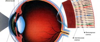
- peripheral paired organ of vision - the eye (with its light-perceiving photoreceptors - rods, cones and light-sensitive ganglion cells of the retina);
- nervous structures and formations of the central nervous system: optic nerves, chiasma, optic tract, visual pathways - II pair of cranial nerves, oculomotor nerve - III pair, trochlear nerve - IV pair and abducens nerve - VI pair;
- lateral geniculate body of the diencephalon (with subcortical visual centers), anterior tubercles of the quadrigeminal midbrain (primary visual centers);
- subcortical (and stem) and cortical visual centers: the lateral geniculate body and the pillows of the visual thalamus, the superior colliculi of the midbrain roof (quadrigeminal) and the visual cortex.
The normal irritant of the organ of vision is light. Under the influence of light in sticks
,
cones
(see below) and
light-sensitive ganglion cells,
the breakdown of visual pigments (rhodopsin, iodopsin and melanopsin) occurs. The rods function in low-intensity light, at dusk; the visual sensations obtained in this case are colorless. Cones function during the day and in bright light; their function determines the sensation of color.
Humans and many other animals have binocular vision, which provides three-dimensional images. Many diurnal animals have color vision.
Content
- 1 Eyes
- 2 Alternative ways to navigate in space
- 3 Evolution of the visual system 3.1 Invertebrates
- 3.2 Vertebrates
- 3.3 Mammals
- 4.1 Invertebrates 4.1.1 Protozoa
- 4.2.1 Visual system of reptiles, birds and some fish
What does the refraction of the image on the retina give?
As a result of this reaction, a nerve impulse is generated, which is transmitted along the nerve endings to the optic nerve, and then to the occipital lobe of the cerebral cortex. It is interesting that the pathways of the visual analyzer have complete and incomplete crossovers with each other. Thus, information from the left eye enters the occipital lobe of the cerebral cortex on the right and vice versa.
An interesting fact is that the image of objects after refraction on the retina is transmitted upside down.
In this form, information enters the cerebral cortex, where it is then processed. Perceiving objects as they are is an acquired skill.
Newborn babies perceive the world upside down. As the brain grows and develops, these functions of the visual analyzer are developed and the child begins to perceive the outside world in its true form.
The refraction system is presented:
- anterior chamber;
- posterior chamber of the eye;
- lens;
- vitreous body.
The anterior chamber is located between the cornea and iris. It provides nutrition to the cornea. The posterior chamber is located between the iris and the lens. Both the anterior and posterior chambers are filled with fluid, which is able to circulate between the chambers. If this circulation is disrupted, a disease occurs that leads to vision impairment and can even lead to its loss.
The lens is a biconvex transparent lens. The function of the lens is to refract light rays. If the transparency of this lens changes due to certain diseases, a disease such as cataract occurs. Currently, the only treatment for cataracts is lens replacement. This operation is simple and quite well tolerated by patients.
The vitreous body fills the entire space of the eyeball, providing a constant shape of the eye and its trophism. The vitreous body is represented by a gelatinous transparent liquid. When passing through it, light rays are refracted.
Eyes
Main article: Eye
In animals and humans, the organs of vision are the eyes. In addition to vertebrates, cephalopods and many arthropods, as well as individual representatives of other types of animals - cnidarians, annelids, flatworms, have highly organized eyes (capable of creating images of objects and providing object vision).[1] The compound eyes of insects have a fundamentally different structure compared to the chamber eyes of vertebrates and cephalopods, but are connected with them by gradual transitions in the comparative morphological series.
A little about the structure of the visual analyzer
The eyeball is located in the orbit on a fat pad, which serves as a shock absorber. With some diseases, cachexia (emaciation), the fat pad becomes thinner, the eyes sink deeper into the eye socket and it feels like they are “sunken”. The eyeball has three membranes:
- V.Rohto eye drops will help restore vision after intense or prolonged work in front of the computer;
- Eliminate itchy eyes and restore clarity of vision;
- Relieve signs of fatigue;
- Moisturize your eyes if they are excessively dry, including those caused by wearing contact lenses.
- protein;
- vascular;
- mesh.
The characteristics of the visual analyzer are quite complex, so they need to be sorted out in order.
The tunica albuginea (sclera) is the outermost layer of the eyeball. The physiology of this shell is designed in such a way that it consists of dense connective tissue that does not transmit light rays. The muscles of the eye that provide eye movements and the conjunctiva are attached to the sclera. The front part of the sclera has a transparent structure and is called the cornea. A huge number of nerve endings are concentrated on the cornea, providing its high sensitivity, and there are no blood vessels in this area. It is round and somewhat convex in shape, which allows for proper refraction of light rays.
The choroid consists of a large number of blood vessels that provide trophism to the eyeball. The structure of the visual analyzer is designed in such a way that the choroid is interrupted at the place where the sclera passes into the cornea and forms a vertically located disk consisting of a plexus of blood vessels and pigment. This part of the shell is called the iris. The pigment contained in the iris is different for each person, and it provides the color of the eyes. With some diseases, the pigment may decrease or be completely absent (albinism), then the iris becomes red.
In the central part of the iris there is a hole, the diameter of which varies depending on the intensity of illumination. Rays of light penetrate the eyeball onto the retina only through the pupil. The iris has smooth muscles - circular and radial fibers. It is responsible for the diameter of the pupil. Circular fibers are responsible for the constriction of the pupil; they are innervated by the peripheral nervous system and the oculomotor nerve.
The radial muscles are part of the sympathetic nervous system. These muscles are controlled from a single brain center. Therefore, the dilation and contraction of the pupils occurs in a balanced manner, regardless of whether one eye is exposed to bright light or both.
Alternative ways to navigate in space
There are other sensory systems similar in function to vision that are used for orientation in space, for example, ultrasonic echolocation of bats and cetaceans, which allows them to detect the smallest objects, electrolocation of some fish and the platypus, thermal location of rattlesnakes.
Also used for orientation in space are the sense of smell (the tongue of snakes is most characteristic in this sense, although dogs are also widely known as an example of orientation by smell), hearing (lateral line in fish), and tactile sensations (perception of pressure and temperature, palpation ).
Evolution of the visual system
Invertebrates
As determined using genetic transformation methods, eyeless
Drosophila and
Small eye
mice, which have a high degree of homology, control the development of the eye: when creating a genetically engineered construct, with the help of which the expression of the mouse gene was caused in various imaginal discs of the fly, the fly appeared ectopic compound eyes on the legs, wings and other parts of the body.[2 ] In general, several thousand genes are involved in the development of the eye, but a single “starter gene” (“master gene”) initiates this entire gene network. The fact that this gene has retained its function in groups as distant as insects and vertebrates may indicate a common origin for the eyes of all bilaterally symmetrical animals.
Vertebrates
The optic cups of vertebrates are formed as outgrowths of the diencephalon, and the primary center for processing visual information is located in the midbrain.
Mammals
It is assumed that during the Mesozoic period, early mammals occupied a subordinate position in relation to the “reigning reptiles” (especially dinosaurs, which predominantly occupied the ecological niches of large predators and herbivores), had small sizes and a twilight lifestyle. Under such conditions, vision for orientation in space becomes secondary to smell and hearing. The chemical feelings that remain emotionally charged to us today are serviced by the forebrain and limbic system. It is assumed that the forebrain becomes more important under these conditions. When the “reigning” reptiles disappeared at the end of the Mesozoic, greater evolutionary opportunities opened up for the “oppressed” mammals. They populated all possible ecological niches of the liberated world; for some groups, vision again became the most important of all senses. However, the newly formed visual pathways headed to the most important part of the brain - the forebrain, which expanded and formed the large hemispheres characteristic of mammals. The retino-tectal pathway remains a relic of the old visual pathway, and the retino-geniculo-striatal pathway is rapidly becoming the most important pathway for transmitting visual information to the brain.
Organ of vision
Everyone knows that the organ of vision in humans and any other living creature is the eye. More precisely, this is its colloquial name. In fact, it consists of the eyeball and an auxiliary apparatus. They have their own functions. And the eyeball does the most important thing, because it is in it that the peripheral section of all analyzers is located.
The pupil is the first thing I would like to pay attention to when talking about the analysis of visual stimulation. The process of light absorption occurs in the pupil. It is through it that the rays enter the eye. There is the retina, which has an extremely complex and fragile structure. And it is on this surface that cones and rods (these are light-sensitive cells) are located. And the nerve itself extends from the cells. It is the beginning of the conductive part of the analyzer itself.
Visual system in different taxonomic groups
Invertebrates
Invertebrates have very diverse eyes and ocelli in terms of structure and visual capabilities - unicellular and multicellular, straight and inverted (inverted), parenchymal and epithelial, simple and complex.
Arthropods often have several simple eyes (sometimes an unpaired simple eye - for example, the nauplial eye of crustaceans) or a pair of complex compound eyes. Among arthropods, some species have both simple and compound eyes: for example, wasps have two compound eyes and three simple eyes (ocelli). Scorpions have 3-6 pairs of eyes (1 pair is the main, or medial, the rest are lateral), the shieldfish has 3. In evolution, compound eyes arose by the fusion of simple ocelli. Close in structure to a simple eye, the eyes of horseshoe crabs and scorpions apparently arose from the complex eyes of trilobite-like ancestors by merging their elements (Beklemishev, 1964).
Protozoa
Some protozoa have poorly differentiated organelles for light perception (for example, the stigma in Euglena viridina).
Insects
The eyes of insects have a facet structure. Different species perceive colors differently, but in general, most insects are good at distinguishing not only the rays of the spectrum visible to humans, but also near ultraviolet. This depends, in addition to genetic factors (the structure of the receptors), and on less absorption of UV light - due to its shorter path in the optical system of the eye. For example, bees see an ultraviolet pattern on a flower.
Vertebrates
Visual system of reptiles, birds and some fish
It has been established that reptiles, birds and some fish have a wider range of perceived optical radiation. They perceive near ultraviolet (300-380 nm), blue, green and red parts of the spectrum. In some amphibians, for example, the crested newt, as shown by R. Mattei in 1925, vision can be restored after cutting the optic nerve [3].
The visual apparatus of birds has features that are not preserved in human vision. Thus, bird receptors contain microspheres containing lipids and carotenoids. It is believed that these microspheres - colorless, and also colored yellow or orange - act as specific light filters that form a “visibility curve”.
In many birds, their binocular vision, due to the specific location of the eyes, does not provide such a large field of stereoscopic vision as in humans.
Mammal vision
A mutation, once realized in one of the ancestors of mammals and fixed throughout the class, reduced the number of types of cone color receptors to two. It is believed that the ancestors of mammals - small rodents - were nocturnal and compensated for this loss by the significant development of twilight vision (with the help of receptors - rods).
Later, however, in primates (including humans), another mutation caused the appearance of a third type of cone - color receptors. This was caused by the expansion of the ecological niche of mammals, the transition of some species to a diurnal lifestyle, including in trees. The mutation was caused by the appearance of an altered copy of the gene responsible for the perception of the middle, green-sensitive region of the spectrum. It provided better recognition of objects of the “day world” - fruits, flowers, leaves.
The human eye consists of the eyeball and the optic nerve with its membranes. Humans and vertebrates each have two eyes located in the eye sockets of the skull.
Human eye
Stereoscopic vision
In many species whose lifestyle requires a good estimate of the distance to an object, the eyes look forward rather than to the sides. Thus, mountain sheep, leopards, and monkeys have better stereoscopic vision, which helps assess the distance before jumping. The person also has good stereoscopic vision (see below, section Binocular and stereoscopic vision
).
An alternative mechanism for estimating the distance to an object is implemented in some birds, whose eyes are located on different sides of the head and the field of three-dimensional vision is small. Thus, chickens make constant oscillatory movements with their heads, while the image on the retina quickly shifts, inversely proportional to the distance to the object. The brain processes the signal, which allows it to catch small prey with its beak with high accuracy.
Each person's eyes appear identical in appearance, but are still functionally somewhat different, so they distinguish between the leading and trailing eyes. Determining the dominant eye is important for hunters, videographers and other professions. If you look through a hole in an opaque screen (a hole in a sheet of paper at a distance of 20-30 cm) at a distant object, and then, without moving your head, alternately close your right and left eyes, then for the dominant eye the image will not shift.
Physiology of human vision
Main article: Human vision
Due to the large number of stages in the process of visual perception, its individual characteristics are considered from the point of view of different sciences - optics, psychology, physiology, chemistry.
Binocular vision in humans, as in other mammals, as well as birds and fish, is ensured by the presence of two eyes, information from which is first processed separately and in parallel, and then synthesized in the brain into a visual image. In distant phylogenetic predecessors of humans, the eyes were located laterally, their visual fields did not overlap, and each eye was connected only to the opposite hemisphere of the brain - contralaterally. During the process of evolution, in some vertebrates, including the ancestors of humans, due to the acquisition of stereoscopic vision, the eyes moved forward. This led to an overlap of the left and right visual fields and to the emergence of new ipsilateral connections: left eye - left hemisphere, right eye - right. Thus, it became possible to have visual information from the left and right eyes in one place, for comparing them and measuring depth.
Ipsilateral connections are evolutionarily younger than contralateral ones.
During the development of stereoscopic vision, as we move from animals with laterally oriented visual axes to animals with frontally oriented eyes, the proportion of ipsi fibers increases (table).[4] Number of uncrossed and crossed fibers in the optic nerve in a number of mammals
| Kind of animal | The ratio of the number of non-cross fibers to the number of cross fibers |
| Sheep | 1:9 |
| Horse | 1:8 |
| Dog | 1:4,5 |
| Opossum | 1:4 |
| Guinea pig | 1:3 |
| Cat | 1:3 |
| Ferret | 1:3 |
| Toque | 1:1,5 |
| Human | 1:2; 1:1,5; 1:1[5] |
Most features of human binocular vision are determined by the characteristics of neurons and neural connections. Using neurophysiological methods, it has been shown that binocular neurons of the primary visual cortex begin to decode the image depth specified on the retinas by a set of disparities. It has been shown that the most important requirement for stereoscopic vision is differences in the retinal images of the two eyes.[6]
Due to the fact that the visual fields of both eyes of humans and higher primates overlap to a large extent, humans are better able than many mammals to determine the appearance and distance (the accommodation mechanism also helps here) to close objects, mainly due to the effect of stereoscopic vision. The stereoscopic effect persists at a distance of approximately 0.1-100 m. In humans, spatial-visual abilities and three-dimensional imagination are closely related to stereoscopy and ipsi-connections.
Elementary visual sensations and perception of complex features
Description
We must pay tribute to the judge who quite rightly rejected the claim: at the very beginning of the 19th century, when this case was heard in a small German city, it was very difficult to substantiate the inconsistency of the Claimant’s statements.
The doctrine of vision at that time was reduced to a rather primitive theory of the structure of the eye, based on some data from anatomy and geometric optics. “An adequate irritant? And what is it?" - even a specialist physiologist of that time would have asked if you had tried to explain to him the difference between the sensation of a flash of light that occurs when you press on the eye in the dark, and genuine light perception. The natural sympathy that I feel for the judge who made a fair decision forces me to admit that he substantiated the verdict experimentally, for example, by making sure that by pressing on the eye, he “sees the light” even under tightly closed eyelids, and through the eyelids, as is known , and in the clear sun you can’t see anything.
But this experiment, strictly speaking, would not be decisive. Well, let the “own light of the eye” be visible when nothing can be seen because the eye is closed. This does not yet prove that with such “light” open eyes will not see anything. However, this can also be verified experimentally. But here’s what you can’t check: even if you yourself can’t do it, maybe others are able to see in this light?.. Well, at least some. And so on.
In a word, do you know how things stand in our time with the so-called supersensible perception? In much the same way, until 1826, it was possible to “prove” identification using light generated by a blow to the eye. This date is significant not only for the science of vision, but also for the entire physiology of the sense organs: in fact, from this moment, marked by the appearance of the works of Johannes Müller, the physiology of the sense organs
began as a science.
Every perceptive organ is adapted to perceive a certain natural physical agent (it is the latter that we now call an adequate stimulus), but all sense organs have one common property - irritability
. Anything that can irritate their nervous tissue can cause sensation; the nature of the sensation depends not on the nature of the stimulus, but on the structure of the irritated organ. That's the point. The eye is adapted to perceive light. Only light can realize the ability to see inherent in the organ of vision. But the eye (more precisely, the nervous apparatus of the organ of vision) is capable of communicating to the brain only sensations of light, and not heat, touch, taste, or smell. The eye “sees light” under any irritation - thermal, mechanical, electrical, x-ray, etc. This “light” is as incapable of serving vision as the “sour taste” of weak direct current (have you ever tried to apply the contacts of a battery to your tongue ?) cannot replace pickled cucumber or a portion of vinegar for dumplings.
When studying the visual apparatus, both adequate and inadequate stimuli are used. When studying the naturally occurring process of vision, they use only an adequate stimulus - light
.
In order for a natural visual sensation to arise, it is necessary that the light source is located within the field of view, and the radiation remains within the boundaries of the visible spectrum (wavelength from 700 to 420 millimicrons) and the radiation intensity is not lower than the threshold.
Threshold concept
- one of the most important sense organs in physiology. You met him in experiment 16. The true (absolute) threshold of photosensitivity is found when the observer has been in complete darkness for a sufficiently long time; This time is usually about 60 minutes. The minimum brightness of a small spot of light sufficient to produce a visual sensation is determined. This brightness will be the threshold.
Once the threshold is passed, not only the presence of light is felt, but also the direction towards the light. Thus, the simplest visual sensation is characterized by two elementary properties: directional light is felt
.
Photosensitivity function
in itself does not yet provide the possibility of vision.
Contrast sensitivity is also needed - a sense of differences (differences) in brightness. Contrast sensitivity
in humans is very well developed, for example, an observer notices the difference in the brightness of the illumination of two areas, one of which is illuminated by a lamp of 100 candles, and the other by 102 candles;
On an evenly lit fan, a person can distinguish a black thread, the transverse diameter of which is only 4-6 arc seconds. Distinguishing the boundaries of the difference in brightness, highlighting contours, is already a transition from elementary sensations to the perception of complex features, in particular to the perception of shape. The simplest form of this function, closest to contrast sensitivity, is the discrimination of brightness gaps between small, closely located objects; We are talking about visual acuity. When exploring this function in experiment 15, you used black objects on a white background (the contrast was about 80%). Consider the same letters on a light gray background, then on a dark gray background: the darker the background, the lower your visual acuity will be. Try to show not black, but gray letters on a white background - the same thing will happen: the lower the contrast, the worse the discrimination. Under natural conditions, a person sees the world because it is a world of contrasts. When there are few objects in the surrounding space that sharply contrast with the background, visual orientation is impossible. A uniformly illuminated field of view—physically quite defined, of course—appears to the observer as “empty,” shapeless, and indefinite. Scattered light for vision is the same darkness. The only thing that can be “considered” in such a field—balls, droplets, threads, bubbles—are particles that disrupt the optical uniformity of the tissues of your own eye (experiments 11 and 12). If you have noticed these “flies” before, you probably noticed that they are especially clearly visible against a uniform, bright background (sky, snow, white paper). The point here is not only in lighting, but also in the fact that, without receiving normal, sharply contrasting images, the eye, as it were, catches in an empty field everything that is at least somewhat different from emptiness, formlessness. If a large black bird or a red plane flies across the sky, at which you look through a pinhole, observing particles that violate the optical uniformity of the transparent media of your eye, the balls, threads, etc. will immediately fade or disappear completely. Here another very important property of the organ of vision (we will simply say “eyes”) is manifested - the eye not only reacts to contrast, but also “hunts” for it, highlights the most contrasting areas of the visual field, and emphasizes the boundaries of the difference in brightness. This property is so essential for the process of vision that under certain conditions (remember experiments 20, 21, 36, 37) vision even “creates” contrasts that are absent in the physical characteristics of the stimuli
.
The visual process unfolds over time, and the nature of sensations also changes over time. The appearance, change, and disappearance of a stimulus are the main sources of visual sensation
. The eye gets used to constant and constant irritants; these stimuli very quickly cease to cause sensation; for example, the blood vessels of the retina, which always cast shadows on the same areas of the retina, are not perceived under normal conditions (experiment 12).
Physical stimulus
can appear and disappear almost instantly: within one four-hundredth of a second, the brightness will jump from zero to maximum and drop back to zero. A visual sensation will arise when the stimulus is no longer in the field of view. Depending on the strength of irritation and subsequent conditions, it will last from several seconds to tens of minutes, and both the brightness and color of the perceived trace of irritation of the retina will periodically change.
Thus, the elementary visual sensations are the sensation of the presence and direction of light and the sensation of contrast. These sensations are characterized by a certain intensity (strength), depending in a certain way on the intensity of the physical stimulus, and a duration, depending both on the time of action of the stimulus and on trace processes unfolding over time in the visual apparatus itself (experiments 15-20, etc.).
We haven't said anything here about color perception.
, in particular about the sensations arising from the action of chromatic rather than white light. The threshold for the perception of color is significantly higher than the threshold for light sensitivity, but with sufficient intensity of colored light, vision appears to function as well as with white light. At low intensities, color is not felt at all (“at night all cats are gray”). If you slowly increase the brightness, the color blue is perceived first, then green, and lastly red. Do the Purkinje experiment: holding a sign with red, blue and green sectors in front of you, plunge the room into dense twilight. Gradually, as you adapt to the darkness, you will first be able to distinguish one sector - the lightest (later it turns out that it is green), then another - darker (but you will probably understand its color first - blue); red will look black and will be visible later than everyone else. You can observe this in the field: at dusk, when the poppies seem completely black, the cornflowers still turn bright blue in the rye.
Issues of color vision are complex and poorly developed. Therefore, here we will touch on them no more than is necessary for the main topic. It is important, for example, that not only brightness, but also color contrast (provided that it is perceived by the vision of a given observer) is sufficient for the visual perception of form. Therefore, we will also consider the sensation of color “elementary.”
The transition from elementary sensations to complex signs is not abrupt, since the very concept of “complex sign” cannot be defined precisely enough. We will limit ourselves to a conventional division: we will classify as elementary sensations the subjective perception of light emitted or reflected by external objects (thus, this will include sensations of brightness, direction, contrast and spectral composition of light), and as complex perceived features we will classify the subjective perception of the objects themselves - their shape, relief , distance, movement and so on.
The need for such a division arises because the mechanisms of perception of complex signs cannot be reduced to the sum of elementary sensations. It is not difficult to verify this.
The book lying in front of you has the shape of a rectangle - this is, in the literal sense of the word, obvious. But why you see it as a rectangle is unclear: after all, on the retina of the eye, the image of a book has the shape of a trapezoid with the base downwards (the angle between the visual axis and the plane of the table on which the book lies is not 90 degrees, but only about 60). Look at a book from the side, from a distance, from below, you will always perceive it as a rectangle. This reveals one of the main properties of visual perception - constancy (constancy, invariance) of a complex feature
, in this case the visible shape of a familiar object.
In experiment 33, you were convinced that the size of an object is perceived quite stable, although with increasing distance the image on the retina steadily decreases. Unexpected evidence of the independence of apparent size from the size of the image on the retina was also obtained in experiment 18, when you “projected” a sequential image either onto your palm, then onto a wall or onto the ceiling; in the palm of the hand the bird was small, on the wall it was large, but the source of both images was constant, since the area of irritation on the retina did not change; only the distance to the plane onto which the image was projected changed.
These experiments clearly revealed an important pattern of visual perception of complex features—the synthesis of sensations, leading to the formation of a holistic picture of the visible world.
It is not the apparent size of an object that is invariant, but the ratio of the size to the perceived distance to the object. If you misjudge the distance, you will have a misconception about the size of the object, and vice versa. An excellent example is given by S.I. Vavilov: “... for a short moment the cat was visible the size of a cow; it seemed as if this cat was walking along a distant fence; in fact, she was walking along the roof, near the window through which she could be seen. The result was approximately a twenty-fold error in estimating the distance
».
The illusion did not last very long: the size of the animal, precisely known to the observer, is such a powerful factor in the perception of the correct relationship of things in space that the visual system immediately overestimated the distance.
But the size/distance ratio is far from an isolated factor in the perception of real objects in the external world. After all, the assessment of distance has its own “instruments” of perception, independent of the apparent size, of a mono- and binocular nature. Under normal conditions, the main tool for assessing the location of objects in the three-dimensional world is binocular stereoscopic vision (experiments 25-31), which we have already mentioned in the section “The Eye and Ray Optics.” Let us only recall that the physiological mechanism of stereo vision is disparity
- a certain asymmetry in the shape and position of two images belonging to the same object on the retina of both eyes. This is a very thin and precise mechanism. Suffice it to say that the difference in the distance of two objects (or two parts of one object - relief) is perceived by binocular vision, starting with the asymmetry of the images, which is only 10-12 arc seconds. The mechanism operates at the same time throughout the entire visual field, common to both eyes, but most accurately in relation to objects located close to the observed object. At the same time, the position of all visible objects in space relative to the fixed object is felt. A person does not need to move his gaze from one object to another in order to perceive their distance relative to each other. It is felt by the disparity of the images of these objects, by their physiological doubling (experiment 26). Even with a fixed gaze, the entire visible world is a three-dimensional field-space.
The integrity of the visual field, the interconnection of all objects located in it, is a pattern that is so important for the functioning of vision that all interference with holistic perception is artificially suppressed in the visual apparatus. You know one example: the blind spot (experiment 14) cannot be observed in natural conditions, there is no “hole” in the visual field, the brain seems to fill the part of the visual field corresponding to the position of the blind spot.
There is an even more striking way of demonstrating this fundamental feature of vision. Roll up a narrow tube (at least from newspaper) and place it on one eye; Place your own palm at the end of the tube in front of the other eye so that it obscures the center of the field of vision of that eye. Thus, you “turn off” the entire periphery of the visual field for the eye with which you are looking through the tube, and the entire center of the visual field for the other eye. Look straight ahead. This experience is called a “hole in the palm.” A rather strange field of vision is formed: its periphery is objects in the room and the palm, and in the palm, through which distant objects are visible - and all this makes up a single picture. In the visual apparatus, the part of the palm image that covers the center of the visual field of one eye is completely suppressed.
Suppressing Image Details
, interfering with the formation of an interconnected visual picture, is a process as natural and necessary for visual perception as the excitation of nerve cells from which the formation of an image begins. Both processes occur automatically, without the participation of consciousness.
Do another simple experiment; let's call it " transparent hand"
"
Bend your arm at the elbow, raise it and hold it vertically so that your wrist is 25-30 centimeters from your eyes. Look at your hand. The subject is quite material, isn’t it? Now just look into the distance without changing the position of your hand and head. The flesh of the hand seemed to “dissolve”, the hand became “transparent”. In fact, there was simply an almost complete suppression of that part of the image of the external world that interferes with the formation of a holistic picture. As a result, only those objects to which your attention is directed are clearly visible. binocular vision
works . Check: as before, without changing the position of your hand and head and looking into the distance, close one eye - you will see both the hand and the distant object; the same thing will happen if you cover not the right, but the left eye. You can, of course, observe not a hand, but, say, an opaque strip of cardboard. At a certain width of the strip (greater than the diameter of your wrist), it will not become completely “transparent”, although its apparent width will decrease. The fact is that complete or almost complete suppression of the image of an object that covers part of the binocular field of vision occurs only in those cases when the object does not cover the central parts of the field of vision of both or at least one eye. Notice that the hand appears transparent even when you place it directly opposite one eye (looking, of course, with both eyes).
Central vision
plays the role of the main axis around which the picture of visible space is formed. You know that it is central vision that has the greatest acuity (experience 15). In binocular vision, the visual axes of both eyes are directed to the same area of the observed object. Remembering the merging of “identical” images into a single image (experiments 25-29) and the natural decrease in visual acuity from the center to the periphery, you can easily imagine visual space, reflecting the three-dimensional world of real objects, as a kind of functional pyramid. At its apex there is an object, the images of which are projected onto the central fovea of the retina of both eyes; these images are the most detailed. Images of other, non-fixed objects are projected onto the “ledges” of the pyramid; the further a given ledge is from the top of the pyramid, the less detailed the image lying on it.
The observer's attention is usually directed precisely to the object viewed by central vision. But central vision and attention are different things. You know that, looking at the teacher, you happened at the same time to concentrate all your attention on some other person or object, which at that time you saw only out of the corner of your eye. This task is not easy, but it can be done: it can be done because, I repeat, central vision and attention are different things; not easy, since it is central vision that brings maximum information about the object of observation to consciousness, and therefore, under natural conditions, the “axes” of vision and attention coincide.
Looking motionlessly in front of you, you see binocularly three-dimensional space in a solid angle with an angular diameter of approximately 120 degrees. But, wanting to examine in detail all the objects located in this space, you move your gaze from one object to another, since accurate perception of shape, color, relief, distance requires the work of central vision.
The periphery of the visual field gives an approximate idea of space and objects, and the center checks and refines it. The periphery allows the brain to form a prediction, to make a forecast about complex features of objects located in space, and central vision serves to control this forecast. Therefore, good visual orientation in space requires not only high acuity, but also a normal visual field (experiment 13).
In order for an accurate idea of three-dimensional objects viewed in real space to form in the observer’s mind, his visual system must perform a huge number of very diverse operations, coordinate assessments of various features of these objects into a single coherent picture. Let us list the main operations and estimates involved in the synthesis.
Highlighting contours and distinguishing between brightness changes
(based on the contrast sensitivity of the eye) allow, with the help of mechanisms on which visual acuity depends, to distinguish individual objects and the details of these objects. Synthesis of characteristic features of the shape of an object allows them to be identified (very little is known about the physiological content of this operation). In accordance with the apparent size of the identified object, the assessment of its distance from the observer, prepared by other assessments that do not depend on identification, is checked. Among the latter: linear perspective (that is, a gradual decrease in the size of all visible objects as they move away, the convergence of parallel lines, the convergence of the earth and sky at the horizon), aerial perspective (the gradual loss of visible relief details by objects as they move away, a decrease in the intensity of shadows and colors on their surfaces, the desire of objects to merge with the background), the overlap of distant objects with nearby ones; This is by no means an exhaustive list.
All of the above operations and assessments are performed not only with binocular vision, but also with one eye; therefore they are called monocular perceptual factors
. It is thanks to monocular factors that “illusions that truly reflect reality”, which are very interesting to us, are possible. This definition, despite its paradoxical sound, literally expresses the essence of the matter, since we are talking about graphics, painting, cinema, in a word, about those well-known cases when a person perceives “depth” while looking at a two-dimensional plane, paper, canvas or screen. Having carefully studied the landscape painted by a good realist artist, you will find that the impression of three-dimensionality arises precisely on the basis of the above-mentioned patterns of perception. This impression is sharply enhanced if you limit the field of view to the “field” of the picture (viewing the picture with one eye “through a fist,” that is, a narrow tube). It is better to keep your head still.
When observing real space, head movements relative to stationary objects (or vice versa) help - even with monocular vision - to accurately perceive the relative distance of objects based on the speed and direction of movement of their images across the retina.
The effect caused by limiting the field of view when viewing a painting is explained by the fact that this excludes, firstly, the surrounding three-dimensional objects (frame, wall, people) that contradict the perception of depth in the painting and, secondly, the binocular mechanisms of perception of distance, which In the case of an unlimited field of view, they come into conflict with the monocular factors of perception (at a constant angle of convergence to the picture plane, the binocular mechanism testifies: “There is no difference in distance - there is no depth!”, and the monocular factors repeat theirs: “There is depth!”).
The possibility of conflict between different perceptual mechanisms is a very important point. In the holistic act of vision, binocular and monocular factors of visual perception serve not only for different, but also for the same assessments, partially overlapping each other. As a result, the space visible to a person contains all the elements that do not contradict a coherent, holistic, meaningful picture.
In all cases, eye movements are necessary to check, clarify, and rearrange what is perceived, since thanks to these movements, different parts of the visual field alternately fall on the “edge” of the central axis of the monocular cone or binocular pyramid.
Vision
- an almost continuous analyzer-synthetic process. We distinguish in it the levels of elementary sensations and complex perceptions of objects, highlight special features and mechanisms, and list the operations and assessments performed by the higher apparatus of vision. This is legal, otherwise the research cannot be conducted. But the listed components, considered individually or in groups, still speak about the fundamental organization of the visual process as much as blocks of multi-colored stones removed from a mosaic fresco tell about its content. Even in order for each pebble to be appreciated, you need to know the whole picture, you need to understand its purpose and its meaning.
After all, this is exactly the goal we set for ourselves from the very beginning - to find out how human vision works. We expected to get an answer to the question of how reliably reality is perceived through vision.
Let’s summarize everything we’ve learned and see if we have received answers to the questions that interest us.
—-
Article from the book: Experiments with vision at school and at home | Gregg J.
Notes
- Beklemishev V.N.
Fundamentals of comparative anatomy of invertebrates. - M., Nauka, 1964, vol. 2, p. 143—159 - glava 14.1.p65 Archived April 19, 2009.
- R. Matthey (1925). "Récupération de la vue après résection des nerfs optiques chez le Triton." Comptes rendus des séances de la Société de biologie et de ses filiales 93
: 904-906. - Blinkov S. M., Glezer I. I. The human brain in figures and tables. - L., 1964. - 180 p.
- Data from different authors.
- Bishop PO (1981) Neural mechanisms for binocular depth discrimination. In: Advances in Physiological Sciences. Sensory Functions (Eds. Grastian E., Molnar P.), v. 16, p. 441—449