Nephrostomy: indications, results
Nephrostomy is a special drainage tube that is installed in the renal pelvis. Intervention is most often resorted to in case of blockage of the ureter, when there is no other option to restore the outflow of urine.
Organ dysfunction is associated with pain and causes a number of pathologies.
The purpose of the drainage tube is to normalize the movement of fluid and relieve pain. The drainage tube also helps treat inflammatory processes.
Most often, such surgery is indicated for patients with cancer. This is very important, because the abnormal movement of urine from an organ is a threat of non-stop damage to its tissues, and this, in turn, leads to their complete irreversible destruction.
A drainage tube can be installed temporarily; it is applicable to approach the upper tract of the urinary system. In this case, it is removed as soon as the functions of the organ are restored. In other cases, drainage is installed for life.
Sometimes the procedure is vital, but not all patients agree to have it performed.
Meanwhile, modern drainage tubes prevent urination, and now there are numerous substances that help care for them without much difficulty.
Shown
This surgical intervention is widespread in urology. The main indication for it is the delay in the movement of fluid from the kidney. This delay can be caused by the following pathologies:
- Adenoma of the corresponding organ;
- Cancer diseases;
- Urolithiasis;
- Hydronephrosis;
- Pyelonephritis;
- Urethral strictures;
- Genetic diseases of the urinary system.
Endosonography, endosonography
1.General information
Of all the terms denoting narrowing, partial or complete closure of the lumen in any vessel or duct (obstruction, obstruction, stricture, embolism, etc.), the most “alarming” is the diagnosis of “obliteration”. This means that there is no patency at all, that the lumen is either initially closed or completely blocked. In urology, speaking about disorders of the urinary tract, including the ureter, the term “stricture” is used much more often, but in international terminology there is also an extreme option: complete stricture, complete obstruction, which is called obliteration of the ureter (not to be confused with the urethra, i.e. with the distal, lower part of the urinary tract).
The ureters are two relatively thin and relatively elastic, due to the presence of muscle tissue in the walls, connecting “tubes” from the kidneys to the bladder. There is urine in the collector bladder, i.e. The liquid used in the process of renal filtration with “production waste” accumulates to a certain volume, and in an adequate (socially acceptable) situation is evacuated through the urethra to the outside. The thickness of the ureter in different areas is not the same and varies from 3 to 10 mm; the length depends on specific individual characteristics and can range from 20 to 30 cm. Due to anatomical reasons, female ureters are, on average, shorter than male ones.
A must read! Help with treatment and hospitalization!
Preparation
First, the patient must tell the doctor about the medications he is taking. This is especially true for blood thinning drugs. You should stop taking them a week before surgery. We also stop taking NSAIDs 2-3 days before, and diuretics 24 hours before.
The night before your procedure, eat dinner earlier than usual.
In the morning you can drink a little (up to 250 grams of liquid) but no later than 2 hours before the operation.
You must refrain from eating approximately 2-3 hours before arriving at the clinic.
Don't forget to take the medications prescribed by your doctor.
Do not apply makeup or cream to your skin.
Remove piercings and jewelry, and remove contact lenses if you have them.
4.Treatment
Obliteration of the ureter is an emergency life-threatening condition and, accordingly, assistance should be provided on an urgent or emergency basis (depending on the situation). Attempting medicinal resolution or, worse, self-medication poses a fatal risk. Catheterization for the purpose of mechanical restoration of patency in most cases does not bring results; Currently, this approach, also quite risky, is not the method of choice. The most common method of intervention is abdominal urologic surgery to remove the obliterated area with subsequent connection of the ends. Today, minimally invasive endoscopic techniques are being actively developed to completely eliminate the problem with minimal surgical trauma to the urinary tract (which is very important in terms of preventing relapses of fibrous stricture) and a minimal risk of intraoperative infection, complications in the rehabilitation period, etc. However, the clinical picture is not always leaves time for at least emergency preparation of endoscopy (and any intervention on the urinary tract requires high qualifications, and sometimes even jewelry equipment). Therefore, it is critically important for any, especially rapidly developing, urinary problems to seek help as soon as possible.
Peculiarities
Today, a doctor can install a drainage tube in two ways:
- Nephrostomy performed in an open manner;
- Nephrostomy puncture.
The first option is more traumatic and requires general anesthesia. The surgeon makes an incision into the skin, underlying tissue and kidney. Today this option is rarely used.
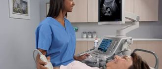
The second method is less traumatic. No incisions are needed to establish drainage. It is installed through a puncture, which the doctor performs under ultrasound control. Naturally, general anesthesia is not required here, and the intervention itself is easier to tolerate and the recovery period is shorter.
What to expect after
The patient remains in bed until the end of the sedative medication. Then he returns to the ward.
The end of the tube that protrudes from the body is attached to a urinal bag, usually located on the leg. Urine will begin to flow there immediately after installing the drainage tube.
It is important to empty the urine bag on time, otherwise, when it is full, it will become heavy. If he becomes unfastened, he can easily pull out the tube.
Excrement should flow freely into the urine bag.
You should contact the clinic if:
- The excrement contains blood;
- They became cloudy;
- An unpleasant odor appeared.
Hydronephrosis
It is important to remember that the expansion of the collecting system may not be associated with urinary tract obstruction (pyelectasia), but may be functional in nature. Most often, pyeelectasis due to functional reasons is observed in children under the age of one year and does not require surgical correction. To confirm the diagnosis of hydronephrosis caused by organic obstruction in the area of the PUS (ureteropelvic segment), kidney scintigraphy is performed after the administration of a diuretic and the Whitaker test.
Atrophy of the renal parenchyma during hydronephrosis has its own peculiarities, namely that the kidney, even with complete obstruction, continues to excrete urine. With complete obstruction in the LMJ area, soluble substances and water are absorbed through the renal tubules into the lymphatic system. The pressure in the pelvis is thus reduced (less than 6 mm Hg) and glomerular filtration is resumed. It has been noted that with unilateral hydronephrosis, kidney function decreases faster than with bilateral damage of the same severity. As hydronephrosis increases, the healthy kidney becomes larger (hypertrophied). The experiment showed that kidney function can be restored even after four weeks of complete obstruction. But meanwhile, it has been proven that irreversible changes in the kidney parenchyma can develop even after 7 days of obstruction. It is especially difficult to assess the likelihood of restoration of kidney function in case of partial obstruction, as is most often the case in the case of congenital anomalies (obstructive ureterohydronephrosis, hydronephrosis). It has been established that with unilateral hydronephrosis, the function of the second, healthy kidney increases by one and a half to two times; Accordingly, the volume of the pelvis of a healthy kidney increases. This is important to consider in order not to perform unnecessary plastic surgery on a healthy kidney.
Diagnosis of hydronephrosis in children.
The clinical picture depends on the age at which the defect was discovered. Some patients report pain, pain without clear localization, most often noted in the abdominal area in the form of pressure. In children, such pain is often confused with gastrointestinal disorders. If the diagnosis is not made in a timely manner, the intensity of the pain decreases due to the death of the renal parenchyma and a decrease in pressure in the renal cavity. Obstruction caused by the accessory inferior polar vessel most often, according to our observations, causes similar complaints. Since it develops somewhat later and is practically not diagnosed in early childhood. As it progresses and, especially with the addition of pyelonephritis, the clinical picture becomes clearer. Intense paroxysmal pain in the lumbar region and fever appear. When prescribing diuretics or infusion therapy, the pain symptom intensifies. Many pathogens of urinary tract infections (Proteus, Staphylococcus) secrete urease and alkalinize the urine; an alkaline environment promotes the formation of stones.
What diagnostic methods are used to detect hydronephrosis?
The first and most effective diagnostic method, used prenatally, is ultrasound. The method is harmless and is optimal for screening urodynamic disorders. Ultrasound examination (ultrasound) makes it possible to determine disturbances in fetal urodynamics already at the second prenatal screening study. This contributes to the early detection of hydronephrosis and allows us to develop the correct tactics for managing patients with obstructive uropathy. The ultrasound method is also effective in the neonatal period during examination for hydronephrosis and subsequently after surgical correction, it allows you to monitor the result of the treatment. Methods have been developed to assess the degree of obstruction based on ultrasound data using a diuretic (furosemide). This method, along with the isotope method (nephroscintigraphy), allows you to correctly set the indications for surgical treatment and avoid unnecessary surgical treatment in the absence of an organic barrier (obstruction) to the outflow of urine from the renal pelvis.
In addition to the ultrasound method, radioisotope diagnostics are used (static nephroscintigraphy and dynamic nephroscintigraphy).
With obstruction of the urinary tract, the isotope is poorly captured by the kidney, slowly passes through the parenchyma and remains in the pelvicalyceal system longer than normal. If there is doubt about organic obstruction, a test with a diuretic can be performed.
Excretory urography has not lost its relevance. This method of examination for hydronephrosis clearly demonstrates the degree of expansion of the renal cavity system, deformation of its calyces as a result of sclerotic processes supported by chronic pyelonephritis. Also, by the degree of filling of the pyelocaliceal system with the contrast agent, as well as by the rate of its evacuation at equal time intervals, we can judge the degree of obstruction and indirectly about the excretory function of the kidney. Excretory urography helps to correctly differentiate between hydronephrosis in the intrarenal pelvis and megacalycosis. Megacalycosis is a congenital renal anomaly associated with dysplasia of the medulla of the renal parenchyma. As a result, during ultrasound examination, as well as excretory urography, the kidney has normal dimensions and an even contour. Its cups are significantly expanded and deformed, the papillae are flattened. But the number of cups is of fundamental importance in this case. If in a normal kidney the number of cups is 7-8. Then in a kidney with congenital megacalycosis, there may be more than twenty of them. Uncomplicated megacalycosis does not require surgical treatment.
Negative results
Sometimes they arise. Displacement of the tube or its protrusion sometimes occurs.
This happens much more often if an open operation is performed.
Negative consequences may include bleeding, hematomas, etc.
As medical practice shows, these consequences are extremely rare.
Troubles may appear some time after the operation. Among them:
- Violation of the tone of the pelvis;
- Malfunction of the ureter;
- Pyelonephritis;
- Urolithiasis disease;
- Necrosis of kidney tissue.
Proper care of the drainage tube greatly reduces the risk of unpleasant results.
2. Reasons
Ureteral stricture, i.e. partial, incomplete closure of the lumen can occur for various reasons, and the number of such factors is quite large (trauma, including surgical, congenital anomalies, deformations, radiation injuries, etc.). As for obliteration as a complete blockade, the main cause is post-inflammatory fibrosis - the process of scarring, the replacement of wall tissue with connective tissue, which is much more dense, rigid and rough. The walls of the ureter gradually thicken and may eventually come into contact, resulting in complete loss of patency of the urinary tract.
Probable causes of ureteral obliteration also include migration of a renal calculus (stone): in some cases, a stone, reaching the narrowest place, can “fit” tightly and block the lumen completely. Finally, obliteration can be caused by a growing malignant tumor.
In general, ureteral obliterations are, fortunately, a fairly rare condition, and always fatal.
Visit our Urology page


