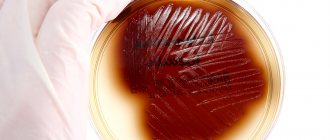
Bacterial culture is by far the most informative study that allows us to identify the causative agents of the disease, as well as determine the sensitivity of pathogenic microflora to antibiotics. All biological fluids of the human body are suitable for bacterial culture - urine, feces, blood, semen, mucus from the oral cavity or genitals. But in the vast majority of cases, this research technique is used by urologists and gynecologists. That is, we are talking about urogenital smears.
When is an appointment for a bacterial culture test issued?
The attending physician may give the patient a referral for a urogenital smear if a bacterial infection is suspected. Another reason for analysis is routine examination and prevention of the development of diseases of a bacterial nature. Bacterial culture is the most informative research method for identifying pathogens of sexually transmitted diseases. Biological material for research is taken directly from the urethra, vagina, cervix or rectum. It all depends on the specific situation and patient complaints.
A microflora study is mandatory when planning pregnancy; in this case, a referral is given to both partners. A pregnant woman regularly undergoes a culture test, since pathogenic microflora can cause abnormal development of the fetus and provoke premature termination of pregnancy.
Content:
- When is an appointment for a bacterial culture test issued?
- Preparing for the study
- Who is responsible for deciphering the bacterial culture analysis?
If a patient consults a urologist with complaints of discomfort during urination, pain in the lower back and lower abdomen, in order to exclude infection, a referral is given for a urogenital smear.
The reason to visit a gynecologist or urologist and get a referral for bacterial culture is a rash on the genitals and in the groin area, ulcers, the appearance of purulent discharge or discharge with a distinct odor.
This diagnostic method is also used to determine the nature of many other diseases, in particular ailments of the ENT organs. For example, to determine the cause of purulent plaque in the mouth or excessive mucus discharge from the nose, this test is also prescribed.
The purpose of cultivating microorganisms and its significance for diagnosis
In addition to water, air, soil, which contain various microorganisms in varying concentrations, including those that bring disease (pathogenic), many branches of medical science are interested in microbes living on the skin and mucous membranes of the human body, which can be represented by:
- Permanent inhabitants who do not pose any danger to humans, that is, the normal microflora of the body, without which we simply cannot live. For example, the disappearance of bacteria living in the intestines and participating in the digestion process leads to dysbiosis, which is not easy to treat. The same thing happens with the disappearance of vaginal microflora. It is immediately populated by opportunistic microorganisms, gardnerella, for example, which causes bacterial vaginosis (gardnerellosis);
- Opportunistic pathogenic flora, which causes harm only in large quantities under certain conditions (immunodeficiency). The above-mentioned gardnerella is a representative of this type of microorganism;
- The presence of pathogenic microbes that are not present in a healthy body. They are alien to the human body, where they enter accidentally through contact with another (sick) person and cause the development of an infectious process, sometimes quite severe or even fatal. For example, an encounter with the pathogens of syphilis - no matter what, it is treated at first, but (God forbid!) it will unleash cholera, plague, smallpox, etc.
Fortunately, many of them have been defeated and are currently kept under seal in special laboratories, but humanity must be prepared at any moment for the invasion of an invisible enemy capable of destroying entire nations. Bacteriological culture in such cases plays, perhaps, the main role in identifying the microorganism, that is, determining the genus, species, type, etc. (toxiconomic position), which is very important for the diagnosis of infectious processes, including sexually transmitted diseases.
Thus, sowing methods, like nutrient media, are different, however, they have the same goal: to obtain a pure culture without foreign impurities in the form of microbes of other classes that live everywhere: in water, in the air, on surfaces, on humans and inside it.
Preparing for the study
Bacterial culture with determination of sensitivity to antibiotics requires compliance with certain preparation rules. Only in this case will it be possible to say that the data obtained correspond to reality and make it possible to establish the correct diagnosis.
Preparation depends on what biological material is needed for the study. For example, if we are talking about venous blood, the only recommendation is to take the test on an empty stomach. And, if possible, a day before the procedure, give up alcohol and fatty foods.
If you need to donate urine, the conditions change slightly. In the absence of pathologies, the fluid in the bladder is absolutely sterile, and its saturation with microflora occurs already at the moment of passage through the urinary canals. A small content of pathogenic microorganisms in the liquid is considered normal, but exceeding them indicates the development of the disease.
Therefore, it is important to correctly collect material for analysis. To do this, patients should adhere to the following recommendations:
- preliminary hygiene of the genital organs;
- collection of “average” urine;
- Only sterile containers are used to collect the analysis;
- submitting urine to the laboratory no later than 2 hours after collection.
Feces for analysis are collected immediately after defecation into a sterile container.
If biological material for analysis is taken from the oral or nasal cavity, vagina, rectum or urethra, the collection takes place directly within the walls of the medical institution.
It is forbidden to carry out hygiene procedures using antiseptics in advance, as this will distort the results of the study.
A pregnant woman is given a referral for bacterial culture twice - at the initial stage (during the registration process) and at 36-37 weeks. A swab is taken from the vagina, cervix and nose. The main goal is to control the microflora of the mucous membranes. If abnormalities are detected, immediate therapy is indicated, especially if we are talking about the detection of E. coli in late pregnancy - during natural childbirth, the child may become infected.
It should be understood that a smear on the flora and bacterial culture are two different research methods. In the first case, biological material is simply studied in order to identify bacteria there. In the second, the resulting biomaterial sample is placed in a nutrient medium. After a certain period of time, the “grown” microorganisms are examined and the causative agents of the disease are identified.
Bacterial culture results should be expected within 3 to 7 days. In addition, this is the only way to determine the sensitivity of pathogenic microflora to antibiotics and select the most effective treatment.
When is tank sowing prescribed and how to understand the answers?
Name of microorganism and its quantity
Patients do not prescribe bacteriological analysis to themselves; this is done by the doctor if he has suspicions that the problems of a patient presenting various complaints are associated with the penetration of a pathogenic pathogen into the body or with the increased reproduction of microorganisms that constantly live with a person, but exhibit pathogenic properties only in certain conditions. Having passed the test and after some time received an answer, a person gets lost and sometimes gets scared when he sees incomprehensible words and symbols, therefore, to prevent this from happening, I would like to give a brief explanation on this issue:
- The first point of conclusion, as a rule, is the name of the pathogen in Latin, for example, Escherichia coli. This is E. coli, it is a natural inhabitant of the intestines and in acceptable quantities does not cause any harm;
- The next point is the concentration of the microorganism. E. coli – abundant growth (1x10^6 or more) norm – less than 1x10^4;
- Next – pathogenicity: the flora is opportunistic.
When examining biological material for the presence of pathogenic microorganisms, the answer can be negative or positive (“bad tank culture”), since the human body is only a temporary shelter for them, and not a natural habitat.
Sometimes, depending on what material is to be inoculated, you can see the number of microorganisms expressed in colony-forming units per ml (one living cell will grow a whole colony) - CFU/ml. For example, urine culture for bacteriological examination under normal conditions gives up to 103 CFU/ml of all identified bacterial cells, in doubtful cases (repeat the analysis!) - 103 - 104 CFU/ml, in case of an inflammatory process of infectious origin - 105 or higher CFU/ml. About the last two options in colloquial speech, sometimes they are simply expressed: “Bad tank sowing.”
How to “find control” against a pathogenic microorganism?
Simultaneously with the inoculation of the material in such situations, the microflora is inoculated for sensitivity to antibiotics, which will give a clear answer to the doctor - which antibacterial drugs and in what doses will “scare” the “uninvited guest”. There is also a decryption here, for example:
- The type of microorganism, for example, is the same E. coli in the amount of 1x10^6;
- The name of the antibiotic with the designation (S) indicates the sensitivity of the pathogen to this drug;
- The type of antibiotics that do not act on the microorganism is indicated by the symbol (R).
Bacteriological analysis is of particular value in determining sensitivity to antibiotics, since the main problem in the fight against chlamydia, mycoplasma, ureaplasma, etc. remains the selection of effective treatment that does not harm the body and does not impact the patient’s pocket.
Table: Alternative example of tank culture results identifying effective antibiotics
Study of vaginal microbiocenosis with determination of sensitivity to antibiotics
Bacteriological examination of material obtained from the vagina, which allows you to assess the quantitative composition of the microflora, the ratio of microorganisms, identify a decrease in the number of lactobacilli, an increase in the growth of facultative microorganisms or the appearance of atypical microorganisms and determine their sensitivity to antibacterial drugs.
Synonyms Russian
Culture for vaginal dysbiosis with sensitivity to a/b, culture of a vaginal smear on a tank. biota and sensitivity to a/b.
English synonyms
Vaginal Culture with Antibiotic susceptibility testing.
Research method
Microbiological method.
What biomaterial can be used for research?
Urogenital smear.
How to properly prepare for research?
- Women are recommended to take a urogenital smear before menstruation or 2-3 days after it ends.
General information about the study
The normal microflora of the vagina, due to the stability of its quantitative and species composition, prevents the colonization of the vagina by pathogenic microorganisms and suppresses the excessive proliferation of opportunistic pathogenic microorganisms (OPM), which are included in small quantities in the normal microcenosis. The vaginal microecosystem consists of permanently inhabiting (obligate) and transient (occasional) microorganisms. The main representatives of the normal vaginal microflora are:
- gram-positive obligate anaerobic and microaerobic bacteria (lactobacteria, bifidobacteria, peptostreptococci, clostridia, propionobacteria, mobiluncus),
- gram-negative obligate anaerobic bacteria (bacterioides, prevotella, porphyromonas, fusobacteria, veillonella),
- facultative anaerobic microorganisms (gardnerella, corynebacteria, mycoplasma, staphylo- and streptococci, enterobacteria, yeast, fungi of the genus Candida).
At different periods of a woman’s life, depending on the activity of the reproductive function and hormonal balance, the vaginal microcenosis has certain characteristics. Before the first menstruation and the formation of menstrual function against the background of non-functioning ovaries, the microflora of the vagina is dominated by gram-positive cocci - epidermal and other coagulase-negative staphylococci, micrococci, non-hemolytic streptococci. Non-pathogenic Neisseria and Corynebacteria are less common, and Escherichia and Enterococcus are even less common. As puberty progresses, the number of lactobacilli increases, and in sexually mature girls the microflora is almost entirely represented by lactobacilli.
In healthy women of reproductive age, the total number of microorganisms in vaginal discharge is 107-109 CFU/ml (colony forming units per milliliter) and consists of more than 40 different species. The predominant species are Doderlein bacilli – Lactobacillus spp. (95-98%) - a large group of bacteria, mainly microaerophiles. Despite the diversity of the species composition of lactobacilli isolated from the vagina of healthy women (more than 10 species), it is not possible to identify a single species that would be present in all women. Most often it is possible to isolate the following lactobacilli: L. acidophilus, L. brevis, L. jensenii, L. casei, L. leishmanii, L. Plantarum. Among transient vaginal microorganisms, the most common are coagulase-negative staphylococci, primarily S. epidermidis, Corynebacterium spp., Streptococcus spp., Bacteroides, Prevotella spp., Mycoplasma hominis, which are usually present in moderate quantities (up to 104 CFU/g). Micrococcus spp., Propionibacterium spp., Veillonella spp., Eubacterium spp. are found just as often, but in smaller numbers. Among the relatively rare microorganisms (less than 10% of those examined) are Clostridium spp., Bifidobacterium spp., Actinomyces spp., Fusobacterium spp., Ureaplasma urealyticum, Staphylococcus aureus, Neisseria spp., E. coli and other coliform bacteria, Mycoplasma fermentans , Gardnerella vaginalis, Candida spp.
A decrease in the number of lactobacilli and excessive growth of opportunistic microorganisms leads to dysbiotic disorders, which can clinically manifest as inflammation of the vaginal walls - vaginitis, accompanied by severe itching, burning, and abnormal discharge. Pathological changes in the vaginal microcenosis can occur during treatment with antibiotics (local or systemic), cytostatics, hormones, radiation therapy, especially against the background of endocrinopathies (primarily diabetes), with anemia, congenital malformations of the genital organs, with the use of contraceptives and disorders in the immune system.
A study of vaginal microbiocenosis (bacteria culture) helps to diagnose dysbiosis (bacterial vaginosis), identify the causative agent of nonspecific bacterial vaginitis, fungal infection, pelvic inflammatory disease or sexually transmitted infections.
This research method is based on the ability of microorganisms to multiply on artificial nutrient media, which makes it possible to identify the types of bacteria and fungi living on the mucous membrane, determine their concentration and sensitivity to antibacterial drugs. When a probable causative agent of an infectious-inflammatory process is identified, it is incubated with antibiotics or diffusion disks impregnated with a drug are used - a drug to which the isolated microorganism is sensitive - this prevents the growth of bacteria.
The bacteriological method is indispensable for infections caused by opportunistic microorganisms (urogenital mycoplasmas, yeast fungi, Entrobacteriaceae, Streptococcus spp., Staphylococcus spp., etc.), since only with its help can the amount of the pathogen be assessed. Determining sensitivity to antibacterial drugs allows you to avoid the prescription of useless but unsafe drugs and select adequate therapy.
What is the research used for?
- For the diagnosis of dysbiotic disorders of the vaginal microflora;
- to identify the microorganism that caused the development of the infectious and inflammatory process of the vagina and pelvic organs;
- to determine drugs to which the causative agent of the infectious-inflammatory process is sensitive (to select effective antibacterial therapy);
- for the diagnosis of nonspecific bacterial vaginitis/vulvovaginitis, bacterial vaginosis, vulvovaginal candidiasis.
When is the study scheduled?
- With clinical signs of inflammatory diseases of the vagina and pelvic organs (itching, burning, leucorrhoea);
- when infectious and inflammatory changes are detected by microscopy of a vaginal smear;
- if treatment for vaginitis is ineffective;
- when selecting antibacterial drugs for the treatment of inflammatory diseases.
What do the results mean?
The result is interpreted by the attending physician taking into account the patient’s complaints, medical history, clinical manifestations of the disease and the exclusion of sexually transmitted infections.
Sensitivity to antibiotics is determined by identifying diagnostically significant growth of opportunistic biota.
Normally, lactobacilli predominate in the culture; opportunistic microorganisms are absent or detected in small quantities - less than 104.
With bacterial vaginosis, the growth of lactobacilli is sharply reduced or absent, and the number of opportunistic microorganisms is increased. When cultured, anaerobic microorganisms, Gardnerella vaginalis, Mycoplasma hominis, Mobiluncus spp., Bacteroides spp., peptostreptococci, can be isolated.
With vulvovaginal candidiasis, against the background of a decrease in the number of lactobacilli, an increase in Candida spp is observed. more than 104. With this pathology, microscopy of the material should reveal pseudomycelium of the fungus.
Nonspecific bacterial vulvovaginitis is characterized by the growth of one or more opportunistic microorganisms in a diagnostically significant titer with a decrease in the number or absence of lactobacilli in the culture.
Important Notes
- It is recommended to combine the study with microscopy of genital discharge, Gram-stained. If there are clinical signs of infectious and inflammatory diseases of the genital organs, it is necessary first of all to exclude sexually transmitted infections.
- It should be remembered that all opportunistic microorganisms can also be found in healthy women and exhibit their pathogenic properties only at significant concentrations. The results of the study should be interpreted taking into account the symptoms of the disease and the patient’s complaints.
Also recommended
- Microscopic examination of discharge from the genitourinary organs of women (microflora), 3 localizations
- Analysis of vaginal microbiocenosis. 16 indicators, DNA quantitative [real-time PCR]
- Analysis of vaginal microbiocenosis. 8 indicators, DNA quantitative [real-time PCR]
- Culture for Gardnerella vaginalis with determination of titer and sensitivity to antimicrobial drugs
- Neisseria gonorrhoeae, DNA [real-time PCR]
- Trichomonas vaginalis, DNA [real-time PCR]
- Ureaplasma species, DNA quantitative [real-time PCR]
- Culture for Ureaplasma urealyticum with determination of sensitivity to antibiotics (with a titer of 1x10^4 and higher)
- Chlamydia trachomatis, DNA [real-time PCR]
- Culture for Chlamydia trachomatis with determination of sensitivity to antibiotics
Who orders the study?
Obstetrician-gynecologist.
Literature
- Daniels R. Delmar's Guide to Laboratory and Diagnostics Tests // Cengage Learning – 2009 – 1003 p.
- Larsen B., Monif G. Understanding the bacterial flora of the female genital tract. // Clin Infect Dis. – 2001 – 32 (4) – p.69-77.
- Infections in obstetrics and gynecology / Ed. O. V. Makarova, V. A. Aleshkina, T. N. Savchenko. – M.: MEDpress-inform, 2007. – 464 p.
