Lacrimal organs: a little anatomy and physiology on the topic
Tears are not just liquid that flows abundantly down the cheeks while crying. And they are needed not to express emotions, but for the normal functioning of the eyes. After all, if there were no tear fluid, the eyes would simply dry out.
Tear fluid is constantly secreted. The lacrimal glands do this. Within a day, up to a milliliter of tears accumulates.
Where do they go?
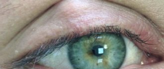
If you look closely, in the inner corners of the eye there are tiny papillae on the tops of which dots are visible. These points are nothing more than the beginning of the tubules that drain tears. They suck in tear fluid and prevent it from leaving the eye.
When a person cries, too many tears are released and they do not have time to go away in the usual way; they flow out and run down the cheeks. This can be compared to the situation when you pour water into a funnel. If you pour slowly, the water easily passes through the narrow hole, if quickly, it does not have time and splashes out beyond its limits.
The lacrimal canaliculi, starting on the surface of the conjunctiva, end in the lacrimal sac, where tears are collected to then enter the nasal cavity through the nasolacrimal duct.
Tear fluid not only moisturizes the cornea, but also cleanses it. If a speck of dust gets into our eyes, we cry. If we come into contact with sharply smelling substances (for example, cutting onions), we cry. Tears wash away irritants from the surface of the eye, just as you wash dirt off your hands.
Causes of dacryocystitis in a child
All causes of dacryocystitis in adults and children are divided into congenital and acquired.
Congenital dacryocystitis
A newborn baby is born with a violation of the patency of the lacrimal ducts (narrowing, complete blockage of the lacrimal canals or the presence of folded areas on the mucous membrane of the lacrimal sac), preservation of the so-called gelatin plug (it protects the lower part of the nasolacrimal canal during intrauterine development and ruptures with the first breath ).
Acquired dacryocystitis
A child and an adult can acquire the disease due to the presence of foreign bodies in the nasolacrimal canal (cilia, dust, etc.), purulent conjunctivitis or other infectious diseases of the eyes and nasal mucosa, the consequences of eye injuries, inflammation of the maxillary sinus or other sinuses.
Dacryocystitis - why does it appear?
In every third child, at the time of birth, the nasolacrimal duct is blocked by a thin membrane - a membrane. She should break through with the first cry, but in 5% of babies this does not happen. And when the lacrimal apparatus works at full capacity, the tears cannot leave as they should and look for workarounds - they flow down the cheeks.
In older children, dacryocystitis can be caused by trauma, conjunctivitis, or the consequences of infectious diseases (complications of ARVI and other diseases associated with inflammation of the nasal mucosa).
Sometimes the outflow of tears is disrupted by grains of sand that get into the nasolacrimal duct or lacrimal canaliculi during strong winds.
Symptoms
Diagnosis is made based on clinical signs. When you press on the area of the lacrimal sac, its contents (purulent or mucous) begin to separate. Symptoms of dacryocystitis in newborns:
- Teariness - the presence of tears is noticeable along the edge of the lower eyelid, but the child is at rest (children begin to cry with tears only by 1 month of life).
- Tearing.
- The presence of copious discharge in the conjunctival cavity, the conjunctiva itself without signs of inflammation.
The patency of the tear ducts is checked with a special test. A dye (collargol) is injected into the conjunctival cavity. After insertion, a cotton swab is placed into the nasal sinus. After 5 minutes, the doctor assesses the presence of collargol on a cotton swab.
How to suspect dacryocystitis in a child?
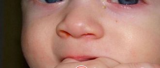
The disease does not begin immediately, but 2-8 weeks after birth, when the lacrimal apparatus begins to work at full capacity. The tear fluid stagnates, creating excellent conditions for the growth of bacteria. As a result, the mother notices that the baby constantly produces tears, and then pus in the corner of the eye. Conventional treatments for conjunctivitis are useless. It is necessary to contact an ophthalmologist.
Dacryocystitis can be either unilateral or bilateral.
Surgical methods
If after the first stage of treatment signs of the disease persist, surgical methods are prescribed.
Probing
Parents are wary of this method, but they should remember that the procedure is painless and is performed only under local anesthesia .
The essence of the procedure:
- A special probe is passed along the natural course of the lacrimal duct, pushing out the membrane and the resulting plug.
- The lacrimal ducts are washed with an antiseptic solution.
- The procedure takes up to 3 minutes.
- Local antibacterial therapy is prescribed for 6–7 days.
- There are cases when repeated probing is performed (the hole is closed with a lump of pus or mucus).
The procedure is performed on children aged 2–3 months according to the indications of a specialist. Delay can cause complications (the film thickens over time), relapses and the formation of adhesions in the lacrimal sac are possible.
Expert opinion
Ermolaeva Tatyana Borisovna
Ophthalmologist of the highest category, Candidate of Medical Sciences
The baby will not feel any pain at all, but may cry due to the bright light aimed at the open eye. After the procedure, the child will not experience discomfort.
Physiotherapy with probing
Massage, rinsing, baths are accompanying procedures that help quickly get rid of canal blockage.
Is it possible to be cured without this procedure?
In 90 cases out of 100, regular massage and drops give the desired result - the ducts are freed from the plug. If symptoms persist for a month, it is advisable not to delay probing.
Bougienage
Bougienage is a type of sounding. The procedure is safe, performed with anesthesia, lasts 5–6 minutes . It is prescribed only when at the first stage of treatment (rinsing, massage) the gelatin plug could not be broken, the canal is still clogged.
The operation is performed in the first year of the baby’s life (up to 5–6 months). Blood tests for coagulation are taken first. During the procedure, the walls of the lacrimal canal are moved apart with a special instrument (bougie), the canal is pierced, and then washed with a special solution.
Operative surgery
Expert opinion
Ermolaeva Tatyana Borisovna
Ophthalmologist of the highest category, Candidate of Medical Sciences
These methods are used in severe (chronic or acute) forms of the disease, taking into account the child’s age, neglect or repeated signs of canal blockage. Surgical operations completely restore the function of tear secretion.
Using a laser
A special laser endoscope is used to make a hole in the nasal bone connecting the nasal canals with the lacrimal sac.
Using an endoscope
A similar procedure involves using an endoscope to make a passage between the nasal cavity and the lacrimal canaliculus (the site of the blockage is cut).
Fracture of the nasal bone, without displacement of fragments
The method is rarely used (in 1 - 2% of cases). During the operation, the patency of the lacrimal canaliculus is restored by destroying the nasal bone (if pathologies are identified).
Balloon dacryocystoplasty
During the operation, a guide with a special balloon is attached to the tear duct through an opening in the corner of the eye. The balloon is filled with liquid and expands, clearing the tear duct.
Dacryocystorhinostomy
Conducted for children aged 10 years and older. During the operation, a temporary (for 1.5 - 2 months) prosthesis is installed - a tube through which tears flow into the nose, bypassing the blocked passage. The prosthesis is installed through the lacrimal sac, and a hole is made in the nasal bone.
What if you don't go to the doctor?
If there is already purulent discharge, then dacryocystitis will not go away on its own. Every day it will develop, which threatens:
- redness and swelling of the eyelids;
- swelling of the periorbital tissue;
- development of phlegmon;
- transition of inflammation to the orbit and further to the meninges;
- damage to the cornea with subsequent loss of vision.
Therefore, if you suspect dacryocystitis, you need to make an appointment with an eye doctor. Otherwise, instead of a simple and quick treatment for dacryocystitis, you will have to fight its complications for a long time and hard.
Indications for use
The drug is indicated for the treatment of infectious pathologies caused by bacteria sensitive to ofloxacin. Prescribed for:
- purulent inflammation of the eyelids;
- inflammation of the lacrimal sac;
- conjunctivitis of bacterial origin;
- keratitis;
- creeping ulcer of the cornea;
- blepharoconjunctivitis;
- eye damage with chlamydia;
- barley;
- meibomite.
Floxal is also recommended to be taken as a prophylactic agent to prevent the development of acute inflammatory pathologies of the organ of vision.
How will the doctor confirm the diagnosis?
The ophthalmologist will look at the child’s eyes, ask the mother how the disease began and progressed, and perform a Vesta test.
The Vesta test is performed simply and painlessly for the baby. A solution of collargol is instilled into the eye, and a cotton swab is placed in the nasal passage on the same side. If the nasolacrimal duct is passable, then the tampon is stained within 2-5 minutes. If not, the diagnosis of dacryocystitis is confirmed.
Modern ophthalmologists perform ultrasound to not only recognize dacryocystitis, but also to determine the exact location of the barrier to tears.
special instructions
If the child wears contact lenses, they need to be removed during instillation and put back on after about 15 to 20 minutes. Due to the risk of photophobia, the use of sunglasses is recommended. It should be borne in mind that during the treatment period, the child’s visual acuity may temporarily decrease, so he needs to be careful when playing sports.
If the treatment of acute bacterial conjunctivitis requires the use of several drugs, then they are used at five-minute intervals. The drops always drip first, and then after 5 - 10 minutes you can use the ointment, incl. and Floxal.
The use of ointment can negatively affect the state of attention and reaction. If these violations are pronounced, then the child needs to be exempted from classes, especially physical education lessons.
Dacryocystitis confirmed. What to do?
The ophthalmologist acts gently, prescribing treatment in stages. Antiseptic solutions must be prescribed to prevent the occurrence and spread of infection. The main goal of treatment is to restore the patency of the nasolacrimal duct. There are several methods for this.
Massage of the lacrimal sac
It is carried out 5-6 times a day, when the baby is calm. The mother places her index fingers downwards from the inner corners of the baby's eyes. The thumbs rest on his chin and the middle fingers on his eyebrows. Then the following movements are performed:
- the index fingers move down, following along the wings of the nose, and then, vibrating, rise up - to the eyes, and then to the eyebrows.
This is repeated 10 times. It is very important that the doctor shows you the massage technique, since the strength with which the described movements must be performed also matters. If you act weakly, you may not achieve the effect; if you act too strongly, you may damage the lacrimal sac.
After ten days of massage, the ophthalmologist looks at the child again and checks whether the patency of the nasolacrimal duct has been restored. If it has not recovered, they move on to the next procedure - bougienage.
Bougienage of the nasolacrimal duct
Bougienage or probing is considered a surgical intervention, although not very complicated. It is best to carry it out before the age of three months, since in this case it is most effective and is better tolerated. Before the operation, you need to take tests, and the intervention itself is carried out in the morning, on an empty stomach. The doctor selects a bougie (probe) of the required size and carefully guides it through the nasolacrimal duct. As a result, all obstacles to the tear fluid are eliminated. After bougienage, the canal is washed with antiseptics. A few more days you will need to drip some antiseptic crumbs into the eyes - and that’s it, the problem is solved.
The effect of bougienage is visible immediately, and the mother and child are sent home after several hours of observation.
Please note: if bougienage is performed at a later date, a repeat procedure may be required.
Did your baby have dacryocystitis? Did you have to resort to bougienage or was one massage enough?
Floxal: instructions
The drug Floxal refers to antibacterial (antimicrobial) drugs used in ophthalmology to treat eye diseases.
The action of the drug is based on ofloxacin, which belongs to a large group of fluoroquinolones and destroys staphylococci, E. coli, streptococci and other pathogens that are sensitive to this antibiotic.
Floxal drops are distinguished by their rapid action (10-15 minutes after instillation) and long-lasting therapeutic effect (within 4-6 hours).
They are produced on the pharmaceutical market in the form of eye drops and eye ointments. Eye drops are more widely used because they are convenient to use.
Indications for use:
- conjunctivitis
- barley
- keratitis
- dacryocystitis
- chlamydial infection in the eye
- bacterial corneal ulcer.
Side effects from the use of the drug are rare, but may include discomfort, burning, itching, flushing, lacrimation, and dizziness.
Before using the drops, wash your hands thoroughly. Next, the bottle is shaken and opened. The drops need to be dripped into the area of the lower conjunctival sac of the eye - to do this, the lower eyelid is pulled back. By pressing on the bottle, you drop the drops into your eyes.
After using eye drops, vision will be impaired for some time. Therefore, you do not need to immediately get behind the wheel of a car or engage in work that requires visual stress.
The instructions for Floxal should be studied in detail before you begin treatment, and any questions you may have should be discussed with your doctor.
How to understand that dacryocystitis has completely resolved?
If all the doctor’s instructions have been followed correctly, you will see that the child begins to produce tears naturally. All signs of the inflammatory process and dacryocystitis, such as swelling and redness, disappear.
After you see these signs, you should contact your doctor again to conduct a study of the patency of the nasal passages using the Bugaev or West test. If, after the procedure, the cotton swab that is inserted into the nose is colored, we can talk about your child’s complete recovery.
But, still, even with successful treatment, it is worth paying special attention to the hygiene of the child’s eyes. After complete recovery, it is recommended to regularly see a doctor, and also have in your first aid kit all the necessary drops for washing the tear ducts.
Symptoms of blocked tear duct in newborns
Clinical signs of dacryocystitis and lacrimal duct obstruction in infants are:
stagnation of tear fluid (lacrimation);
- lacrimation;
- conjunctival hyperemia (not always);
- soreness and swelling of the eyelids;
- “glued” eyelashes after sleep;
- the presence of purulent discharge (the symptom may be “blurred” due to the use of antibacterial drugs).
Please note: in most cases, unilateral obstruction of the lacrimal duct is diagnosed, but sometimes the pathology can affect both eyes of the newborn.
A characteristic symptom of this disease is the release of mucous or purulent contents of the lacrimal sac into the conjunctival cavity when pressure is applied to its projection.
Signs of the development of a complication (progressive purulent inflammation) are restless behavior of the child, frequent crying and an increase in general body temperature.
