Renal nephroscintigraphy is an effective type of kidney examination through the introduction of a special radioactive label into the body of an adult patient or child. This drug, in combination with a gamma camera, provides a broadcast of a complete image of the condition of the kidneys. The method is considered absolutely harmless, since the process involves a minimal radioactive load on the body, and the drug used is quickly removed from the body without additional support.
Renal nephroscintigraphy has a significant advantage over x-rays, ultrasound or tomography, since it studies not only the detailed structure of the kidneys, but also thoroughly demonstrates their individual characteristics and degree of activity. The altered tissue cells respond unnaturally to this irradiation, and therefore the camera easily records them if they are present in the organ.
Types of nephroscintigraphy
The study is highly effective in detecting kidney diseases at their initial development, during a period when tissues have not yet had time to significantly change their structure and appearance, but functionally have already begun to demonstrate negative changes in their functioning.
For the most effective examination in different cases, as well as for different tasks, they resort to one of two main research schemes.
- Statistical nephroscintigraphy of the kidneys. Recommended for obtaining accurate data regarding the physiological characteristics of the kidneys. Depending on the required tasks and complexity, the process can take from 30 minutes to 2.5-3 hours. First, a special radioactive agent penetrates into the patient’s body, and only after 30-60 minutes the main manipulations begin.
- Dynamic nephroscintigraphy of the kidneys. It involves not a sudden, but a gradual introduction of the diagnosed medical product into the body and an examination some time after this manipulation. Using this method, the doctor receives the most detailed information about the nuances of the passage of the radionuclide through the body along with urine. This guarantees a detailed study of the functioning and condition of the patient’s kidney blood vessels, the degree of their saturation with blood and the organ’s performance.
The final decision about which method to use in a particular case is made by the attending physician based on the characteristics of the patient's condition.
Free consultation with a urologist
Determination of kidney scintigraphy (nephroscintigraphy)
Renal scintigraphy or radionuclide scanning of the kidneys (renoscintigraphy, nephroscintigraphy ) is a diagnostic research method that involves introducing a small amount of radioactive medicine (radioactive tracer) into the body and obtaining images of the kidneys using a gamma camera. The resulting images can help in the diagnosis and treatment of various kidney diseases.
Purpose of kidney scintigraphy (nephroscintigraphy)
While most tests—such as X-rays, ultrasound, or computed tomography (CT)—provide information about the structure of the kidneys, radionuclide testing provides an opportunity to study kidney function. Candidates for renal scintigraphy may include patients with acute or chronic renal failure, urinary obstruction, renal artery stenosis, kidney transplant, renal trauma, reflux nephropathy, renal vascular disease and/or hypertension, or congenital anomalies. .
Precautions when performing kidney scintigraphy (nephroscintigraphy)
Renal scintigraphy requires the use of radioactive material; Therefore, in pregnant women or women who suspect that they are pregnant, kidney scintigraphy is performed only when absolutely necessary. Women should tell their doctor if they are breastfeeding. The doctor recommends that the woman stop breastfeeding for a period of time, which depends on the type and dose of the radioactive drug.
Description of kidney scintigraphy (nephroscintigraphy)
Renal scintigraphy is performed in the nuclear medicine department of a hospital or clinic. The patient is positioned in front of or under the gamma camera. A gamma camera is a special piece of equipment that detects radiation (gamma rays) emitted by radioactive medicine that has accumulated in the patient's body and produces an image. A radioactive drug is injected intravenously. Immediately after the injection, the study begins - the blood flow in each kidney is assessed. A sequence of images is obtained at specific time intervals, which depend on the radioactive drug used. A kidney scan is performed to determine the patient's glomerular filtration rate. Kidney scintigraphy uses a radioactive drug called technetium DTPA (Tc99m DTPA). This radioactive medicine may also reveal a blockage in the urine collection system in the kidneys.
The radioactive drug technetium, DMSA (Tc99m DMSA), is used to study renal tubular function.
Kidney scintigraphy takes from 45 minutes to three hours, depending on the purpose of the study. Most often, the duration of kidney scintigraphy ranges from an hour to an hour and a half. It is important to understand that renal scintigraphy can detect renal dysfunction, but cannot always determine the nature of this disorder. Radionuclide studies of the kidneys are useful for obtaining information about how different structures of the kidneys work, which in turn can help make the correct diagnosis.
Typically, images are obtained in a direct projection, but images can be obtained at oblique angles. If necessary, the patient can be positioned to obtain renal motility data, i.e. sitting or lying down while images are obtained. If obstruction (blockage) or kidney function is assessed, a diuretic (a drug to induce urination) such as Lasix is given. If hypertension or renal artery stenosis is assessed, captopril or enalopril (ACEIs, angiotensin-converting enzyme inhibitors) are administered.
Preparation for kidney scintigraphy (nephroscintigraphy)
No special preparation is required for kidney scintigraphy. For some types of tests, the patient must drink extra fluid and empty their bladder before the test. If the patient has recently undergone another radionuclide study, then it is necessary to refrain from repeat studies for a certain period of time so that residual radioactivity does not accumulate. The patient must remove all metal objects from the examination area.
After kidney scintigraphy (nephroscintigraphy)
Patients can return to their normal lifestyle immediately after kidney scintigraphy. Most radioactive drugs are eliminated through the urinary system, so increasing fluid intake after a kidney scintigraphy will help clear the radioactive drug from the body more quickly.
Complications of kidney scintigraphy (nephroscintigraphy)
Nuclear medicine research is safe. Unlike some contrast agents used in kidney x-rays, radioactive medicines rarely cause side effects. There are no long-term effects of radioactive drugs, since they quickly decay and do not have any immediate functional effects on body tissues. When radioactive drugs are administered, your blood pressure may temporarily rise or fall, or you may feel the urge to urinate.
Results of kidney scintigraphy (nephroscintigraphy)
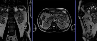
Renal scintigraphy shows normal renal function depending on the patient's age and health status, as well as the relative position, size, configuration and location of the kidneys. Primary blood flow images reflect the circulation in both kidneys. For patients in whom renal scintigraphy suggests renal damage or obstruction, other diagnostic methods, such as CT (computed tomography) or ultrasound, are required to obtain additional information. In addition, if the kidneys are the wrong size, have an unusual contour, or are unusually positioned, other imaging techniques may be required.
Health care roles
Renal scintigraphy is performed by a technologist from the nuclear medicine department, who has been trained in working with equipment and radioactive materials and processing data obtained during the study. The technologist must explain to the patient how the study is performed and administer the radioactive drug.
All collected data, the patient’s medical history for description (interpretation) are transferred to a radiologist who is a specialist in the field of nuclear medicine. Patients receive test results from their primary care physician or the physician who referred them for kidney scintigraphy.
The article is for informational purposes only. For any health problems, do not self-diagnose and consult a doctor!
Author:
V.A. Shaderkina is a urologist, oncologist, scientific editor of Uroweb.ru. Chairman of the Association of Medical Journalists.
‹ Postcoital test Up TRUS of the prostate ›
Indications for examination
The urologist refers the patient to detailed nephroscintigraphy of the kidneys if he has the following indications:
- suspicion of the presence of tumor formations or metastases in the area of the kidneys and other urinary organs;
- the need for an additional and more accurate determination of the characteristics and nature of education (often for this purpose the method is used in conjunction with other studies);
- detection of deformations, changes in volume, location or structure of the kidneys;
- preparatory manipulations for planned surgery on this organ;
- the need to monitor the condition of the kidneys after a course of radiation or chemotherapy;
- assessment of the degree of functionality and capabilities of the kidneys and other organs of the urinary system.
What is scintigraphy?
Kidney scintigraphy is a radionuclide scan of the urinary system. Diagnostic manipulation involves the introduction of a radioactive substance into the patient’s body, which makes it possible to obtain images and photographs using a gamma camera. The procedure helps to identify kidney pathologies (one or two) in the early stages, when clinical signs and additional research methods do not allow a diagnosis.
A distinctive characteristic of scintigraphy is that it is a study that allows you to evaluate not only the structure of a paired organ, but also its function. Unlike ultrasound, MRI, CT and X-rays, which show dimensions and internal structure, radionuclide scanning provides more detailed information. Diagnosis is necessary for patients with acute renal failure, stenosis of the ureter or urethra, trauma of a paired organ, vascular pathologies in it, or congenital anomalies.
Renal nephroscintigraphy can be performed using several methods:
- dynamic;
- static;
- radionuclide angiography.
Contraindications for diagnosis
Kidney nephroscintigraphy is not always prescribed for a child or an adult. Despite its safety and reliability, it has several contraindications.
Patients are not referred for diagnostics in the following situations:
- high severity of the patient’s condition: due to health reasons, not all people can lie freely without moving on the diagnostic table for about half an hour;
- a course of chemotherapy or radiation has just completed, the body needs rehabilitation for some time;
- a surgical operation undergone in the recent past: nephroscintigraphy of the kidneys can affect the accumulation of fluid in the just operated area of the body, which will adversely affect the rehabilitation period.
Dynamic or statistical nephroscintigraphy of the kidneys is also undesirable during pregnancy (however, it can be prescribed as an exception in particularly serious situations) and if the patient is found to have an individual intolerance to the drug injected into the body.
Degree of danger to the body
The contrast does not cause any particular harm after administration. However, surges in blood pressure and frequent urination in patients may be observed. In order for the substance to leave the body faster and the negative symptoms to disappear, it is important to drink more clean water.
Scintigraphy has no special contraindications and does not cause complications when performed correctly by an experienced doctor. But there are restrictions for seriously ill bedridden patients, for whom it is difficult to withstand up to 2 hours in one position.
Attention! The method is not used in pregnant women, because the contrast contains a small dose of radiation. During static nephroscintigraphy of the kidneys, considerable harm can be caused to the child.
Allergy sufferers should approach the procedure with caution. You should first determine whether a negative reaction will occur after radionuclide administration. In all other cases, the diagnostic method is not dangerous and does not cause discomfort.
Preparation for the diagnostic procedure
Renal nephroscintigraphy does not require special preparation and requires a special diet.
In some cases (based on the characteristics of the examination), the patient must first drink additional clean water or, on the contrary, completely empty the bladder. The attending physician or specialist performing statistical or dynamic nephroscintigraphy of the kidneys will warn about all these actions, if necessary.
Before lying on the doctor's couch, you must remove any metal objects, as well as jewelry and clothing with metal inserts.
Features of kidney scintigraphy:
Static renal scintigraphy: during injection of the drug, radionuclide angiography is performed (within 1-2 minutes), then 2 hours after the administration of the radiopharmaceutical, a static study of the kidneys is performed, which takes 15-25 minutes. The conclusion is issued on the day of the study. Dynamic nephroscintigraphy: the patient is injected with a radiopharmaceutical directly on a gamma camera; the study takes 30 minutes and begins immediately after the injection. The conclusion is issued on the day of the study.
Patients are recommended to have with them ultrasound, urography, CT, MRI of the kidneys and urinary tract, extracts from the medical history or outpatient cards (if available).
Indications and contraindications for renal scintigraphy
Nephroscintigraphy is a valuable diagnostic method for kidney cysts and tumors. In the first case, the scintigram shows a defect in the parenchyma clearly demarcated from healthy tissue. With a neoplasm, the image is blurry, and if the tumor is completely damaged, the organ does not accumulate the radiopharmaceutical at all. The study makes it possible to distinguish malignant changes in the kidneys from those in the abdominal organs.
The scintigram will accurately show abnormal position (dystopia) or prolapse of the kidney (nephroptosis). In this case, it is possible to carry out a differential diagnosis of these two pathologies by examining the patient in a lying and standing position.
Scintigraphy will provide invaluable assistance in recognizing various anomalies in the development of urinary organs. For example, you can obtain an image of a horseshoe kidney and determine the degree of functionality of each of its halves and the isthmus. This information is necessary to determine the extent of surgery.
In the case of doubling of urine-forming organs, the technique makes it possible to accurately determine the performance of both the upper and lower segments, as well as the shape and size of each part.
Nephroscintigraphy is very informative for urolithiasis. The ability to study the structure and functions of various parts of the kidney allows for wider use of organ-sparing surgical interventions for stones and neoplasms. If a stone is present in the ureter, then the contours of the kidney and urinary tract are traced on the scintigram to the level of its blocking by a foreign body.
This study is also prescribed to determine the volume and velocity characteristics of renal blood flow. If hemodynamics are impaired in the urinary organs, the presence of stenosis of large vessels is determined.
Finally, nephroscintigraphy confirms or refutes the presence of such life-threatening conditions as pyelonephritis and chronic renal failure, and detects areas of urinary tract obstruction and vesicoureteral reflux.
The main contraindication to radioisotope studies is pregnancy, since some exposure to radiation, although minimal, still occurs. During lactation, it is not forbidden to carry out kidney scintigraphy, however, breastfeeding must be stopped on the day of the procedure and during the following days so that the radiopharmaceutical has time to completely leave the mother’s body.
Nephroscintigraphy sessions are not recommended for patients who have recently undergone chemotherapy or surgery. In the postoperative period, the body accumulates fluid, which can serve as a reservoir for the radioactive drug.


