What is aphakia?
Reference! In the ICD-10 list, aphakia is designated as H27.0 and is described as a visual pathology characterized by the absence of a lens in one or two organs of vision.
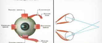
Aphakia is characterized by a significant decrease in visual acuity, a lack of organs' ability to accommodate, and periodic trembling of the iris.
Often such a disorder is the result of exposure to various external physical factors . In rare cases, aphakia is congenital.
There are monocular and binocular forms of pathology. In the first case, the absence of a lens is observed in only one of the organs of vision.
Interestingly, this form is more dangerous, in terms of complications , than binocular (when the lenses are absent in both eyes).
The fact is that healthy and pathological organs of vision perceive visible images differently.
In particular, for each eye, visible objects will have a different apparent size (the pathology is called aniseikonia).
This not only makes it impossible to objectively assess the size of an object and the distance to it, but also leads to a general deterioration in visual acuity, as both eyes begin to “adjust” to each other.
For your information! As for the binocular form, it is basically an acquired form of the disease, which appears after injuries or surgery, as well as after severe cataracts.
In this case, visual acuity is greatly impaired, while the organs of vision cannot focus on objects at different distances, and patients with binocular aphakia see well only objects at a certain fixed distance.
Key Facts
Cataracts of the eye - what kind of disease is it? Sooner or later, every person comes to this question. And this is not surprising. Cataract, like glaucoma, is an age-related disease; its appearance and development is caused by age-related changes that occur in the body and affect all organs and systems, including the visual one. Cataract is not contagious, it is not an infectious disease, it is not transmitted by contact or airborne droplets. In simple terms, cataracts are clouding of the lens.
Ophthalmologists consider lens clouding to be a natural process, so cataracts cannot be fully considered a disease. It is rather a condition of the eye apparatus, which, however, requires specialized medical care and observation by an ophthalmologist. The only method that can lead to a complete cure is replacing the lens of the eye for cataracts.
The risk group for cataracts includes all people over 60 years of age. With each passing year, the likelihood of illness increases, especially with diseases such as diabetes and hypertension. In addition to cataracts, these diseases lead to deterioration of the retina and decreased visual acuity, and are sometimes accompanied by glaucoma. Often, in older patients, glaucoma and cataracts are diagnosed at the same time, and this increases the likelihood of vision loss.
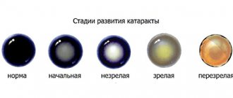
Cataract develops gradually; a number of stages are conventionally distinguished, characterized by a certain set of symptoms and signs of changes in the tissues of the lens. Doctors call the development of the disease “maturation of cataracts.” The duration of the process depends on the cause, age and other circumstances, and can last from several months to 10-15 years.
In the initial stage of cataract, the manifestations are mild, vision practically does not deteriorate.
In the stage of immature cataracts, there are areas of clouding of the lens, which affects visual acuity.
Mature cataracts are characterized by significant visual impairment and complete clouding of the lens.
At the last stage of overripe cataracts, deformation of the lens and loss of vision occurs.
Causes
The acquired form of aphakia develops for the following reasons:
- penetrating injuries to the eye, leading to the loss of its functions by the lens and its subsequent removal. In some cases, spontaneous loss of the lens from its niche occurs due to injury.
- lens luxation;
- resorption of traumatic cataracts;
- lens aplasia.
In some cases, resorption (resorption) of the lens occurs during the period of intrauterine development, as a result of which the child is born with aphakia .
Know! The reasons for this phenomenon can be different - from genetic failures to the negative impact on the fetus of toxins that enter the mother’s body if she smokes or uses drugs or alcoholic beverages during pregnancy.
Symptoms
The disease is characterized by the following signs that can be seen visually or determined during instrumental diagnostics :
- a sharp decrease in visual acuity by 9-12 diopters, which requires the use of strong corrective optics;
- the anterior chamber of the eye, due to the lack of a lens, has greater depth;
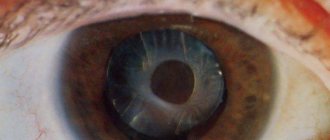
- the lens is not visible during biomicroscopic examination;
- trembling of the iris when the eyeball moves;
- it is possible to detect particles of the lens or its capsule when examining the pupillary area;
- in some cases, scars are found on the cornea.
After surgical removal of a cataract (as a result of which the pathological lens is also removed), the resulting aphakia can be characterized by the spontaneous appearance of strong reverse astigmatism.
The shell of the eyeball has irregularities not in the form of bulges, but in the form of depressions and depressions.
Within two months, this disorder disappears and the condition of the ocular surface stabilizes.
Symptoms and diagnosis
Symptoms of electroophthalmia depend on the severity of the lesion and the individual's personality. They can be mild and almost cause no concern, but they can also be very strong. These symptoms appear only 6 hours after the lesion.
The epithelium is damaged, inflammation occurs, pathological processes begin in the upper layer and symptoms become obvious:
- Redness of the conjunctiva, it can be very strong, or it can be barely noticeable
- Severe tearing
- Pain that gets worse with movement
- Sensation of a foreign body in the eye, as if something is greatly interfering
- Visual acuity decreases to a greater or lesser extent
- Photosensitivity increases, photophobia appears.
The pathological process develops gradually and moves progressively. Progress is replaced by regression. Recovery takes from 36 to 72 hours.
Diagnostics
Note! The following research methods allow specialists to determine this violation:
- Ultrasound of the eyeball. It is performed not so much to determine the absence of the lens, but to exclude retinal detachment developing against this background.
- Visometry. Performed for a general assessment of visual acuity. A sharp deterioration in such indicators over a short period of time may be an indirect sign of aphakia.
- Refractometry. A method for studying refraction, the indicators of which in aphakia can decrease by 10-12 units.
- Gonioscopy. The method allows you to measure the depth of the anterior chamber of the eye (obviously, in the absence of a lens, which should be located in this chamber, the depth will be noticeably greater).
- Tonometry (necessary for a general assessment of intraocular pressure).
To identify various concomitant pathologies, ophthalmoscopy is performed.
Treatment
First of all, for aphakia, ophthalmologists use a conservative method of treatment , which consists of selecting appropriate glasses (for binocular aphakia) or contact and intraocular lenses (in the case of a monocular type of pathology).
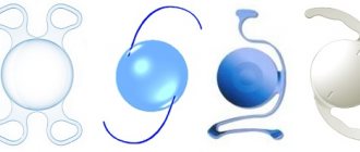
Keep in mind! The optimal treatment method is implantation of an intraocular artificial lens into the posterior chamber of the eye.
Such operations, compared to wearing glasses, are much more effective, and if the patient has no contraindications to surgery, implantation can be performed even on children, starting from the age of two.
Afakia
Aphakia is corrected using glasses, contact and intraocular lenses. The indication for spectacle vision correction is a bilateral form of the disease. For unilateral aphakia, glasses are recommended only if contact methods of correction are intolerant. The choice of glass for an emmetropic eye is difficult, because even glass of +10 diopters is not comparable to the refractive power of the lens, which is 19 diopters. This is due to the fact that the refractive index of the liquid that surrounds the lens is higher than that of the air surrounding the glass.
The optical power of a glass lens depends on the patient's refraction. For hyperopia, it is necessary to choose glasses with stronger optics than for myopia. There is no need to prescribe vision correction methods to patients with high degrees of myopia before lens removal. Due to the lack of accommodation ability, the patient should be prescribed glasses for near vision that are 3.0 diopters stronger than for distance vision.
Contact or intraocular vision correction is indicated for patients with monocular aphakia. Prescribing glasses to patients with this form of the disease will worsen aniseikonia. During surgery (intraocular correction), an artificial lens with individually selected optical power is implanted. The most preferred treatment option is the use of posterior chamber lenses, since localized at the location of the natural lens, they provide high quality vision. Congenital aphakia can be corrected using this technique only after the child reaches two years of age.
Forecast and prevention of aphakia
The prognosis for life and ability to work with proper correction of aphakia is favorable. In the absence of timely treatment, there is a high risk of complete loss of vision, which subsequently leads to disability.
In ophthalmological practice, there are no specific measures to prevent congenital aphakia. To prevent the development of acquired forms of the disease, it is necessary to undergo an annual examination by an ophthalmologist. This will help to timely diagnose those diseases that can lead to surgical removal of the lens. Persons at risk of eye injury due to the nature of their profession must use protective glasses or masks during working hours.
Treatment prognosis
Surgery to eliminate the disease is a relatively simple procedure , and the prognosis for such treatment is always favorable , but only if the patient seeks treatment in a timely manner .
Otherwise, atrophic processes in the structures of the eye progress and the refractive index decreases, which in turn can result in complete and irreversible loss of vision .
There are no preventive measures that could prevent such a violation.
And since this problem always arises due to injuries, damage to the organs of vision or due to cataracts, it is necessary first of all to protect yourself from such negative effects and eye diseases that could lead to the development of various pathological conditions.