Dear visitors of the portal! The “medical consultations” section is suspending its work.
The archive of medical consultations for 13 years contains a large number of prepared materials that you can use. Best regards, editors
Olga asks:
There is a lot of tension in and around the eyes. Feeling of dryness, constant swelling, inflamed, red eyes, slight clouding of the fundus. Worsening in the evening and after sleep. Points +2.0. Thank you.
Krasutsky Viktor Iosifovich answers:
Dear Olga! A more complete examination is necessary, since it is unclear what is meant by “clouded fundus” - this simply does not happen, and demodex must also be ruled out. To do this, an eyelash is taken for analysis. If the problem is severe dry eye syndrome, appropriate long-acting medications are prescribed.
Lucia asks:
Hello...What eye disease does the sudden appearance of a small “midge” spot indicate when reading...What eye drops can you recommend?...Thank you in advance
Answers:
Hello. The appearance of spots in the form of “midges” before the eyes when reading can be evidence of various pathological conditions, most of which require both high-quality diagnosis and qualified intervention. First of all, these symptoms allow one to suspect diseases such as glaucoma or degenerative changes in the retina. Therefore, a necessary step on your part will be to seek help from an ophthalmologist, perform a gonioscopy, measure intraocular pressure, examine the fundus using ophthalmoscopy, check the peripheral visual fields, after which it will be possible to prescribe appropriate treatment.
Alexander asks:
When I wake up in the morning, there is a pain in my eyes, as if sand had been poured into my eyes. After a while, all this goes away.
Answered by the Medical Consultant of the website portal:
Hello, Alexander! Most likely you are dealing with chronic conjunctivitis - chronic inflammation of the mucous membrane of the eyes. It is necessary to identify the nature of the disease and obtain recommendations for treatment. An ophthalmologist, whom you can visit at the clinic at your place of residence, will help you with this. Take care of your health!
Larisa asks:
What is retinal ANGIOPATHY and how is it treated? I am 56 years old, a teacher, hypertension, impaired glucose tolerance.
Oksana Sergeevna Averyanova answers:
Angiopathy is a certain condition of blood vessels (in this case, the retina), associated with changes in vascular tone, blood viscosity, blood flow speed and the condition of the inner wall of blood vessels. Angiopathy is a manifestation of all of the above components. In your case, this is obviously the result of hypertension. so hypertension needs to be treated.
Gennady asks:
I am 60 years old, a few days ago the symptoms of a feeling of veils before my eyes became more frequent, today all day long, with short intervals between these states, after a lunchtime nap - these symptoms have not yet appeared. Sometimes there is slight dizziness, arterial. blood pressure and pulse are within normal limits. Thank you!
Maykova Tatyana Nikolaevna answers:
Gennady, you need to look at the fundus of the eye, because the manifestations you described indicate a violation of visual perception by the retina. This can be a manifestation of diseases of the eyes, the nervous system, blood vessels, and metabolism. Therefore, you cannot do without an examination - you need to either calm down or act.
Nadezhda asks:
Hello! Recently, my dad’s right eye began to enlarge; when he sleeps, the upper eyelid does not close completely, he suffers from frequent headaches, and his blood pressure fluctuates. You can’t drag him to the hospital, but at least tell him what to do. Please tell me this could be related.
Vasily Vladimirovich Kislitsyn answers:
Hello, Nadezhda. The symptoms your father has are too serious to be ignored. You need to immediately be examined by an ophthalmologist, neurologist, or endocrinologist. It is necessary to perform an ultrasound of the orbit, MRI or CT. Possible formation in the orbit (tumor) or manifestations of endocrine ophthalmopathy (Graves' disease).
Ekaterina asks:
The left eye began to see very poorly in the distance, everything is blurry, but up close it sees well, what it could be, and the right eye sees well both near and far. For 22 years, the work is connected with a computer; I spend quite a lot of time near it for 6 - 8 hours
Anna Ivanovna Turchin answers:
Dear Ekaterina. Judging by the fact that you see poorly in the distance, but well up close, you have myopia in one eye. Constant work on the computer only aggravates the situation. Therefore, I recommend that you contact an ophthalmologist, undergo a comprehensive diagnosis, after which the doctor will select a correction method that suits you.
Sofia asks:
Lately, small balls have begun to appear in the corners of my eyes. Then a mole, and behind it a venous star. Could this be related to liver disease? If not, what could it be? Thanks in advance for your answer)))
Olga Stepanovna Dubenyuk answers:
It is necessary to see what color the “balls” are; if white, then it is a millium (“blockage” of the sebaceous gland) which is removed by a cosmetologist; spider veins can appear with a hereditary predisposition, excessive insolation, with rosacea, as well as with liver diseases. These formations are removed using a laser.
Vera asks:
In the mornings, the right eye opens poorly. you have to open it by hand.
Answered by the Medical Consultant of the website portal:
Hello! The symptom you indicated of deterioration in the opening of one eye may be evidence of neurological disorders accompanied by damage to the nerves innervating the levator palpebral muscle. The cause of such disorders can be completely different processes with local damage to the nerves or changes in the brain (neoplasms, structural changes). To find out the etiology of your disease, you need to contact a neurologist and undergo a series of additional studies (MRI of the head, electromyography). Without identifying the causative factor, it is not possible to prescribe any treatment. Be healthy!
Pavel asks:
Hello! I have this question: about 10 years ago, or more, suddenly interference in vision appeared - when you move your gaze, black dots, streaks and spots appear. All this happens on an ongoing basis, but is most noticeable against a light background, like a computer screen, for example. I was examined for a long time. They said that everything was fine, perhaps it was the vitreous body, the gel. The fact is that as a child I had a head injury - a fall. Doctors linked the vision problem to that injury, such as eye deformation. Do you think this could be a consequence of that childhood trauma, and if so, why did it manifest itself after so much time (more than 20 years have passed)? Could my condition get worse? Are there any treatment methods? Thank you very much, Pavel P.
Elena Stanislavovna Prokhvachova answers:
Dear Pavel, what do you mean when you talk about eye deformation? The eye may be elongated if myopia occurs. In this case, destruction of the vitreous body is a common occurrence. A head injury could trigger the development of myopia, but not the appearance of floaters 20 years later. Drip potassium iodine drops 1-2 drops 2 times a day for 1 month, then take a 1 month break and repeat the course, do 3 cycles. From traditional methods, a mixture is used internally: aloe juice 1 part + honey 1 part + Cahors 2 parts, drink 30-50 grams before each meal for at least 1 month. (store the mixture in the refrigerator). I wish you health!
Anastasia asks:
Hello, I recently noticed in my eyes, namely on the whites, darkish spots that look like small bruises, there are also a lot of burst blood vessels, the eyes are constantly red, there are no other symptoms. I work as a manicurist, my eyes are always under a lot of stress, is it possible that these spots appeared from work, or is there another reason?
Prayer Oksana Vasilievna answers:
Good afternoon. If you see something unclear in your eye or anywhere, why not see a specialist?! Do you really think that your description is enough to make a diagnosis?! Well, at least because, in principle, there cannot be bruises in the eye, since this is a subcutaneous formation, and the hemorrhage that we can see in the eye is red, then yellowish when reabsorbed. There are not many burst vessels in the eye at the same time; then blood would simply flow from the eyes; they can be dilated, for example, as a manifestation of an inflammatory reaction. Severe visual strain can also lead to vasodilation.
Alexandra asks:
Hello, I have the following problem: I slept in the lenses at night (I forgot to take them off), when I opened my eyes in the morning, tears were running from both eyes, I took off the lenses, now my right eye is terribly watery because of the light, I can’t look at the computer, TV, telephone, the eye is red, swollen, behind the upper eyelid, it feels as if something is in the way, a runny nose has appeared. I rinse it with tea, drop cypromed. Previously, I could not lie down at all, because the eye began to water terribly and ache (pinches) as soon as I’ll decide to lie down. Could you tell me what it is and what should be dropped into the eye?
They will talk about the reasons why people cannot open their eyes. Doctors take this problem seriously. Drooping eyelids are always a worrying sign. Drooping of the upper eyelid - ptosis - is a symptom of the disease. Myasthenia gravis
may be the cause of drooping eyelids.
This disease is characterized by a condition of the immune system
, which begins to harm nerve impulses.
This disease results in muscle weakness.
Muscles lose the ability to contract normally.
Nerve impulses cannot transmit a signal
from one nerve to another.
There are also signs of myasthenia gravis
, this is double vision and strabismus.
This disease is dangerous. A person may stop breathing
due to muscle weakness. This condition needs to be treated.
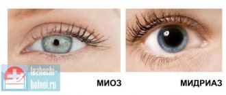
Another cause of ptosis may be Horner's syndrome.
It is characterized by drooping of one eyelid, a narrow pupil and
the absence of tears in the eye
. The body has a system of wakefulness and sleep.
It happens that the tumor is in the apex of the lung
we compress the nerves that go to the eyeball,
the parasympathetic system
. We are talking about cancer of the apex of the lung. The tumor must be removed.
The cause of drooping eyelid may be conjunctivitis with swelling of the eyelids.
Infection can lead to swelling,
and allergies can also lead to swelling.
It may also be unpleasant for a person to look at the light; he closes his eyes.
This disease is treatable.
can form
on the eyelids Such wen often appears in older people.
We are talking about high cholesterol levels.
The carrier of fat is cholesterol.
If cholesterol levels are elevated, fatty deposits begin to appear. Sometimes this is a hereditary increase in cholesterol
that is not associated with obesity.
High cholesterol levels are dangerous for blood vessels.
Cracks appear in the vessels, fat becomes clogged in them, plaques appear, and then
there may be a blood clot and a stroke
or heart attack. Atherosclerosis leads to narrowing of blood vessels.
If one eyelid does not lift, this may indicate a serious illness
.
One of the manifestations of this disease is one eye that is slightly closed. Ptosis may be accompanied by constriction of the pupil and retraction of the eyeball
.
These symptoms are characteristic of a number of neurological diseases.
But today we will talk
about a tumor in the upper part of the lung,
which is connected to the neck by nerves. A spasm of the muscles that open the eyelids may occur.
It seems that the person is squinting very hard and cannot open his eyes.
If the conduction system is disrupted, for example, with multiple sclerosis.
The immune system attacks its organs
in this disease.
The immune system attacks the nerve sheaths, the nerves cannot conduct impulses, and a spasm occurs.
The eyes may become sticky due to the discharge of pus. This is conjunctivitis. The eyeball is covered by a membrane, it is white or blue, this is the protection of the eye. The conjunctiva is washed with tears.
There should be no visible vessels in it.
If there is inflammation, the eye turns red. Bacteria or viruses enter the eye. There is a burning sensation, lacrimation, and photophobia. The eye may stick together
in the morning, and pus may ooze from the eye.
At night, you can put antibiotic ointment behind your eyelids.
If the inflammation is very acute, you can even give a hormonal drug.
There are antihistamines if you have allergies. If inflammation is caused by a virus
, then there are interferon solutions. You should not instill tea infusion. It's useless.
For conjunctivitis, drops are used,
but they need to be dripped correctly, they must linger in the cavity for them to act.
You need to raise your head, pull back the lower eyelid,
apply drops,
hold the eyelid for a while
.
For bacterial conjunctivitis, antibiotic drops are used.
For viral conjunctivitis
antiviral drugs and interferon-based drugs are used
for immunity
.
a bacterial infection
is often concomitant . Therefore, it is necessary to use antibacterial drops.
For allergic conjunctivitis
you need to
exclude the allergen,
do not use mascara that caused the allergy.
At home, you need to follow the rules of hygiene
if someone is suffering from viral conjunctivitis.
Wash your hands
and use antibacterial hand gels.
Eyes may turn red when a foreign body enters the eye. Is it a chemical effect or physical. Corneal erosion may occur
. A laser pointer or beam can damage your eyes. Doctors can see such damage during examination. The doctor will put drops in the eye and see the erosion.
The pain is severe with a corneal ulcer. Tears flow, the person cannot open his eyes. If something gets in, you need to remove it with a cotton swab or rinse with water.
If you try to remove the foreign body with something, you may scratch the eye further.
The choroid of the eye may suffer. In this case, the eyes are red. Uveitis is a serious condition that can make you blind. The iris is supplied with blood. The vascular network envelops the eyeball from the inside. If a virus, including the herpes virus
gets inside.
The choroid of the eye becomes inflamed. Vision becomes blurred. 25% of blindness in the world is due to uveitis.
It is important to quickly consult a doctor and treat the disease. There is no need to mindlessly drop eye redness drops. If you don't identify the causes of red eyes, you risk going blind. A person may have blepharospasm. It all starts with one eye, and then can move to the second. Attacks can increase in duration and are often a sign of multiple sclerosis. The brain cannot work due to cholesterol plaques. It is important to do an MRI of the brain.
anonymously
Hello. I am 55 years old. Sometimes it happens to me that I look at a person and see the right side of their face, but not the left. I thought the pressure was dropping at this moment, but when I measured the pressure it was normal. After 30 minutes it goes away. When I start to get nervous about something or under mental stress, as they say, “my brain is boiling,” this is what happens to me, as if there is boiling water in my head. After a short while, it goes away. There is also a time when the jaw slams shut, as if it is uncontrollable. I also open my eyes in the morning with my hands, don’t think it’s from pus, no, they’re clean, they just don’t open and that’s it. And during night sleep my hands go numb. Please answer: is it related to blood vessels or nerves? What needs to be treated and by which doctor? Perhaps also sore knees, feet and convulsive stretching of the calves are also associated with all this?
Consultation with a neurologist on the topic “Eyes do not open in the morning” is given for informational purposes only. Based on the results of the consultation received, please consult a doctor, including to identify possible contraindications.
About the consultant
Details
Neurologist, Candidate of Medical Sciences, medical experience: 17 years. Author of more than 50 publications and scientific papers, active participant in conferences, seminars and congresses of neurologists in Russia.
Area of professional interests: -diagnosis, treatment and prevention of neurological diseases (vegetative-vascular dystonia, discirculatory encephalopathy, consequences of strokes, arterial and venous disorders, memory impairment, attention, neurotic disorders and asthenic conditions, panic attacks, osteochondrosis, vertebrogenic radiculopathies, chronic pain syndrome). — Patients with complaints of migraines, headaches, dizziness, tinnitus, numbness and weakness of the limbs, disorders of the autonomic nervous system, depressive and anxiety states, panic attacks, acute and chronic back pain and herniated discs. — Functional diagnostics of the nervous system: electroencephalogram (EEG), ultrasound Dopplerography of the carotid and vertebral arteries (USDG), transcranial Dopplerography (TCD), rheoencephalography (REG), echo-encephalography (ECHO-EG). — Anti-stress mesotherapy for the back. — Shock wave therapy. - Hirudotherapy. — Mistletoe therapy.
Ask a Question
Similar questions
You work at the computer all day and watch your favorite movies in the evening. If you are very tired, go to bed without washing off your eye makeup. And in the morning, again without sleep, you get ready for work. Don't be surprised if your eyes look tired. But it will take very little time to restore their freshness and beauty.
Exercise for the eyes
To help your eyes rest after working at the computer, you can do a few simple exercises; they can be performed in any position:
- Bend your elbows so that your palms are slightly below eye level.
- Open your fingers.
- Make smooth turns with your head left and right, while looking through your fingers, into the distance, and not at them.
- Let your gaze glide without lingering on one thing.
- If you do everything correctly, your hands will “float” past you: it should seem to you that they are moving.
Make alternately three turns with your eyes open and three with your eyes closed (even closed eyes should not “linger” on anything). Do the exercise 20-30 times, while breathing freely, without straining.
For what reasons does ptosis of the upper eyelid develop and how to treat it?
Ptosis (ptosis - fall) of the upper eyelid is drooping of the eyelid, provoked by external factors or a congenital anomaly.
Depending on the cause, it manifests itself with various symptoms and can be complicated by infection. Ptosis does not cause harm to health, but it does cause discomfort. Therefore, it is better to start treatment from an early age.
In case of an acquired defect, you need to contact an ophthalmologist and also undergo a therapeutic course.
There are several types of ptosis depending on the severity and side. Let's take a closer look at what can cause the defect and how to get rid of it?
Causes of drooping eyelids in children
Considering congenital ptosis, we can identify causes associated with underdevelopment of facial muscles and nerve damage. This occurs in the womb and is due to the autosomal dominant type.
This means that if one parent had ptosis, it will be inherited by the child. Other causes of congenital drooping eyelid:
- Dysfunction of the oculomotor nerve - this nerve is responsible not only for the function, but also for the correct position of the eyelid. Weakness of the rectus muscle of the organ of vision does not affect the ability to see, but simply does not allow the eyelid to fully open.
- Blepharophysm is a congenital pathology, against which ptosis appears. This genetic anomaly is characterized by insufficient development of the palpebral fissure; it does not open completely. In this case, the ptosis will be bilateral, and the muscles of both eyelids will be underdeveloped. With blephorophimosis, the lower eyelids may also be affected.
- Palpebromandibular syndrome is a rare pathology and is a precursor to ptosis. When chewing, the eyelid rises, but without the action of the masticatory muscles, the eyelid droops.
Advice! Such anomalies are easily diagnosed, after which the vision organ is corrected and congenital ptosis is eliminated. Parents should know this and not waste time.
Causes of acquired ptosis
The acquired anomaly develops more often in adults and is divided into several types, depending on the underlying cause:
- Neurogenic - paralysis of the oculomotor nerve due to diabetes or oncology leads to compression of important structures of the eye. Some treatment of ophthalmic diseases requires artificial disruption of nerve function, which is why ptosis occurs.
- Myogenic – common causes of acquired anomaly that develops with myasthenia gravis. The eyelid cannot withstand increased loads; it droops and doubles. Short-term correction of the anomaly is possible when the symptoms are relieved by the effects of endorphins.
- Mechanical – occurs due to pressure on the structures of the eye from a tumor or scar after surgery.
- Aponeurotic - most often occurs in older people, when the muscle tendon moves away from the plate responsible for raising the eyelid. The age factor complicates treatment, as the muscles are weakened.
In terms of severity, congenital ptosis, like acquired ptosis, has 3 forms:
- partial - the edge of the eyelid in a relaxed state is lowered to a third of the pupil;
- incomplete - the edge of the eyelid is lowered to half the pupil;
- full – the eyelid covers the pupil completely.
The last stage can affect the quality of vision, reduce it, and lead to strabismus.
How does ptosis manifest?
You can determine ptosis of the upper eyelid visually, based on its main symptoms. The main manifestation of the anomaly is a drooping eyelid. At the same time, the person throws his head back and strongly raises his eyebrow to see better. The eyelid does not lift on its own and the sensations are different for everyone.
Often people with a congenital or acquired anomaly complain of tearing, constant irritation of the eye, discomfort due to the fact that they want to open the eye completely, but to no avail. Children may experience increased eye fatigue as they have to keep it open all the time.
In rare cases, double vision and strabismus are observed. Not only the aesthetic appearance suffers, but also the psychological comfort of such people.
Correction must be carried out in any case and restoration of normal function is guaranteed in 100% of cases.
Only the approach differs: some patients try to cope on their own using exercises, others turn to a surgeon, after which surgery is prescribed.
Additional diagnostics
If a visual examination of the patient is not enough for the ophthalmologist and there are doubts, additional diagnostics are performed. It includes determination of eye pressure, measurement of eye symmetry. During the diagnostic process, it is very important to distinguish acquired ptosis from a congenital anomaly, because their correction is different.
The presence of an inflammatory process near the eye may alert the ophthalmologist, so laboratory diagnostics of the material of the inflammatory focus is carried out. If an infection is present, antiviral treatment is carried out first, and only then correction.
Advice! Under no circumstances should you do exercises or other therapeutic procedures until the infectious source has been eliminated. Otherwise, such treatment will end with eye infection.
For those who find it difficult to open their eyes in the morning, you can do the following exercises:
- Stretch well, roll several times from side to side. Don't hold your breath while doing this. On the contrary, breathe deeply and calmly.
- Open your eyes and mouth wide several times.
- Close your eyes tightly (6 times), do 12 light blinks.
- Exercise for tired eyes “writing with your nose”: close your eyes. Imagine that the tip of your nose is a pen with which you can write. Now write whatever you want in the air with your pen.
- Eyebrow exercise: Raise your eyebrows as high as possible, while observing the sensation that appears at the top of your ears. In the future, this sensation will need to be done without raising the eyebrows. Take your time, this is difficult to do the first time.
- Palming: Sit up straight and relax. Close your eyes so that the middle of the palm of your right hand is opposite your right eye, the same with your left hand. The palms should lie softly, there is no need to press them forcefully to the face. The fingers can cross on the forehead, they can be located side by side - as is more convenient for you. The main thing is that there are no “slits” that let light through. Close your eyes. Now place your elbows on the table. The neck and spine should be almost in one straight line. Check that your body is not tense, and your arms, back, and neck should be relaxed. Breathing should be calm.
Now try to remember something that gives you pleasure. You can do this exercise to music. It is very difficult to consciously relax your eyes. Therefore, there is no need to try to control your condition - this will only harm the purpose of the lesson; instead, think about something pleasant.
Eye exercises will help relieve fatigue, and to make your eyes look rested, you can make special lotions and masks:
Eye toning lotions
Lotions can be done:
- from steep black tea;
- from herbs (chamomile, mint, sage, dill, parsley, cornflower);
- you can prepare a mixture:
- chamomile
- sage.
Mix in equal proportions. Take one tablespoon of the mixture, one tablespoon of boiling water. Let it brew for 15 minutes, strain, and cool.
All lotions are made using cotton pads. They need to be moistened in the resulting product, squeezed out a little and applied to the eyelids for about 10-15 minutes.
You can alternate cold and hot lotions. To do this, take sage, make two containers: hot and cold, and alternate the lotions one minute at a time for 10-15 minutes. This will relieve puffiness, fatigue, and restore smoothness and elasticity to the skin.
To relieve swelling, you can use used tea bags without additives, after keeping them in the refrigerator for 10-15 minutes.
Masks
All masks should be applied to a gauze pad and kept for 10-15 minutes.
From raw grated potatoes
Add flour, sour cream or fresh milk to raw grated potatoes. Stir until creamy.
From greenery
Add sour cream to the greens and stir.
Oil masks
It is best to make them from any kernel oils, such as apricot, peach, almond. It is necessary to moisten a cotton pad and apply it to the eyelids.
After the mask, you need to wash your face with cool water and apply eye cream along the massage lines. After 20 minutes, blot the cream with a napkin.
Vegetables
Raw vegetables (potatoes, cucumber) also bring great benefits. They can be cut into circles and applied to the eyelids. As soon as the circle warms up, it needs to be replaced with another - and so on for 15-20 minutes.
- You should not put too much strain on your eyes with computers and TV;
- do not sleep with your face in the pillow;
- do not drink or eat much at night.
Remember, your eyes are your most important source of communication with the world. Take care of them.
The eyes are considered the most sensitive human organ. That is why, as a reaction to some pathological process or external influence, pain occurs. If it is observed for a long period, then this is a signal to contact a specialist. Eyes can hurt in the morning for many reasons. These include inflammatory or infectious processes, fatigue, and an allergic reaction. Only an ophthalmologist can figure out why discomfort occurs in the morning.
When determining a particular pathology, it is important to take into account the side symptoms. In addition to severe pain in the morning, the patient may experience:
- redness;
- discharge from the eyes;
- burning sensation, pain;
In some cases, blurred vision or blurred vision may occur.
One of the common reasons why eyes hurt in the morning is lack of sleep, and fatigue can also affect it. Throughout the day, the organs of vision are constantly strained, which is why they need a long rest. If the patient suffers from insomnia and sleeps less than 5-6 hours, in the morning he feels discomfort because the eyes do not have time to recover.
However, the causes of eye pain in the morning can be more serious. Among them are:
- dry eye syndrome;
- incorrect selection of contact lenses;
- infectious diseases;
- pathologies of blood vessels, optic nerves;
- inflammatory processes.
Causes
Often the initial stage of eye diseases is asymptomatic, which makes it difficult for a person to diagnose the pathology and begin treatment in a timely manner. When discomfort appears in the eyes, most people attribute it to ordinary fatigue. Then, after a certain amount of time, you can see liquid or semi-liquid white discharge from the organs of vision. At night, they accumulate in the eye area and harden, forming crusts. Without effective therapy, the amount of discharge increases and purulent impurities appear.
Possible reasons why your eyes stick together in the morning:
- application of low quality decorative cosmetics to the eye area;
- obstruction of the lacrimal canal (a problem mainly characteristic of newborns);
- conjunctivitis of various etiologies (bacterial, infectious);
- blepharitis (inflammation of the eyelids).
- allergic reactions (for example, swimming in a pool with chlorinated water, visiting a bath with aromatic oils).
When the eyes stick together and fester, the disease is infectious in nature. To identify it at the initial stage, it is necessary not to leave the first symptoms unnoticed: increased tearing, burning, a feeling as if a foreign object has entered the eye.
Inflammatory diseases
A special place among the pathologies in which the eyes hurt badly in the morning is inflammation. They occur in adults and children of all ages and can be caused by infection, injury, or allergies. The most common inflammatory disease is conjunctivitis. It can be contagious and requires immediate treatment.
A patient suffering from conjunctivitis feels severe pain, the eyes stick together, and crusts form on the eyelashes. During the day, the discomfort may subside, and the amount of purulent discharge decreases. It should be noted that in the absence of proper therapy, conjunctivitis can become chronic.
My eyes hurt a lot in the morning, what should I do?
The effectiveness of visual treatment directly depends on correct diagnosis. What to do if your eyes hurt badly in the morning, only a specialist can tell you after conducting a series of examinations.
If your eyes hurt every morning, when you visit an ophthalmologist
, an examination will be performed, the doctor will listen to all your complaints and symptoms. If there are no external manifestations of the disease, you may be advised to conduct additional diagnostic tests.
The cost of diagnostics depends on the complexity of the pathology and varies across Moscow between 1000-5000 rubles. You can understand how serious your symptoms are by using the self-diagnosis service on our website. To do this, you need to answer several questions.
Which doctor treats eye pain?
If you experience pain in your eyes, you should contact an ophthalmologist, who will conduct an examination and make the correct diagnosis.
After the examination, the doctor will prescribe the necessary diagnostics in your case. Some diseases are difficult to diagnose, as they say “by eye”. Therefore, you need to trust your doctor when prescribing tests. After all the tests, the doctor will be able to formulate the correct course of treatment. Remember: accurate diagnosis and correct diagnosis are already 50% of success in treatment!
After sleep, it is difficult to open your eyes; the eyelids seem to stick to the eyeball. I have cataracts. Could there be another reason and how to treat it? The doctor did not say anything specific during the appointment.
Svetlana, Lipetsk
ANSWERED: 06/28/2012
Hello! This may be due to a subacute inflammatory process with discharge, which, when dried, can create a feeling of stickiness of the eyelids. In addition, there may be a violation of tear production - dry eye, which can also be accompanied by similar symptoms. The treatment of these processes is different, so it is difficult to recommend something specific remotely. Sincerely, Chief Physician of the JSC Clinic of Doctor Kurenkov Zhemchugova Alena Vladislavna www.visus-novus.ru
Clarification question
Related questions:
| date | Question | Status |
| 21.11.2018 | I have a strong fear of death before going to bed because I stop feeling my body and can hardly breathe when my eyes are still open. (Sometimes it happens in the middle of the night, sometimes after sleep, but it doesn't bother me.) Can you say with 100% certainty that I won't die from sleep paralysis? And how can you convince yourself not to be afraid to “go to sleep”? Every time I understand that this has already happened and everything will be fine, but it’s still a very unpleasant situation. | |
| 06.03.2013 | We are one month old today! In the maternity hospital we slept well, we woke up to eat (BG) after about 3 hours, but even then the baby showed interest, she could just look around for about an hour. Upon arrival home (5 days after giving birth), the baby began to wake up in an hour and look around about 2 times a day for an hour or two. In the third week, we started swaddling only her legs (she slept well with her arms open, in her clothes during the day), and we started having colic... For 2-3 days she slept poorly, as she was tormented... | |
| 21.04.2015 | I have a headache very often. I've been wearing glasses -0 for a year now. 5 vision. There are weaknesses, but if you eat something, it goes away immediately (similar to diabetes). Sometimes dizziness. There is no vomiting or cramps. The temperature is normal. There is no sleep disturbance. Previously, the top of my head hurt when I touched it. There is also pain in the left ear. I also have ascariasis and giardiasis. I haven't started treatment yet. Sometimes I see double. And it’s as if I see something floating. Weight 42 kg. 160 cm. 10/22/1998. Blood tests are not bad, but leukocytes are 9.6. What to do? | |
| 20.11.2013 | Hello! I’ve been a sleepwalker since childhood, but it didn’t bother me before and in general, I tried not to pay attention to it, it didn’t interfere with my sleep, and I didn’t do anything dangerous to my health. And then, by the time I was 18, it seemed to have gone away, but after a long break, I started walking again and more often than before. And I also began to see myself walking in a dream, but I couldn’t do anything, just a few seconds and I passed out again. After this, hallucinations began after sleep, I suddenly woke up and saw some outlines, completely... | |
| 22.01.2014 | Good afternoon I am 20 years old, I started having sex 5 months ago. There had never been an abortion or childbirth, but my husband and I decided to have a child. But alas, a month ago I had a curettage of the uterine cavity, due to an incomplete miscarriage during pregnancy. The histology results came back: in the studied material there were fragments of a glandular polyp, fragments of gravid endometrium with a picture of reverse development. Can you please help me understand this conclusion? Did they remove it for me or not?... | |
| 06.10.2016 | TODAY A CANCER PATIENT IN SEVERE PAIN IS NOT USED TO BE HELPED BY TRAMADOL INJECTIONS, AN ATTEMPT TO STRENGTHEN THEM WITH DIMEDROLUM, ANALGIN, SIBAZONE HAS NOT BROUGHT SUCCESS DURING THE DAY. I HAD TO GET AN INJECTION OF OMNOPONE 1 ML AT 5:00 PM, THE PATIENT FELL ASLEEP, CONTINUING TO SLEEP FOR 3 AND A HALF HOURS WITH EYES Slightly OPEN. IS THIS NORMAL OR SHOULD I TRY TO WAKE UP? |
Ptosis of the upper eyelid - causes in adults
One of the pathologies of the human body affecting the facial area is ptosis of the upper eyelid. This disease in medical science is called “blepharoptosis”. It manifests itself as a drooping of the upper eyelid, covering the palpebral fissure.
In addition to the aesthetic problem of changing the appearance of the face, the pathology affects the person’s health and can lead to the development of other diseases associated with the quality of vision.
These include astigmatism, double image, amblyopia, and a decrease in the sensitivity threshold of the cornea.
In a healthy person, the upper eyelid normally covers the pupil by no more than one and a half millimeters. If this norm is exceeded or with two eyes open there is an asymmetry in the covering of the pupil, in this case drooping or ptosis of the upper eyelid occurs.
The pathology can be acquired during a person’s life, or congenital. In this regard, it occurs both in childhood and in adults.
Nature of the pathology
Congenital pathology occurs in infants as a result of underdevelopment of certain facial muscles that are responsible for the movement of the upper eyelid, or as some feature of the genetic structure.
One of the forms is myasthenic ptosis as a congenital feature.
It manifests itself in rapid fatigue of the facial muscles, as a result of which the disease does not manifest itself in a person after waking up, and after a few hours one can observe drooping eyelids and overlap of the palpebral fissure.
In some patients, myasthenic ptosis occurs after exposure to certain provoking factors. In this case, the pathology is classified as acquired.
Aponeurotic ptosis, the occurrence of which is caused by stretching or weakening of the facial muscles, also acts as an acquired disease. It can also appear as a result of aging of the body and a decrease in the elasticity of muscle fibers.
The state of the body can be influenced by external factors such as gravitational force. This is a natural factor that affects all organisms equally. In humans, the influence of gravity can cause the formation of gravitational ptosis. The soft tissues of the facial surface sag and begin to sag under their natural mass, covering the opening of the eyes.
Acquired pathology can be a neurogenic form of the disease (it manifests itself when nerve endings are damaged), or a traumatic one (the upper eyelid can sag as a result of head injury).
Causes of congenital ptosis
The development of congenital pathology most often affects both eyes. Unilateral eyelid ptosis in humans develops due to exposure to external factors and is an acquired disease.
The causes of the development of the disease at birth can be:
- presence of autoimmune diseases;
- birth injury;
- development of an additional fold on the eyelid;
- underdevelopment of the ocular sac due to genetic characteristics;
- manifestation of the Marcus-Gunn phenomenon (involuntary movement of the eyelids during chewing movements);
- development of tumor elements on the facial part, including in the eye area.
Diagnosing the congenital form of the disease can be quite difficult. Breasts often blink during feeding, which makes it almost impossible to recognize ptosis.
Acquired pathology can be caused by the following factors:
- the presence or development of diseases of the central nervous system that cause paralysis of the optic nerves;
- natural aging;
- presence of chronic diseases;
- mechanical head injuries;
- medical manipulations on the eye area and face in general (plastic surgery).
Upper eyelid ptosis: degrees
The pathological condition of the eyelid can affect one eye or both at the same time. In this case, it is worth talking about the presence of unilateral or bilateral ptosis.
The manifestation of the disease can be observed to varying degrees, so it is customary to divide the pathology into three stages:
- partial covering of the palpebral fissure (when the pupil is covered by no more than 1/3) – first degree;
- incomplete drooping of the eyelid (the palpebral fissure is open within half of the pupil) – second degree;
- complete ptosis of the eyelid (the pupil is completely covered by the upper eyelid) – third degree.
The presence of the disease in the last stage without timely treatment often leads to decreased vision - amblyopia.
Diagnostics
Early diagnosis of any disease allows it to be cured at the initial stage with minimal consequences. Only a specialized ophthalmologist can make a diagnosis of blepharoptosis. The main task in diagnosis is to find out the initial causes of the development of pathology.
The doctor conducts a number of studies and measurements:
- eyesight check;
- measurement of intraocular pressure;
- determining the presence of strabismus;
- determination of muscle strength of the upper eyelid;
- measuring the crease of the upper eyelid;
- checking the symmetry of eye mobility;
- assessment of the position of the upper eyelid relative to the pupil and its mobility;
- assessment of eyebrow mobility.
An examination by an ophthalmologist involves collecting an anamnesis, where the patient must indicate the presence of all the necessary information regarding previous diseases, injuries, operations, as well as the presence of hereditary diseases and blepharoptosis in the older generation of relatives.
Along with other types of the disease, the patient may be diagnosed with false ptosis. This is a form of drooping eyelid, which is observed very often in old age due to decreased skin turgor. The resulting skin fold overhangs and covers part of the palpebral fissure.
The development of cosmetic procedures leads to the fact that an increasing number of people using “beauty injections” are becoming victims of drooping eyelids.
The use of drugs containing botulinum toxin to maintain the beauty and youth of the skin leads to temporary tissue paralysis. The facial muscles become stiff and insensitive.
This leads to loss of sensation in the upper eyelid.
The effect of such procedures lasts from six to twelve months. The negative effect on the facial muscles may subside on its own after a certain period of time. Most patients do not wait for spontaneous improvement and seek medical help. During the action of the drugs, vision deterioration may occur, strabismus or myopia may develop.
Treatment options
Treatment of drooping eyelids can be carried out in two ways: conservative and surgical. Conservative therapy is mainly aimed at eliminating the root cause of the disease and restoring the normal functioning of the levator muscle. This is achieved with the help of medicines, physiotherapy and alternative medicine methods.
Non-surgical treatment includes:
- local effect of ultra-high-frequency therapy - exposure to high-frequency electromagnetic pulses on the cornea of the eye;
- drug treatment to restore nerve tissue;
- facial gymnastics;
- physiotherapy;
- therapeutic local massage;
The use of such techniques brings results only in the case of the development of first degree ptosis. The techniques provide a positive tendency towards normalization of vision, but this effect is not achieved in all cases. If conservative treatment does not bring a positive result, surgical intervention is necessary.
An important point is the treatment of the congenital form of the pathology. In childhood, visual acuity develops, which will be hampered by a drooping eyelid.
The use of surgical intervention is relevant if, after identifying the cause of the underlying disease and its treatment, no positive dynamics are observed.
Surgical treatment of this disease does not require much time. The operation lasts no more than an hour. If the form of the disease is congenital, the levator muscle, which is responsible for the mobility of the eyelid, is trimmed. If the ptosis is acquired, its tendon is shortened.
In adults, surgery is performed using local anesthesia. The surgical wound is sutured with a cosmetic suture and heals within a short period of time. A sterile bandage is applied for several hours after the intervention.
After complete healing, the seam is almost invisible to others.
Alternative medicine methods in some cases bring quite good results, provided they are used regularly. These include:
- daily application of compresses with decoctions of medicinal herbs, grated raw potatoes or parsley;
- rubbing the upper eyelid area twice a day with an ice cube from a decoction of chamomile or another medicinal plant;
- the use of cosmetic masks with a lifting effect.
These procedures are especially effective in eliminating false ptosis. To prevent age-related changes associated with sagging skin of the upper eyelid, you should choose the right daily care according to your skin type and perform a simple set of gymnastic exercises for the face and eyes.
Gymnastics for the eyes
Good results with regular use are shown by performing eye exercises. This is a specially designed set of exercises that helps get rid of ptosis of the upper eyelid in the first and even second stages after three to six months of regular training.
The exercises are performed in the following sequence:
- The gaze is fixed on the object, slowly making circular movements with the eyes in a clockwise direction. Repeat 5-7 times.
- Look up and open your mouth wide. In this position, blink frequently for 30 seconds. The time should be gradually increased, adding 10 seconds each and bringing it up to four minutes.
- With your eyes closed and in a relaxed state, you count to five, after which your eyes open and your gaze concentrates on the horizon line. Repeat at least 5-7 times.
- With your eyes open, slightly stretch the skin in the temple area and blink for thirty seconds.
- With your eyes closed, lightly press the skin at the outer corners of your eyes with your fingers. In this position, you must try to open your eyes as wide as possible, overcoming resistance. Repeat 5-7 times.
- The head is thrown back and the eyes are closed. In this position, the count is carried out to ten.
This set of exercises not only helps strengthen the eye and facial muscles, but also improves visual acuity and relieves fatigue during prolonged eye strain, for example when working at a computer. With regular performance of all exercises, a noticeable effect is visible after three to four weeks.
Self-massage is very easy to use and does not require special preparation. Performed without assistance at any time - at home or at work. It includes a list of simple manipulations that are very effective in the prevention and treatment of drooping eyelids.
Self-massage stages:
- Perform only with clean hands after using cleansers and disinfectants.
- A drop of fragrance-free hypoallergenic massage oil is applied to the eyelid area (to avoid an allergic reaction). Massage movements are performed from the inner edge to the outer along the upper eyelid and in reverse order along the lower eyelid for 1.5-2 minutes.
- Light tapping movements in the same sequence are performed with the fingertips for at least 2-3 minutes.
- When the eyelids are completely closed, apply frequent gentle pressure for two minutes using your fingertips.
- After the massage, a compress with chamomile infusion is applied to the eyelids.
The effectiveness of self-massage is achieved mainly due to complete relaxation and distraction from extraneous matters. Emotional relaxation is an important factor.
Prevention
Timely prevention is a guarantee of early detection of the disease.
Experts recommend, as preventive measures, regular examination by specialists and treatment of diseases that can provoke the development of ptosis of the upper eyelid.
To prevent the occurrence of congenital forms of the disease, pregnant women should undergo careful examinations during pregnancy. An active lifestyle and adherence to a regime of activity and rest are often the basis for the health of the body as a whole.
Did you like the post?
Rate it - click on the stars!
articles / 5.
Source: https://www.spacehealth.ru/bolezni-glaz/veki/ptoz-verhnego-veka/ptoz-verhnego-veka-prichiny-vozniknoveniya-u-vzroslyh/