Recently, we have seen a trend towards an increase in the number of cameras on sites. People are now thinking less and less about security tactics, but more and more often about how to install more of these cameras. The more cameras, the supposedly higher the security of the object. But the greater the amount of information that the operator has to work with. If you do not organize a system for managing this information correctly, then there is a risk that the operator will simply get lost in the video data stream, and a large, expensive video system may turn out to be useless.
Let's try to help the operator cope with all this information, and the customer get an effective video surveillance system. What is needed for this?
- Properly organize the operator's workplace
- Minimize the tasks of identifying alarming situations by transferring them to video analytics
But, before moving on to specific recommendations for organizing the operator’s workplace, let’s decide why an operator is needed at all and what tasks are assigned to him.
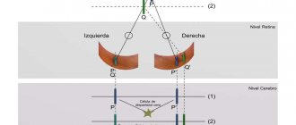
Central and peripheral vision
The human visual system is quite complex. Therefore, it allows you to look at objects, the world around you, navigate in space under different lighting conditions and move around in it. In ophthalmology today there are two types of vision:
- Central. It is an important component of the human visual system. It is provided by the central part of the retina. It is with the help of this vision that you will have a wonderful opportunity to analyze the shapes of the visible and small details. A person's central visual perception will be directly related to the visual angle that is formed between two points located at the edges. The greater the angle reading, the lower the sharpness.
- Peripheral. This type of vision provides a wonderful opportunity to analyze objects that were located around the focal point of the eyeball. It is this that later allows you to navigate in space and darkness. Peripheral vision is much lower in acuity than central vision.
It is important to know! If a person's central vision is directly proportional to the angle of vision, then peripheral vision will directly depend on the field of view.
Use of colors
Depending on the quality of vision, objects of different diameters are used. For normal vision, objects of 3 mm are used, and for low vision - from 5 to 10 mm. At the periphery of the retina there is no light perception; the edge perceives only white. As you approach the center, blue, red, yellow and green appear. All colors are visible in the center.
Boundaries of fields of view when using a white object:
- outward – 900;
- up – 50-550;
- up and out – 700;
- up and in – 600;
- inside – 550;
- down and in – 500;
- down – 65-700;
- down and out – 900.
Fluctuations from 5 to 100 units are possible. Research for other colors is carried out similarly, but with colored objects. The patient notes not the moment of the appearance of the object, but the moment of color recognition. Often changes to white are not detected, but narrowing to other colors is revealed.
What is the optimal field of view indicator?
Each person today has his own characteristics. Therefore, angles and field of view are individual and may differ from each other. The following factors usually influence a person's field of vision in degrees:
- specific signs of the structure of the human eyeball;
- eyelid shape and size;
- features of the composition of the bones of the eye orbits.
Also, a person’s viewing angle will depend on the size of the object in question and its distance from the eyes. The structure of the human visual system, as well as the structural features of the skull, are natural limiters of the angle of vision inherent in nature. However, the angle of limitation of all these factors is insignificant.
It is important to know! Experts have conducted numerous studies and have found that the visual angle of both human eyes is 190 degrees.
The normal field of view for each individual human analyzer will be as follows:
- 50-55 degrees for gradation upwards from the fixation point;
- 60 degrees for the downward and inner side measurement from the nose;
- from the side of the temporal region, the angle can increase to 90 degrees.
If a person's vision examination shows a discrepancy with the norm, then it is necessary to identify the cause, which is most often associated with vision problems. The visual angle allows a person to navigate space much better and receive more information that comes through the visual analyzer.
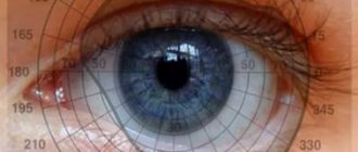
Perimetry norm
A study of the visual analyzer showed that the human eye clearly distinguishes two points when it is focused at an angle of at least 60 seconds. According to many experts, the angle of view will directly affect the amount of information received.
Types of perimetry
Perimetry is carried out using several different methods. The simplest is the Donders test, which allows you to evaluate the boundaries of the visual field. The patient is placed a meter away from the doctor and asked to focus on the examiner's nose. The patient first closes one eye, and the doctor shows a visible object and traces it in one of the meridians.
To study the central field, the Amsler test is used - an even simpler examination method. The test makes it possible to evaluate a zone up to 10° from the center of the visual field. When diagnosing, a grid of horizontal and vertical lines is used, with a dot in the center. The patient must fix his gaze on a point from a distance of 40 cm. Signs of pathology according to the Amsler test: curvature of lines and the appearance of spots. The method is indispensable for the primary diagnosis of macular pathologies.
The central field of vision can be examined using the campimetry method. The patient must close one eye and fixate his gaze on a black board located a meter away. The board (1x1 m) has a white dot in the center. White objects of different diameters (1-10 mm) are moved along the studied meridians until they disappear. Scotomas are first noted on the board, and the results are transferred to a form.
In theory, the results of different methods should match, but in practice, moving objects are viewed better than stationary ones. This is especially noticeable in areas with defects, which is called the Riddoch phenomenon.
Field of vision measurement
Recently, determining visual fields is a really important task. The human visual analyzer is a complex optical system that has been developing over a long period of time. Different color rays are associated with a variety of information components, so the human eye perceives them differently. Peripheral visual analysis ability affects the different color rays that are perceived by our eyes.
The most developed corner has a white tint. Then comes blue and red. The viewing angle decreases the most when analyzing green shades. In most cases, even a slight deviation can indicate serious pathologies in the visual system. Each person has his own norm, but there are indicators by which deviation is determined.
Modern medicine makes it possible to perform a high-quality study of visual fields and quickly identify diseases of the visual system. By determining the angle and identifying image loss, the doctor can quickly determine the location of hemorrhage and the appearance of tumor processes. A good ophthalmologist, as a result of the examination, can identify the following disorders:
- Exudates.
- Retinitis.
- Hemorrhages.
In the presence of such conditions, measuring the visual angle paints a general picture of the condition of the fundus, which is further confirmed using ophthalmoscopy. The study of this indicator and deviation from the norm also gives a picture of the state of the visual analyzer when diagnosing glaucoma. Even in the early stages of this disease, you may notice certain changes.
If during the process of diagnosing the problem a significant part is missing, then this is a serious suspicion of a tumor lesion or extensive hemorrhage in certain parts of the brain.
What you need to know about perimetry
The visual field is the space that a person recognizes when his gaze is fixed and his head is still.
If you look at a certain object, in addition to its clear image, a person sees other objects located around. This is called peripheral vision, and it is not as clear as central vision. Perimetry is an ophthalmological study that allows you to examine the boundaries of the visual fields through a projection onto a spherical surface. There are kinetic and static perimetry. Kinetic research involves using a moving object, while static research involves varying the illumination of an object in one position. The study helps to analyze changes in the visual field and determine the localization of the pathological process (retina, optic nerve, visual pathways, visual centers in the brain). Most often, a narrowing of the visual field and loss of some areas (scotoma) are detected.
Indications for perimetry:
- retinal pathologies (tears and detachments, dystrophy, hemorrhages, burns, tumors);
- diagnosis of macular pathologies, including toxic lesions;
- detection of retinitis pigmentosa;
- diseases of the optic nerve (neuritis, trauma);
- diagnosis of pathologies of the visual pathway and cortical centers in the presence of neoplasms, injuries, stroke, severe malnutrition;
- a brain tumor;
- hypertonic disease;
- traumatic brain injuries;
- signs of cerebrovascular accident;
- confirmation of glaucoma, tracking the dynamics of the process;
- checking patient complaints (aggravation factors);
- preventive examination.
Perimetry is contraindicated if the subject is under the influence of alcohol or drugs, or has mental illness. The procedure does not cause any complications.
What can distort perimetry results:
- drooping eyebrows;
- deep planting of eyeballs;
- drooping eyelid;
- height of the bridge of the nose;
- exposure of the irritant to large vessels near the optic nerve head;
- low visual acuity;
- poor quality correction;
- rim glasses.
False visual field defects can also appear due to the structural features of the face and pupil width. To eliminate false defects, repeat testing is carried out in the same program. In order for dynamic observation to be reliable, the same perimetry conditions must be observed (size of objects, lighting, time and colors).
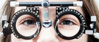
How to measure
With a sharp decrease in the viewing angle, a person will definitely be able to notice this. If the decrease in visual angle occurs gradually, then this process may go unnoticed. That is why many experts recommend an annual examination, which will allow you to quickly detect various deteriorations. Diagnosis and determination of narrowing of the visual field in modern ophthalmology is carried out using an innovative method called computer perimetry. The cost of such a procedure is quite low, and the duration is only a few minutes. However, thanks to computer perimetry, it is possible to quickly determine a decrease in peripheral vision, even with small deviations, and quickly begin treatment.
The diagnostic procedure consists of the following steps:
- Conducting a study to determine the angle of the visual field begins with consultation with a specialist. Before the procedure, the doctor must tell you all the features and rules of the procedure. The patient is examined without optical instruments. Each patient's eye is examined separately.
- The patient must focus his gaze on a static point, which is located on the dark background of the device. During the procedure for measuring the angle of the visual field, bright dots will appear in the peripheral field with different intensities. These are exactly what the patient's eye should see.
- The location of the points is constantly changing, and this allows you to determine with 100% accuracy the moment the site falls out.
- The speed of this examination is quite fast and within a few minutes the program will process the information received and display the result.
Most modern clinics today provide information in printed form. Others provide the opportunity to record the received data on storage media.
What perimetry results indicate pathologies?
Graphically, the field of view looks like a hill. This association makes it possible to explain to the patient in detail the presence of problem areas and their location. Pathologies lead to deterioration not only of peripheral vision, but also to a complete drop in picture clarity. The results of the study are recorded in a specially designed form.
- the presence of more than normal loss in a patient indicates a pathological condition of the visual organs;
- scotomas in the field indicate glaucoma;
- narrowing of peripheral vision signals eye diseases that need to be further investigated.
Deviation of the obtained perimetry data from the norm does not always indicate the presence of eye pathology. The reliability of the technique is affected by patient fatigue, poor room lighting, or difficulties with fixating gaze on stimulators.
Diagnostics determines color disturbances in the retina or its immunity to bright light. Additionally, perimetry allows you to diagnose disorders in the visual apparatus, which subsequently develop glaucoma. To clarify the diagnosis, several types of perimetry can be prescribed.
The main indicators of disturbances during perimetry are narrowing of visual fields and scotomas. Depending on the degree of damage to the visual pathway, the characteristics of field narrowing will differ. Changes can be unilateral or bilateral, as well as concentric and sectoral. Concentric changes are observed along all meridians, and sectoral changes are observed in a specific area with normal boundaries throughout the rest of the length.
Defects that are located in one half of the field in each eye are called hemianopsia. This state is divided into homonymous and heteronymous. Homonymous hemianopsia is a loss from the temporal side in one eye and from the nasal side in the other. Heteronymous hemianopsia is a symmetrical loss of the nasal or parietal halves of the field in both eyes.
Types of hemianopsia according to the size of the prolapse:
- complete (loss of the entire half);
- partial (narrowing of some zones);
- square (changes in the upper or lower quadrants).
A scotoma is an area of loss in the field of view that is surrounded by a safe zone, that is, it does not coincide with the peripheral borders. Such loss can take any shape and be located in any area (center, para- and pericentral zones, periphery).
Scotomas discernible by the patient are called positive. If the prolapse is detected only during the examination, it is considered negative. Patients suffering from migraines note the appearance of a flickering scotoma. It appears suddenly, is short-term in nature and moves into the field of view.
Types of pathological scotomas:
- relative (reduced sensitivity, in which only large and bright objects are detected);
- absolute (complete loss of the field zone).
Paracentral Bjerrum scotomas may indicate the development of glaucoma (increased intraocular pressure). Such a scotoma arcuately surrounds the center of the field, and then enlarges and merges with it. Scotoma appears when intraocular pressure increases, and when it decreases it can disappear completely. At a late stage of glaucoma, two Bjerrum scotomas connected to each other are detected.
How to expand your perspective
A wide field of view allows a person to better navigate space and perceive information more widely. When reading a book, a person with a greater perspective will do it much faster.
Numerous studies have shown that the angle of the visual field can be further expanded with the help of special exercises. Even an absolutely healthy person can develop the capabilities of the visual analyzer. This will significantly improve your perception of the world around you. The scheme of such activities has a name – representation. In simple terms, such exercises will be associated with certain actions during a process such as reading. By doing this regularly you can broaden your perspective.
Many experts today recommend monitoring your health. Therefore, try to visit your ophthalmologist more often. Any disease is much easier to treat in the early stages, and diagnosing fields and angles of vision is an indicative way of early diagnosis of many ailments.
We recommend that you familiarize yourself with: lacrimal duct.
Methods for expanding the angle of view
Increasing the visual angle is called representation. You can make it wider using a set of special exercises. They can be performed not only by patients with any impairments, but also by people with good vision to prevent various diseases of the visual organs.
There are a large number of different techniques that help expand the viewing angle.
Tibetan technique
The Tibetan method of “clear vision” is one of the most common. It consists of several stages:
- You need to take a pencil in each hand and place them together in a vertical position. Pencils are at eye level at a distance of 30 cm from the face. Next, you need to try to focus on any object located behind them. In this case, the image of the pencils will become blurry.
- Next, you should slowly move them to the sides, keeping your hands at the same level. Objects should be moved apart to the maximum visible distance, then returned to their original position. This should be repeated several times. The gaze should be focused on the object behind the pencils. With your peripheral vision you should try to see the movement of objects to the sides and back.
- Then you should change the direction of movement of the pencils. They should be moved up and down. Repeat the exercise 8–10 times. Then change direction again - move the pencils in different directions diagonally. It is important to remain focused on the subject and not on your hands or pencils.
- The last exercise is to return the pencils to their original position and, without moving them, mentally enclose them in a circle. Outline this imaginary circle with your gaze, first clockwise, then in the opposite direction.
The results of these exercises will be noticeable after a month of daily training.
Schulte tables
Ophthalmologists note a good effect after patients regularly work with Schulte tables. They have long been used for teaching speed reading and have an undeniably high effect when working on expanding the viewing angle.
The table is divided into 5 cells, each of which contains numbers from 1 to 25. The patient’s task is to find all the numbers in order as quickly as possible. The sequence can be either direct or reverse.
As the angle of view increases, the time to complete the exercise will decrease.
When using these tables, you should follow some rules:
- The exercise is performed in a sitting position.
- There is no need to pronounce the numbers out loud, just find them with your eyes.
These tables have different options: they can contain numbers from 0 to 100, or even letters of the alphabet; the cells can be colored rather than black and white.
Gymnastics for the eyes
Eye exercises are a simple and at the same time effective means for improving the functioning of the visual organs in general and for expanding the field of vision as well. The exercises take on average 7–10 minutes. They are especially necessary for those people who have eye problems, as well as people with high load on the visual organs.
One of them is blinking for 1 minute. You need to close and open your eyes quickly enough, while trying not to strain your eyelids. Exercise significantly improves blood circulation in the eyes and is especially useful when work requires high concentration.
There are also other simple exercises to improve your peripheral field. They can be performed daily in almost any conditions:
An important condition for the success of any technique is the systematic implementation of exercises. The classes may seem too easy, but they are highly effective. It is very important not to give up exercises, but to do them regularly.