Treatment of varicocele in children
The most highly effective method of treating varicocele, which is recommended by experts, is surgery.
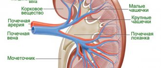
surgery is performed under general anesthesia. The purpose of the operation is to restore normal blood flow. Before surgery, it is necessary to undergo a number of tests that will show the general condition of the patient and the possibility of the operation:
- general blood analysis;
— determination of the Rh factor;
- general urine analysis;
— biochemical analysis;
— ECG.
The following types of operations for varicocele are performed at the First Children's Medical Center:
— Marmara operation (from mini-access). This type of surgery for varicocele has the highest effectiveness rate. The mini-access operation does not involve penetration into the child’s abdominal cavity. The pediatric surgeon makes a small incision in the groin area and excises the affected vessel. This operation virtually eliminates the risk of complications and is characterized by a quick recovery period. Shortening the postoperative period allows children to quickly return to their normal lifestyle.
- Ivanissevich operation. This operation involves ligation of the testicular vein at the retroperitoneal level. the surgeon makes an incision in the anterior abdominal wall in the left iliac region at the level of the anterior superior iliac spine parallel to the course of the inguinal canal. The indication for Ivanissevich's operation is the presence of varicocele of any severity.
We offer surgical treatment of varicocele, after which the child is observed for a day in a day hospital, and then sent home without the risk of complications. In the following days, you need to visit a doctor for dynamic monitoring.
If you have discovered certain signs of the disease in your child, do not delay, make an appointment with a surgeon!
Make an appointment with a surgeon
Choose a doctor
Indications
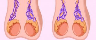
Regardless of the location of the problem, varicose veins go through several stages of development. The veins of the scrotum are affected with severe symptoms only at the penultimate stage. Before this, only a doctor can detect the problem.
First stage. There are no symptoms. The veins are dilated, but only slightly. Only an ultrasound examination will detect varicocele in a 14-year-old teenager. Whether surgery is necessary is too early to say.
Second phase. Here the doctor will notice problems with palpation. First the boy stands. Then he is asked to lie on his back. The doctor must compare the testicles - their size, appearance. He may ask the patient to tense his abdominal muscles. With this action, the veins will become more noticeable. This procedure is called the Valsava maneuver.
Third stage. Varicose veins are already visible as veins protruding above the surface of the skin. Their tortuosity is also noticeable. The diseased testicle decreases in size. Most often, the disease is an accidental discovery by a doctor during a routine examination of adolescents aged 12 to 14 years. In this case, surgery is required.
Everyone knows that any organ requires sufficient blood supply. If vascular pathology is diagnosed, then the testicle may be damaged. Over time, his condition worsens. The changes become irreversible. The following are the consequences of varicocele in a 14-year-old teenager:
- Normal sperm production is disrupted;
- The testicle changes its structure;
- The nature of enzyme production changes and biochemical processes do not proceed as they should (we are talking about the transformation of androgens);
- Blood flow in tissues is disrupted;
- There is a high probability of infertility in the future;
- Inflammatory pathologies of the pelvic organs.
If the doctor says that surgery is needed, it is better to listen to him. This will help avoid such changes.
Varicocelectomy in adolescents – who should be operated on and when?
Sizonov V.V. 1, Makarov A.G. 1, Kogan M.I. 2
1 Children's uroandrology department of the State Budgetary Institution RO "Regional Children's Hospital", Rostov-on-Don
2 Department of Urology and Human Reproductive Health with a course of pediatric urology-andrology, Rostov State Medical University of the Ministry of Health of Russia, Rostov-on-Don Email, [email protected] , dept_kogan_mail.ru
Varicose veins of the spermatic cord are the most common lesion of the reproductive system in adolescents. Before the age of 10 years, the incidence is less than 1%, with the onset of puberty it increases above 15% [1, 2]. The strategy for preventive varicocelectomy in children and adolescents is based on data from the World Health Organization on the identification of varicocele in 20-40% of men seeking help for infertility [3] and the results of studies indicating a decrease in sperm quality in patients with varicocele compared with healthy men [4]. An important circumstance that encourages surgical activity in adolescence is data on the highest degree of restoration of testicular volume after varicocelectomy performed before 14 years of age [5].
On the other hand, there is no direct scientific evidence of the negative impact of varicocele on reproductive function. In addition, there are studies indicating the temporary nature of the improvement in sperm parameters in patients after varicocelectomy [6, 7], which is extremely important when deciding on the timing of treatment for children and adolescents, given the remoteness of the period of reproductive function.
Belgian colleagues published a study [8] in which they examined the feasibility of performing varicocelectomy in children and adolescents based on the likelihood of future paternity. The data they presented demonstrate that in the group of patients who were managed conservatively, 85% of patients managed to become fathers, while among those who underwent surgery, paternity was achieved in 78%. The authors conclude that there is no positive effect on the probability of future paternity of active surgical tactics in the treatment of varicocele in children and adolescents.
The severity of the problem raised is illustrated by the publication in the same issue of two articles containing comments on the work of Belgian colleagues. Kolon [9], pointing out weaknesses in the evidence base of the Belgian study, expresses confidence in the need for further searches aimed at identifying a group of patients in need of early surgical intervention. The editor's commentary expresses fundamental disagreement with the findings about the inappropriateness of surgery in adolescents and expresses confidence in the need to develop perfect criteria for selecting those 20% of adolescents with varicocele who will have problems with reproductive function in the future.
In the modern diagnostic arsenal used when examining a teenager with asymptomatic varicocele, the most important component is the study of testicular volume. The first publications [10-15] devoted to the relationship between the presence of testicular atrophy in adolescents and future reproductive dysfunction were reduced to stating a fact without indicating the degree of decrease in testicular volume. In this case, the study of sperm parameters was not performed, and the criterion for the effectiveness of varicocelectomy was considered to be an increase in testicular volume on the side of the varicocele after surgical treatment.
At the next stage of studying testicular hypotrophy with varicocele, criteria for assessing the degree of hypotrophy were developed based on the study of absolute values of testicular volume [16, 17] and spermogram parameters. Kass et al. published the results of a study of the relationship between the degree of testicular hypotrophy and the deterioration of spermogram parameters, which allowed them to state that a decrease in testicular volume of an ipsilateral varicocele by 2.0 cm3 or more is a level that can be used as a criterion for selecting patients requiring varicocelectomy.
The use of absolute values has a significant drawback, since when assessing the degree of atrophy, age and individual constitutional characteristics are not taken into account. It is clear that a testicular volume deficit of 2.0 cm3 in adolescents at the beginning of puberty reflects a more pronounced atrophic process than in adolescents who have completed puberty. The described obvious shortcomings of absolute values led to the search for criteria using relative indicators reflecting the degree of testicular hypotrophy. The most widely used calculation formula is one that uses the volumes of the right and left testicles in the calculation:
There are many publications in which the threshold values for disproportion between testicles range from 9-20%. Thomas et al. [18] studied the influence of the degree of varicocele on the severity of testicular hypotrophy. The authors concluded that delayed testicular growth develops in every fourth patient with varicocele, regardless of the degree of varicocele. A study of the dynamics of development of testicular volume reduction allowed the authors to recommend establishing indications for varicocelectomy when the atrophy index reaches a level of 15% or more. Alukal et al. [19] published the results of a study that demonstrate the absence of a relationship between the severity of testicular hypotrophy and the degree of varicocele. The editorial commentary points out the inadequacy of selection criteria based on volume studies and the need for direct testing of testicular function in patients with varicocele to determine indications for varicocelectomy.
Two years later, Diamond et al. [20] reported the results of a study of sperm parameters in adolescents with varicocele and varying degrees of testicular volume reduction. The authors conclude that testicular volume disproportion of 10% or more is highly likely to be accompanied by pathospermia. Colleagues suggest that the testicular atrophy index be considered 10% as the limit beyond which continued observation is inappropriate and it is necessary to use active surgical tactics.
In 2009 Kogan M.I. et al. published a series of articles [21-23] devoted to a study very similar in design to the Diamond study. The results of the study led to a proposal to consider the threshold value of the atrophy index >8%, since according to the authors, in all cases when the atrophy index reached a value of >8%, pathological abnormalities in the spermogram were detected in adolescents with varicocele.
Almost simultaneously with works devoted to the significance of testicular asymmetry for determining indications for surgical treatment, a number of publications appear in which the authors demonstrate the possibility of disappearance or reduction of testicular volume asymmetry during the growth of an adolescent. There are studies whose authors consider it possible to observe patients with an atrophy index of more than 20% for three years, since 71% of the observed adolescents showed a reduction in the difference in testicular volumes during observation [24].
The possibility of conservative management of patients with a decrease in testicular volume on the side of the varicocele was expressed by Preston et al. [25] based on data they obtained in a retrospective review. The authors identified a high probability of disappearance of growth asymmetry during follow-up, which allowed them to assume that a single ultrasound examination is not enough to formulate indications for varicocelectomy.
Other studies support the possibility of spontaneous reduction of the imbalance, but report a significantly lower incidence of the phenomenon [26], while noting the possibility of increasing atrophy during follow-up in 35% of patients.
Van Batavia et al. [27] studied the influence of the Tanner stage of puberty on the severity and likelihood of detecting testicular volume asymmetry. The results of the study demonstrate that in children in Tanner stages I-III, the likelihood of detecting a decrease in testicular volume is higher than in patients in stages IV-V. In adolescents with Tanner stages IV-V, the likelihood of a decrease in the severity of testicular volume asymmetry during follow-up is higher than in boys with pubertal stages I-III.
Thus, a decrease in testicular volume on the side of the varicocele is an important criterion for selecting patients in need of varicocelectomy, but the scientific data accumulated today do not allow using this criterion as the only one. The main reasons for the lack of reliability of the decrease in testicular volume in children and adolescents are the possibility of spontaneous restoration of volume during growth and the lack of a generally accepted value for the testicular atrophy index. The accumulated experience and initial understanding by researchers of the lack of specificity of decreased testicular volume as a criterion for damage to testicular tissue in adolescents led to the search for alternative tests that could reflect the severity of testicular alteration in varicocele.
In the mid-90s, the attention of pediatric urologists was attracted to the study of testicular blood flow parameters using ultrasound, with the aim of studying their effect on spermogram parameters. Paduch et al. [28] presented the results of a study of sperm in 74 adolescents aged 17 to 19 years, who were divided into two groups - a control group (healthy adolescents) and young men with varicocele. The authors revealed a negative feedback between spermogram parameters and the value of maximum retrograde blood flow velocity (MRBF). However, in addition to stating the fact of the existence of this connection, the authors did not provide quantitative indicators of MSRC that could be used as markers for selecting patients for varicocelectomy.
Only 10 years later Kozakowski et al. [29] examined the combined effect of MSBC and testicular atrophy index on the prognosis of further testicular development. The authors demonstrated that in the presence of MSB above 38 cm/s and an atrophy index of 20%, there is no likelihood of a decrease in the atrophy index with continued follow-up. These results allowed the authors to suggest performing varicocelectomy in adolescents when an atrophy index of 20% or more and an MVR above 38 cm/s were detected by ultrasound.
In 2013, Van Batavia et al. presented the results of observation of adolescents, who were studied the influence of different values of the atrophy index against the background of MSBC of 38 cm/s and higher on the prospects for spontaneous disappearance of testicular volume asymmetry. The authors came to the conclusion that even in the presence of an atrophy index of 15% and an MSB of 38 cm/s or higher, continued dynamic observation is inappropriate [30]. The authors, in their next publication, studied the effect of MSRC, determined in the postoperative period, on the likelihood and severity of recurrence of varicocele and testicular asymmetry. Van Batavia et al. [31] found that, when detected after varicocelectomy, MSRC above 20 cm/s is highly likely to determine the persistence of testicular volume asymmetry and the development of relapse.
An analysis of the literature devoted to the problem of varicocele in children and adolescents demonstrates the enviable consistency of the tasks that researchers have set and are setting for themselves today and half a century ago. Almost all researchers begin with phrases dedicated to the lack of reliable data on the direct negative impact of varicocele on reproductive function, but they express confidence in the need in some cases to perform varicocelectomy for preventive purposes in children and adolescents. This determines the relevance, from their point of view, of searching for markers that could be used to select adolescents in need of surgery in conditions where direct research into the state of reproductive function is significantly limited.
Another important feature of adolescent varicocele is the significant gap between the timing of surgery and the time when it will be possible to evaluate the final result in terms of preserving fertility.
The described problems are complicated by the heterogeneity of the study group in terms of the adolescent’s sexual development at the time of detection and treatment of varicocele.
Waalkes et al. [32] analyzed existing modern approaches to the treatment of varicocele in children and adolescents. According to their data, today indications for varicocelectomy are associated with the presence of testicular hypotrophy of 20% or more on the side of the varicocele during the year and symptomatic varicocele. The combination of MSRC 38 cm/s and testicular hypotrophy equal to 20% tilts the treatment tactics towards surgical treatment. If an MSB of less than 30 cm/s and testicular asymmetry of less than 20% are detected, dynamic observation for a year is necessary, and only if these indicators worsen is varicocelectomy indicated. In other cases, it is advisable to observe until the age when it is possible to study the spermogram parameters.
Against the backdrop of a huge array of accumulated data on the advisability of using a wide range of studies to resolve the issue of tactics for managing patients with varicocele, the results of studying the practical approaches used by American pediatric urologists look surprising. Coutinho et al. [33] surveyed 74 practicing pediatric urologists. According to the responses received, only 49% of respondents used ultrasound examination of the scrotal organs in patients with varicocele, 38% used a Prader orchidometer to determine testicular volume. Almost a third of respondents suggest varicocelectomy immediately after identifying testicular asymmetry. 57% of pediatric urologists have never referred their patients for semen analysis. The data obtained demonstrate a significant gap between the accumulated scientific potential and its practical use in the treatment of children and adolescents with varicocele.
It seems to us advisable to continue the active search for optimal combinations of markers of testicular tissue damage when determining patient management tactics depending on the stage of puberty. On the other hand, it is advisable to develop a method for monitoring the dynamics of changes in reproductive function in the long term in adolescents.
Literature
1. A Doppler-based study on the prevalence of varicocele in German children and adolescents / D. Pfeiffer, J. Berger, C. Schoop et al. //Andrologia. – 2006. – Vol.38. – P.13.
2. Tekgül S, Riedmiller E, Gerharz P, et al. In: Guidelines on Pediatric Urology. Arnhem, The Netherlands: European Association of Urology, European Society for Paediatric Urology // 2009. – P.23.
3. World Health Organization. The influence of varicocele on parameters of fertility in a large group of men presenting to infertility clinics // Fertil Steril. – 1992. – Vol.57. – P.1289.
4. Sakamoto, H. Relationship between testicular volume and varicocele in patients with infertility / H. Sakamoto, Y. Ogawa, H. Yoshida // Urology. – 2008. – Vol.71 – P.104.
5. The effect of varicocele repair on testicular volume in children and adolescents with varicocele / S. Cayan, E. Akbay, M. Bozlu et al. // J Urol. – 2002. – Vol.168. – P.731.
6. Testis volumes, semen quality, and hormonal patterns in adolescents with and without a varicocele / LC Haans, JS Laven, WP Mali et al. // Fertil Steril. – 1991. – Vol.56. – P.731.
7. Update on treatment of varicocele: counseling as effective as occlusion of the vena spermatica / E. Nieschlag, L. Hertle, A. Fischedick et al. //Hum Reprod. – 1998. – Vol.13. – P.2147.
8. Bogaert, G. Pubertal screening and treatment for varicocele do not improve the chance of paternity as adult / G. Bogaert, C. Orye, G. De Win // J Urol. – 2013, Jun. – Vol.189(6). – P.2298-2303.
9. Kolon, TF The adolescent varicocele – a Shakespearean tragedy or much ado about nothing? / TF Kolon // J Urol. – 2013, Jun. – Vol.189(6). – P.2024-2025.
10. Hösli, PO Varicocele – results following early treatment of children and adolescents / PO Hösli // Z Kinderchir. – 1988, Jun. – Vol.43(3). – P.213-215.
11. Surgical repair of varicocele at puberty: preventive treatment for fertility improvement / A. Okuyama, M. Nakamura, M. Namiki et al. // J Urol. – 1988, Mar. – Vol.139(3). – P.562-564.
12. Hösli, PO Early treatment of varicocele in children and adolescents / PO Hösli // Helv Chir Acta. – 1989, Jun. – Vol.56(1-2). – P.229-233.
13. Mazo, E.B. The role of disorders of mineraloglucocorticoid function of the adrenal glands in the development of infertility in patients with left-sided varicocele / E.B. Mazo, M.V. Koryakin, Yu.V. Kudryavtsev // Urology and nephrology. – 1989. – No. 2. – P.38-45.
14. Kass, EJ Reversal of testicular growth failure by varicocele ligation / EJ Kass, ABBelman // J Urol. – 1987, Mar. – Vol.137(3). – P.475-476.
15. Sigman, M. Ipsilateral testicular hypotrophy is associated with decreased sperm counts in infertile men with varicoceles / M. Sigman, JP Jarow // J Urol. – 1997, Aug. – Vol.158(2). – P.605-607.
16. Kass, EJ Adolescent varicocele: objective indications for treatment / EJ Kass, JE Freitas, JB Bour // J Urol. – 1989. – Vol.142. – P.579-582.
17. Costabile, RA Testicular volume assessment in the adolescent with a varicocele / RA Costabile, S. Skoog, M. Radowich // J Urol. – 1992, May. – Vol.147 (5). – P.1348-1350.
18. Thomas, J. C. Testicular growth arrest and adolescent varicocele: does varicocele size make a difference? / JC Thomas, JS Elder // J Urol. – 2002. – Vol.168. – P.1689-1691.
19. Testicular hypotrophy does not correlate with grade of adolescent varicocele / JP Alukal, D. Zurakowski, A. Atala et al. // J Urol. – 2005, Dec. – Vol.174(6). – P.2367-2370.
20. Relationship of varicocele grade and testicular hypotrophy to semen parameters in adolescents / DA Diamond, D. Zurakowski, SB Bauer et al. // J Urol. – 2007. – Vol.178. – P.1584–1588.
21. Results of 104 adolescents with varicocele randomized to surgery or observation and followed up for 18 months / MI Kogan, A. Afoko, DV Siziakin et al. // 24th Annual EUA Congress, Stockholm Sweden. – 2009. – Abstract 100 Eur Urol Suppl, 2009. – P.145.
22. Adolescent Varicocele: When should we intervene surgically? / M.I. Kogan, A. Afoko, D.V. Siziakin et al. // 24th Annual EUA Congress, Stockholm Sweden. – 2009. – Abstract 100 Eur Urol Suppl, 2009. – P.98.
23. Varicocele: contradictions and problems / M.I. Kogan, A.A. Alvin, A. Afoko et al. // Urology. – 2009. –№6. –P.67-72.
24. 3rd Transient asynchronous testicular growth in adolescent males with a varicocele / TF Kolon, MR Clement, L. Cartwright et al. //Jurol. – 2008. – Vol.180. – P.1111–1114.
25. Conservative management of adolescent varicoceles: a retrospective review / M. A. Preston, T. Carnat, T. Flood et al. // Urology. – 2008,Jul. – Vol.72(1). – P.77-80.
26. Testicular asymmetry and adolescent varicoceles managed expectantly / SA Poon, CK Gjertson, MAMercado et al. // J Urol. – 2010. – Vol.183. – P.731734.
27. Adolescent varicocele: influence of Tanner stage at presentation on the presence, development, worsening and/or improvement of testicular hypotrophy without surgical intervention / JP Van Batavia, SL Woldu, PM Raimondi et al. // J Urol. – 2010, Oct. – Vol.184(4 Suppl). – P.1727-1732.
28. Paduch, D. A. Semen analysis in young men with varicocele: preliminary study / D. A. Paduch, J. Niedzielski // J Urol. – 1996, Aug. – Vol.156 (2 Pt 2). – P.788-790.
29. Peak retrograde flow: a novel predictor of persistent, progressive and new onset asymmetry in adolescent varicocele / KA Kozakowski, CK Gjertson, GJ, Decastro et al. // J Urol. – 2009, Jun. – Vol.181(6). – P.2717-2722.
30. Adolescent varicocele – is the 20/38 harbinger a durable predictor of testicular asymmetry? / JP Van Batavia, G. Badalato, A. Fast, KI Glassberg // J Urol. – 2013, May. – Vol.189(5). – P.1897-1901.
31. Incidence, significance and natural history of persistent retrograde venous flow after varicocelectomy in children and adolescents: correlation with catch-up growth / JP Van Batavia, AM Fast, SN Nees et al. // J Urol. – 2013, Aug. – Vol.190(2). – P.689-695.
32. Waalkes, R. Varicocele in adolescents: a review and guideline for the daily practice / R. Waalkes, IF Manea, JM Nijman // Arch Esp Urol. – 2012, Dec. – Vol.65(10). – P.859-871.
33. Variations in the management of asymptomatic adolescent grade 2 or 3 left varicoceles: A survey of practitioners / K. Coutinho, D. McLeod, K. Stensland, JA Stock // J Pediatr Urol. – 2013, Dec – Vol.11 – P.203-205.
The article was published in the journal “Bulletin of Urology”. Issue No. 1/2014 pp. 41-49
Magazine
Bulletin of Urology No. 1/2014
Comments
To post comments you must log in or register
Surgery for varicocele: methods, indications, performance, rehabilitation
Varicose veins are a problem affecting a huge number of people. This disease has no age: both adults and children can suffer from varicose veins.
Varicose veins are a disease characterized by a pathological change in the venous walls (this can be dilation or elongation). This disease is associated with a violation of the circulatory process. Varicose veins can manifest as swelling of the veins or the formation of subcutaneous nodes.
For the male part of the population, a special form of this disease is relevant - varicocele. This disease, unfortunately, is quite common. Therefore, doctors at the First Children's Medical Center decided to cover issues related to this disease in more detail. In this article we will look at what varicocele is, the main causes of the development of the disease, as well as methods of treating varicocele in children.