What is an aneurysm?
An aneurysm is a protrusion of the wall of an artery (less commonly, a vein) or heart due to its thinning, stretching or damage.
As a result, a so-called aneurysmal sac appears, which can compress nearby tissue. An aneurysm may be congenital. Moreover, when a child is born, this defect is invisible, and the baby develops completely normally. Aneurysms are also caused by diseases that affect blood vessels: hypertension, atherosclerosis, syphilis (at a late stage), sepsis. The risk of developing this disease increases when a blood vessel is injured or injured and when infected blood clots form. You can live with an aneurysm for years, go about your daily activities, and not experience any symptoms. Meanwhile, the aneurysm can quietly increase in size, threatening to explode at any moment - like a time bomb.
Why is an aneurysm dangerous?
Often an aneurysm is discovered incidentally during ultrasound, X-ray or MR examination. If it is detected, urgent treatment is required, since a possible rupture of the aneurysm leads to hemorrhage, which is often fraught with death. When an aneurysm ruptures, a person feels pain, and the patient's blood pressure can rapidly decrease if the bleeding intensity is high.
Smoking has a detrimental effect on heart health and increases the risk of developing many cardiovascular diseases. Source: suriyawut/Depositphotos
It is believed that aneurysm occurs more often in patients over 50 years of age. In young people, it usually develops as a result of injury or congenital malformations.
What a patient may encounter if an aneurysm ruptures
The consequences of a ruptured aneurysm are the most severe. They are more difficult to treat and are accompanied by residual effects:
- difficulties with perception and processing of information,
- decreased visual sharpness, appearance of “blind spots”,
- difficulty moving, convulsions and involuntary movements,
- tingling, numbness, decreased sensitivity of different parts of the body,
- difficulty swallowing food,
- speech disorders,
- epileptic seizures,
- character changes, pronounced apathy or aggressiveness may appear,
- pain syndrome in different parts of the body,
- problems with bowel movements.
If any unpleasant sensations or sudden changes occur, it is necessary to urgently undergo a computed tomography scan.
Classification and course of the disease
In addition to the classification of aneurysms depending on their location, there are others presented in the table below.
| Because of |
|
| By shape |
|
| By structure |
|
Aneurysms do not have a clear stage; the progression of the disease is judged by the size of the aneurysmal expansion, which varies depending on the location. For example, in the case of an abdominal aortic aneurysm, an expansion diameter of up to 6 cm is considered small, while in the case of aneurysms of the cerebral arteries, a size of more than 2.5 cm is already considered gigantic.
Based on the size, doctors predict the risk of complications and decide on surgical intervention.
Rehabilitation after surgery
After the operation, the patient is indicated for special rehabilitation, which is carried out in a specialized sanatorium.
The rehabilitation period can take from 1 to 18 months. At the same time, the patient is re-taught all basic everyday and cognitive skills, and the basic functions of the body (physical and social) are established.
During this period, the patient needs a balanced diet and hygienic care.
Rehabilitation includes physiotherapeutic procedures prescribed by the doctor, therapeutic exercises, balneotherapy, muscle electrical stimulation and acupuncture, which stimulates the necessary functions of the body.
A very informative video about the basics of surgical intervention and rehabilitation for vascular diseases at the Institute of Surgery named after Nikolai Nikolaevich Budrenko:
Types of aneurysm by location
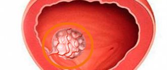
Cerebral artery aneurysm
The most common form of this disease. It is characterized by local dilation of the arteries of the brain. In the case of hemorrhage due to a rupture of an aneurysm, a sharp unbearable headache, loss of consciousness, nausea, vomiting, and convulsions are most often noted. In half of the cases, patients die; many of those who survive risk remaining disabled. However, only about 25 percent of patients with an aneurysm experience a headache similar to a migraine before the critical moment. This disease is often misdiagnosed as a brain tumor.
Aortic aneurysm
This type of aneurysm can develop in different areas of this large blood vessel. According to research results, aortic aneurysm is found in 7 percent of those who die for another reason. In the later stages of the disease, patients complain of pressing pain in one or another part of the body.
There are aneurysms of the thoracic aorta and aneurysms of the aortic arch, the peculiarity of which is that it can develop even within 20 years after a chest injury. There is also an abdominal aortic aneurysm, which is often asymptomatic. However, very thin patients may feel throbbing and pain when they place their hand on their abdomen.
Aneurysm of peripheral vessels (blood vessels of the extremities)
Source: tirachardz / ru.freepik.com
Aneurysms of peripheral vessels are less dangerous in comparison with other types. Most often they develop in the vessels of the lower extremities - the popliteal and femoral arteries. Aneurysms of the vessels of the upper extremities are much less common. The clinical course is often asymptomatic, but sometimes patients note coldness and pallor of the limb, pulsation and pain when palpated.
Such aneurysms are subject to rupture much less frequently, but are often a source of thromboembolic complications.
Heart aneurysm
Characterized by a saccular protrusion of the heart wall. Acquired cardiac aneurysm occurs at the site of a myocardial infarction and is detected in 5-20 percent of patients who have suffered it. Over time, a scar is found at the site of the lesion, which gradually protrudes. An aneurysm can develop either immediately after a heart attack or several months after.
When any form of aneurysm is detected, surgical intervention is most often required. The essence of the operation is to exclude the aneurysm from the bloodstream to prevent life-threatening hemorrhages - endovascularly, by clipping, excision of the damaged section of the vessel and replacing it with a plastic prosthesis or a fragment of a blood vessel from another part of the body.
It is not always possible to prevent the development of an aneurysm, but you can reduce your risk of developing one by quitting smoking and reducing high blood pressure. To do this you need:
- Eat a healthy diet - in particular, eat more fruits and vegetables and less salt;
- drink alcohol in moderation or avoid it altogether;
- maintain a healthy weight;
- devote time to regular physical activity - approximately 150-200 minutes of moderate activity per week;
- reduce caffeine consumption to approximately 400 mg per day, which is approximately 2-3 cups of 250 ml.
Arterial aneurysms
Rice.
1.a. Saccular aneurysm of the right internal carotid artery (angiogram): before opevapia, the neck (indicated by an arrow) and the body of the aneurysm are visible Fig. 1.b. Saccular aneurysm of the right internal carotid artery (angiograms): angiogram after surgery; a clip is visible on the neck of the aneurysm (indicated by an arrow); aneurysm is not performed The vast majority of arterial aneurysms of the cerebral vessels are so-called saccular arterial aneurysms (Fig. 1), which look like relatively small local protrusions of the artery wall. In such aneurysms, it is usually possible to distinguish the bottom, the middle part - the body, and the narrower initial part - the neck. Less commonly, an aneurysm is a large thin-walled cavity as a result of dilatation of the artery over a significant extent (Fig. 2).
Rice. 2.a. Large saccular aneurysm of the bifurcation region of the left internal carotid artery (angiogram): before surgery Fig. 2.b. Large saccular aneurysm of the bifurcation region of the left internal carotid artery (angiogram): the result of surgical exclusion of the aneurysm; clips are visible on the internal carotid artery and aneurysm; with angiography through the right carotid artery, the vessels of both the right and left hemispheres of the brain are well filled
It is believed that arterial aneurysm of cerebral vessels was first discovered by G. Morgagni more than 200 years ago (1761), however, only after the introduction of cerebral angiography into clinical practice [E.Moniz, 1927] this disease of cerebral vessels was well studied and began to be diagnosed intravitally. In the forties of the 20th century, attempts were made to surgically treat arterial aneurysms of cerebral vessels [NM Dott, 1933; Tennis (W. Tonnis), 1936; Dandy (W. E. Dandy), 1944 and others].
During postmortem examination of corpses of people who died from various causes, arterial aneurysms of cerebral vessels are found in 1-5% of cases. However, not all cerebral aneurysms cause certain clinical phenomena. The most common and dangerous manifestation of arterial aneurysms of cerebral vessels is intracranial hemorrhage that occurs when they rupture. According to Pakarinen (S. Pakarinen, 1967), such hemorrhage occurs in approximately one per 10,000 population.
Arterial aneurysms of cerebral vessels are one of the most dangerous diseases of cerebral vessels.
Rice. 3. The most common locations of arterial aneurysms on the arteries of the base of the brain (indicated by circles)
In total, about 70% of patients with ruptured arterial aneurysms of cerebral vessels die from primary or repeated hemorrhages. The vast majority of arterial aneurysms are located on the arteries of the base of the brain (Fig. 3). Thus, according to a study of intracranial aneurysms and subarachnoid hemorrhages conducted by American and English scientists (1966), out of 2672 arterial aneurysms of the cerebral vessels, aneurysms of the intracranial part of the internal carotid artery accounted for more than 40%, and 25% of them were located at the origin of the posterior communicating artery . 28% were in the area of the anterior communicating artery. The third most common location of arterial aneurysms of cerebral vessels is the middle cerebral artery (about 20%), 5.5% of aneurysms are located in the vertebral and basilar arteries (the so-called vertebrobasilar system). In other extremely rare cases, aneurysms occur in the extracranial part of the internal carotid artery, callosal artery, etc. In approximately 20% of cases, arterial aneurysms are multiple.
The cause of the formation of arterial aneurysms of cerebral vessels is still not precisely known. The occurrence of most aneurysms is associated with congenital inferiority of the vascular wall. Factors contributing to the formation of aneurysms may be atherosclerotic changes in blood vessels, hypertension and some other pathological processes. A small isolated group of arterial aneurysms of cerebral vessels consists of the so-called. mycotic aneurysms resulting from the entry of infected emboli into the vessels of the brain and purulent melting of the vascular wall.
Pathological anatomy
The wall of an arterial aneurysm, as a rule, is a thin plate of scar connective tissue. It usually lacks muscle layers and poorly differentiates other layers of the arterial wall. In the area of the bottom of the aneurysm, its wall is usually the thinnest, and ruptures occur more often in this place.
Clinical picture
There are two forms of clinical manifestation of arterial aneurysms of cerebral vessels: apoplectic and paralytic - tumor-like.
In the vast majority of cases, arterial aneurysms are the cause of severe, often recurrent intracranial hemorrhages (apoplectic form).
Ruptures of arterial aneurysms of cerebral vessels are characterized by sudden severe headache, vomiting, and loss of consciousness often occurs, which in severe cases can last several hours or even days. Clinical examination reveals symptoms characteristic of subarachnoid hemorrhage: headache, photophobia, symptoms of meningeal irritation (stiff neck, Kernig's sign, etc.). Symptoms of focal brain damage (paresis of the limbs, speech disorders, mental disorders, etc.) may also be observed, which are caused by hemorrhage in the brain or ischemic damage to the brain as a result of prolonged spasm of the arteries (see Stroke). Depending on the location of the aneurysm, symptoms of damage to individual cranial nerves may occur. For example, rupture of aneurysms of the internal carotid artery located at the origin of the posterior communicating artery is characterized by damage to the oculomotor nerve.
In the most severe cases, against the background of lost consciousness, there is a drop in cardiac activity, respiratory failure, and symptoms such as hormetonia, decerebrate rigidity, etc. can be detected.
Unruptured aneurysms are much less likely to manifest clinically. This is usually observed in paralytic forms in cases where aneurysms reach large sizes and, like a tumor, compress the brain and cranial nerves. Most often, such aneurysms are located in the area of the optic chiasm. They compress the optic and oculomotor nerves, cause endocrine disorders and, in clinical manifestations, can be similar to pituitary tumors, meningiomas of the tubercle of the sella and other basally located brain tumors.
Diagnosis
The occurrence of spontaneous subarachnoid hemorrhage (confirmed by lumbar puncture), especially in relatively young and middle-aged people who do not suffer from systemic vascular diseases, is always sufficient reason to assume rupture of an arterial aneurysm. The reliability of this assumption increases with repeated hemorrhages. In some cases, the combination of the clinical picture of subarachnoid hemorrhage with focal neurological symptoms suggests the localization of the aneurysm in the system of one or another cerebral artery.
The final diagnosis is made only on the basis of angiographic examination. Cerebral angiography (see) is also necessary to resolve the issue of the possibility of surgical treatment. If a rupture of an arterial aneurysm is suspected, a complete angiographic examination with filling of the internal carotid and vertebral arteries is necessary. This study makes it possible to detect multiple aneurysms and study the features of collateral circulation. Preference should be given to the catheterization technique, which allows for a comprehensive study of the vessels of various vascular areas of the brain in one study. To study the blood circulation of the internal carotid arteries, puncture angiography can also be successfully used. To detect an arterial aneurysm, serial angiography is required with the largest number of images in the arterial phase (during the first one to two seconds). In some cases, the recognition of aneurysms is facilitated by the long-term retention of the contrast agent in the aneurysm cavity. Angiography also makes it possible to diagnose intracranial hematomas accompanying aneurysm rupture and identify cerebral circulation disorders caused by artery spasm.
In a small percentage of cases, old aneurysms with calcified walls can be recognized by craniographic examination, which reveals characteristic ring-shaped petrification. Valuable data on the presence of intracerebral hematomas accompanying aneurysm rupture can be obtained using echoencephalography (see).
A contraindication to angiography can only be a very serious condition of patients, excluding the possibility of any surgical intervention.
The prognosis of hemorrhages caused by rupture of arterial aneurysms of cerebral vessels is unfavorable, especially since the hemorrhages are recurrent in nature. More than 20% of patients die from the first hemorrhage. Repeated hemorrhages (there may be 2-3 of them, sometimes more) are especially severe, with a mortality rate of 40-50%. Most often, recurrent hemorrhages are observed within the first 4 weeks after the initial rupture of the aneurysm, but repeated hemorrhages can occur several months and even years after the onset of the disease. In total, about 70% of patients die from primary or repeated hemorrhages. With unruptured aneurysms, including paralytic forms, the prognosis is more favorable.
Treatment
Maintaining bed rest. Coagulant and antihypertensive therapy are ineffective and do not significantly affect the prognosis of the disease. However, in the acute stage of aneurysm rupture, the use of drugs such as epsilonaminocaproic acid, which suppresses the fibrinolytic activity of the blood, reduces the risk of repeated hemorrhages. Therefore, coagulant therapy for aneurysm rupture is certainly indicated, regardless of whether surgical treatment is undertaken or not. The use of these drugs allows, in some cases, to postpone surgical intervention for some time and carry it out under more favorable conditions. In the acute stage of hemorrhage, repeated lumbar punctures are used to reduce intracranial pressure and reduce severe headaches.
The only radical treatment for arterial aneurysms is surgery, the main purpose of which is to prevent repeated hemorrhages from the aneurysm. For hematomas resulting from a ruptured aneurysm, the purpose of the operation is also to remove accumulated blood and eliminate compression of the brain.
To treat arterial aneurysms, two types of surgical interventions are used: intracranial exclusion of the aneurysm and ligation of the carotid artery in the neck.
Intracranial operations
Due to the fact that most recurrent hemorrhages from arterial aneurysms occur within the first 2-4 weeks, early surgery should be attempted whenever possible. However, operations in the acute stage are very dangerous. Most surgeons consider it possible to operate on patients who are in a satisfactory condition (conscious, without gross symptoms of focal brain damage), after the most acute phenomena of hemorrhage have passed (4-5 days after the rupture of the aneurysm). If the patients are in a more serious condition (impaired consciousness, paresis of the limbs, etc.), the operation should be postponed until the patient’s condition improves. The operation is also indicated in the “cold” period, especially if the hemorrhages are recurrent.
For hematomas that cause compression of the brain, urgent removal of the hematoma is necessary for health reasons.
The operation is performed under general anesthesia using dehydrating agents (mannitol, urea) and controlled arterial hypotension (see Artificial hypotension). If a prolonged cessation of blood flow through the vessels of the brain is necessary, the use of hypothermia is justified. Access to aneurysms is determined by their location: during operations on aneurysms of the anterior communicating and anterior cerebral arteries, bilateral frontal access is usually used with the intersection of the sagittal sinus and falciform process. To approach aneurysms of the internal carotid and middle cerebral arteries, frontotemporal trepanation is used. To exclude aneurysms of the basilar artery, an approach under the temporal lobe is usually used, and in some cases, dissection of the cerebellar tentorium is used.
In modern conditions, many surgeons isolate and disable arterial aneurysms using an operating microscope or magnifying glass and appropriate microsurgical instruments. Most often, the aneurysm is switched off by clipping (see Clipping of vessels) or ligation of the neck or the aneurysm itself. Less often, if collateral circulation conditions allow, the arterial aneurysm is turned off along with the artery section. In some cases, they resort to clipping of the afferent artery. If it is impossible to turn off the aneurysm, its walls are strengthened, for which pieces of muscle and quick-hardening plastic substances are used.
Some surgeons turn off an aneurysm by thrombosing it using electrodes inserted into the aneurysm, placing a powerful magnet next to the aneurysm, which retains a thrombosing mixture containing iron in the aneurysm. Several other methods are also used. A necessary condition for assessing the effectiveness of the operation is control angiography.
Ligation of the carotid artery in the neck. The purpose of this operation is to weaken the blood flow in the internal carotid artery distal to the ligation site and in its peripheral branches, to create favorable conditions for thrombosis of the aneurysm. Due to the success of intracranial operations, ligation of the carotid artery in the neck for arterial aneurysms has become less common. This operation is most justified for aneurysms of the internal carotid artery in the cavernous sinus that are not yet accessible to direct intervention, as well as for large aneurysms of the intracranial part of the carotid artery that do not have a neck.
The common carotid artery is often ligated, since in this case the risk of cerebral circulation disturbance is somewhat less than when only the internal carotid artery is turned off, due to the fact that while maintaining blood flow in the external carotid artery due to collaterals, weakening of the blood flow cannot be achieved.
To determine the admissibility of switching off the carotid artery, a careful study of the conditions of collateral circulation is necessary. For this purpose, angiography is used through the opposite carotid or vertebral arteries with simultaneous clamping of the affected carotid artery, test compression of the artery in the neck for 10 minutes, registration of EEG, REG and retinal pressure at the time of test occlusion of the artery, determination of changes in the regional brain using radioactive xenon blood flow in the pool of the switched off artery and some other methods. Switching off the carotid artery, as well as switching off some aneurysms, is possible by catheterizing the artery and its internal occlusion using special balloons.
Assessing the results of surgical treatment is difficult because the outcome of the operation depends on the severity of the patient’s condition and the time elapsed after the rupture of the aneurysm. As the condition of patients improves after hemorrhage, the risk of adverse postoperative outcomes decreases, but at the same time the risk of recurrent hemorrhages decreases. Despite these difficulties, when comparing large homogeneous groups of patients who underwent surgery and were treated conservatively, it was found that the outcomes in the first case are undoubtedly more favorable.
Treatment of large tumor-like aneurysms represents a complex surgical challenge. Due to their large size, frequent thrombosis, and wide connection with large arteries of the brain, such aneurysms are often inoperable. Therefore, determining the indications for surgery should be strictly individual and based on a thorough study of all the features of the case. In this case, it is necessary to take into account the relatively lower risk of hemorrhages in paralytic forms of aneurysms.
On average, mortality during surgical treatment of all groups of patients with arterial aneurysms is still high and amounts to about 30%. When treating patients in satisfactory condition, the outcomes are more favorable. Postoperative mortality in these cases reaches 5-8% [Pool and Potts (JL Pool, DG Potts), 1965; A.I. Arutyunov, 1971, etc.].
Diagnostics
Source: MART PRODUCTION: Pexels
Asymptomatic course and imitation of the clinical picture of other diseases can hide the fact of the presence of an aneurysm until complications develop. Often an aneurysm becomes an incidental diagnostic finding. It is easiest to diagnose a peripheral artery aneurysm symptomatically, since its symptoms are quite obvious.
The doctor can detect large aneurysms of the aorta and peripheral arteries during examination using palpation (feeling) and auscultation (listening). Damage to the arteries of the brain can manifest itself as symptoms of transient ischemic attack, sudden headaches, eye pain, fainting and other symptoms.
A cardiac aneurysm can manifest itself as symptoms of heart failure: shortness of breath, swelling, bluishness of the skin.
The following instrumental diagnostic methods are the most informative for aneurysmal vascular lesions:
- Ultrasound is a fairly universal technique used to diagnose aneurysms of various locations (except for cerebral vessels in adults). There are several options for ultrasound, used depending on the part of the body being examined and the pathology being sought.
- X-ray examination - allows you to identify aortic aneurysms, as well as the displacement of neighboring organs caused by them.
- CT and MRI are the most accurate diagnostic methods and allow you to visualize aneurysms of any location and size, determine the presence of thrombotic masses in them, and assess the condition of the walls.
- Angiography is a highly informative method that can be used in the diagnosis of any aneurysms. Unlike the above, this procedure is invasive, as it is necessary to deliver a contrast agent through a catheter into the vessel of interest. It is difficult to perform, and therefore is used to clarify cases of questionable diagnosis and controversial indications for surgical intervention.
Cerebral aneurysm: causes
Most often, this pathology is congenital, i.e. the walls of blood vessels at birth already have certain defects, or a cerebral aneurysm occurs in a person with the following genetic disorders: - renal polycystic disease; — connective tissue diseases; - congenital defect of arteries and veins; - circulatory disorders... There are other, more obvious, causes of cerebral aneurysm: - various head injuries; — infections; - high blood pressure (hypertension); - atherosclerosis; - brain tumors; - smoking and drug addiction; - high cholesterol; - taking oral contraceptives. By and large, any disease of the cardiovascular system can lead to cerebral aneurysm and this must be known, especially when diagnosing and treating this pathology.
Treatment
Aneurysms are treated exclusively by surgery. Conservative therapy takes place only during the period of dynamic observation of the development of an aneurysm and as a prevention of postoperative complications. The type of intervention depends on the location of the aneurysm, its size, the age of the patient and the presence of concomitant diseases.
Open operations
Traditional interventions are usually used for aneurysms of the aorta and cerebral arteries. To gain access to various parts of the aorta, laparotomy (opening the abdominal cavity) or thoracotomy (opening the chest cavity) is performed. After which the altered section of the vessel is usually excised, and a prosthesis is installed in its place.
In the case of intracranial localization of the aneurysm, a small craniotomy is performed. Then they find the aneurysm and apply a clip to its base, thus turning it off from the blood circulation.
Surgeries for cardiac aneurysms are performed infrequently, since their course is often asymptomatic or the operational risk outweighs the benefits of the operation. But in cases of severe heart failure and severe symptoms, surgery is warranted.
The access, as in the case of aneurysm of the thoracic aorta, is thoracotomy. The essence of the intervention is plastic surgery of the altered heart wall using resection of the aneurysmal sac, application of tightening sutures and synthetic patches.
During the operation, the blood clot that inevitably forms in the aneurysm cavity is also completely removed. This restores the normal geometry of the heart wall, eliminating severe symptoms of heart failure and preventing thromboembolic complications.
Open operations are very complex and traumatic. They are characterized by a fairly high operational risk and a long rehabilitation period. In this regard, in recent years, preference has increasingly been given to less traumatic endovascular operations.
Endovascular surgeries
Stents come in different types, but their essence is the same - to allow blood to move freely inside the vessel.
Source: Open-i (NonCommercial 3.0 Unported) (CC BY-NC 3.0) Widely used in the treatment of aneurysms of any location, with the exception of cardiac aneurysms. Access to the affected vessel during such interventions is carried out by introducing a catheter to it through a peripheral artery (for example, the femoral artery).
Thus, of the external defects after the operation, only a small incision in the projection of the peripheral artery will remain. Using a catheter in the area of the aneurysm, it is possible to install a prosthesis directly inside the vessel to turn it off from the blood circulation.
Also, for aneurysms of the cerebral arteries, stenting and embolization of the aneurysmal sac are performed. The essence of the latter is to fill the aneurysmal expansion with a ball (microcoil) of synthetic material, which is subsequently organized by connective tissue and thus turns off the aneurysm from the blood circulation.
Due to low morbidity and relatively easy rehabilitation, endovascular operations can be used in older patients and patients with severe concomitant diseases, for whom open intervention is contraindicated.
Sources
- S. Z. Amrakhov, A. S. Vischipanov. Surgical tactics and principles of performing operations in patients with coronary heart disease with post-infarction aneurysm of the left ventricle of posterobasal localization. Bulletin of the Scientific Center for Agricultural Sciences named after. A.N. Bakulev RAMS. 2013; 14 (1): 26-33.
- Koon K. Teo, MBBCh, PhD. Aneurysms of peripheral arteries. // MSD Disease Directory - 2021.
- Mark A. Farber, MD; Thaniyyah S. Ahmad, MD. Aneurysms of the thoracic aorta. // MSD Disease Directory - 2021.
- Brain aneurysms - Research Institute of Emergency Medicine named after. N.V. Sklifosovsky.