Leptospirosis is an infectious disease with an acute course. Its causative agent is Leptospira bacteria, and the main points of “impact” are muscles, central nervous system, capillaries, kidneys and liver. You may know this disease as water fever.
It is distributed throughout the world, and most of the pathogens are found in swampy areas and very humid regions. The causative agent of leptospirosis is most often transmitted by rodents, pigs, horses, dogs, as well as representatives of cattle. Among animals, mortality from this disease can reach 90%. It also has a very serious effect on humans, and therefore most often requires immediate hospitalization.
Infection, incubation period, stages
Typically, the infection enters the body through mucous membranes, wounds, the gastrointestinal tract and the genitourinary system - it comes from contaminated water, food, and soil. The incubation period most often takes from four days to two weeks, but in some cases it extends to a month. Most cases of the disease occur at the end of summer and beginning of autumn. Agricultural workers and veterinarians are at risk.
At the first stage, leptospirosis is accompanied by high fever, chills, weakness and nausea. The infection affects the surfaces of cells. The second stage is characterized by the penetration of infection into the capillaries and their damage. The liver, adrenal glands, kidneys, lining of the brain, and central nervous system are affected. To the general signs of malaise and intoxication, pain in the muscles and headaches are added.
At the third stage, antibodies to the pathogen appear in the blood, and the main signs of the disease begin to dull. The fourth stage is associated with the formation of an immune response to infection and recovery.
Since there are several types of pathogen, immunity is formed only to a specific one - in the future, infection with other variants of Leptospira is possible.
Treatment of leptospirosis:
Liquid probiotics Bifidum BAG, Trilact and Ecoflor sorbent effectively remove toxins, half-life products of bacterial cells, and eliminate intoxication
“Bifidum – liquid concentrate of bifidobacteria ” BAG is an effective consortium, which includes 5 strains of bifidobacteria species B.bifidum and B.longum in an active state (titer 1,000,000,000,000 = 10 in 12 trillion). The formula of the drug does not contain lactose and casein (but there are enzymes that break them down, produced by bifidobacteria). By forming a biofilm, it strengthens the protective barrier of the intestinal mucosa, reduces its permeability, preventing the penetration of exo- and endotoxins, allergens, potential pathogens into the body (especially effective against Staphylococcus aureus), helps restore the integrity and structure of the mucous membranes, improve the functions of the immune system, reduce the toxic load on liver and kidneys, improving liver function (confirmed by test results).
“Trilact ” (liquid concentrate of lactobacilli) is a consortium of 5 physiologically and antagonistically active strains of lactobacilli of the species Lactobacillis acidophilus, Lactobacillis casei, Lactobacillis plantarum (Titer 10,000,000,000 = 10 in 10 ten billion).
Also contains metabolites of lactobacilli. It is more active than bifidoconcentrate in regulating lipid metabolism, histamine levels in the blood, influencing the synthesis of immunoglobulins A and E, stimulating the secretory activity of the gastrointestinal tract, and has more pronounced antagonistic activity, especially against pyogenic staphylococci.
“Ecoflor ” (complex immobilized probiotic) - 10 strains of bifidobacteria and lactobacilli of the species B.bifidum, B.longum, L.acidophilus, L.casei, L. plantarum, immobilized on a carrier together with a nutrient medium (vitamins, amino acids, etc.). P.). Titer 108-1010 CFU/g. The carrier used is a carbon-mineral sorbent (SUMS-1) with antacid properties, a developed meso- and macroporous structure, which is characterized by a high selective adsorption capacity for products of incomplete metabolism, endo- and exotoxins of various origins (sorption range 200-1000 kDa) and grams -negative flora.
Probiotic course for removing toxins and immunostimulating
leptospirosis
Course duration 40 days for adults
| Stages of treatment | Duration | Time and dosage of medications | Need for the course |
| 1st stage | 1-10 day during active fever | In the morning – dose Ecoflor 2 packs 25 minutes before meals During the day - dose of Bifidum BAG 0.5 bottles 1 hour after meals In the evening – dose Ecoflor 2 packs 25 minutes before meals In the evening - dose of Bifidum BAG 0.5 bottles 1 hour after dinner | Ecoflor 40 pack Bifidum 25 bottles Trilact 15 bottles |
| 2nd stage | 11-40 day | In the morning – dose of Trilact 0.5 bottles In the evening – dose of Bifidum 0.5 bottles |
| Bifidobacteria B. They form the basis of the microbiocenosis of the large intestine: 90% of the gram-positive flora are bifidobacteria, they normalize the environment of the gastrointestinal tract and other cavities (nasopharynx, respiratory tract, genitourinary tract). Bifidobacteria form a biofilm, strengthen the protective barrier of the colon mucosa, and reduce it permeability, prevent the penetration of exo- and endotoxins into the internal organs. Operate on 3 levels: -on the wall of the mucous membrane; -in the lumen; -at the immune level; Bifidobacteria: - have a positive effect (normalize) on the environment of the gastrointestinal tract and other cavities (nasopharynx, respiratory tract, genitourinary tract); - indirectly affect the improvement of kidney function in renal failure and inflammatory diseases of the genitourinary system; more active than lactobacilli in carbohydrate metabolism, reduce blood sugar levels. Effective in treatment regimens for diabetes mellitus and pancreatic diseases; - participate in water-salt and mineral metabolism, enhance the absorption processes in the wall of the large intestine of calcium ions (formation of the skeleton and teeth, prevention of osteoporosis, etc.), iron (prevention and treatment of anemia), other micro- and macroelements; — supply the body with vitamins: participate in the synthesis and ensure absorption and assimilation of B vitamins, vitamin K, folic and nicotinic acids (prevention and treatment of rickets, vitamin deficiencies, diselementosis); - reduce the toxic load on the liver and improve its functions (accompaniment of hormonal therapy); - quadruples the content of natural killer cells in the large intestine - reduce intoxication and allergization of the body due to the action of biofilm (reducing the permeability of protective mucous barriers), destruction and removal of toxic metabolic products, allergens, toxins; - reduce carcinogenesis (inhibitory effect on colon carcinogens); -anti-aging effect (people with healthy intestines are young, beautiful and energetic); | Lactobacilli L. They are most abundant in the oral cavity, nasopharynx, upper respiratory tract, small intestine, genitourinary system, and skin. Lactobacilli make up 90% of the normal vaginal flora of women of reproductive age and ensure the cleanliness of the vagina. Operate on 3 levels: -on the wall of the mucous membrane; -in the lumen; -at the immune level; Lactobacilli are: -more active influence on the general and humoral immunity of the whole organism - a mild natural immunomodulator; - lactobacilli are more active than bifidobacteria - from pyogenic streptococci (priority for ENT diseases, diseases of the respiratory tract and lungs: sinusitis, otitis, adenoids, rhinitis, tonsillitis, tonsillitis, bronchial asthma, bronchitis, pneumonia) and local use for skin problems; -priority for gynecological diseases (vaginosis, candidiasis); -elimination of Helicobacter pylory (gastric and duodenal ulcers, atrophic gastritis); -pronounced antiviral properties prevent the replication and multiplication of viruses, the formation of virus carriers (viral intestinal infections, herpes viruses, ARVI, FLU, Epstein-Barr virus, viral hepatitis); more active than bifidobacteria in lipid metabolism (control the level of serum cholesterol and reduce its level in the blood, protect blood vessels from atherosclerotic changes, reducing the risk of heart attack or ischemic stroke and other cardiovascular complications). This determines the earlier and longer use of Trilact for allergic diseases and diseases of atherosclerotic origin compared to the classical regimen; -positive dynamics in atopic dermatitis, food and drug allergies, allergic bronchitis, bronchial asthma); -improving milk absorption - utilize lactose, reduce bloating and gas formation, abdominal pain caused by lactose intolerance; - eliminate neurotic disorders (increased fatigue, irritability, sleep disturbances, depression, anxiety); -anti-cancer effects; - support of antibacterial therapy; -reducing the risk of caries; -anti-aging effect (people with healthy intestines are young, beautiful and energetic); |
Probiotic course to accompany antibiotic therapy from the 1st day of taking antibiotics for legionellosis.
Space out antibiotic and probiotic intake at intervals of 2-3 hours.
Restoration of microflora after a course of antibiotics.
Course duration 40 days for adults
| Stages of treatment | Duration | Time and dosage of medications | Need for the course |
| 1st stage | 1-10 day while taking an antibiotic, double doses of a probiotic | In the morning – dose Ecoflor 2 packs 25 minutes before meals During the day - dose of Bifidum BAG 0.5 bottles 1 hour after meals In the evening – dose Ecoflor 2 packs 25 minutes before meals In the evening - dose of Bifidum BAG 0.5 bottles 1 hour after dinner | Ecoflor 40 pack Bifidum 25 bottles Trilact 15 bottles |
| 2nd stage | 11-40 day | In the morning – dose of Trilact 0.5 bottles In the evening – dose of Bifidum 0.5 bottles |
Leptospirosis in humans is an acute zoonotic infection characterized by signs of capillarotoxicosis, damage to the kidneys, liver, central nervous system, skeletal muscles, accompanied by intoxication, fever, severe myalgia and often jaundice. Etiology The causative agents of the disease, Leptospira interrogans, are spiral-shaped microorganisms adapted to life in water. Currently, there are more than 200 leptospira serovars, which are grouped into 23 serological groups. Among them: Canicola, Grippotyphosa, Icterohaemorrhagiae, Hebdomadis, Tarasovi and others. Leptospira are aerobes and quickly die when boiled, dried and exposed to direct sunlight. Sensitive to acids, penicillin, tetracycline, streptomycin and resistant to low temperatures, remain viable during long-term freezing. They persist for a long time in the external environment (water, moist soil), on food products - from several hours to several days. Epidemiology The source of infection is animals. In natural foci - rodents and insectivores (voles, rats, mice, shrews, hedgehogs), in which the infection is asymptomatic, and leptospira is excreted in the urine for a long time. In anthropourgic (synanthropic) foci - large and small cattle, rats, dogs, pigs, which can also carry Leptospirosis without any clinical manifestations. In recent years, the epidemiological significance of dogs and gray rats in the transmission of disease pathogens to humans has increased. Human infection occurs in various ways - percutaneous (through the skin and mucous membranes) and alimentary. In natural foci, a person becomes infected, as a rule, in the summer-autumn period, during agricultural work (mowing wet meadows, hay harvesting, etc.), hunting, fishing, irrigation work, hiking, while swimming, drinking water from random stagnant reservoirs, etc. Sporadic incidence is recorded throughout the year. Workers at livestock farms, meat processing plants, dog breeders, veterinarians, livestock specialists, and livestock owners on individual farms are often infected. A sick person does not pose a danger to others. Pathogenesis The pathogen enters the human body through the skin, mucous membranes of the mouth, eyes, nose, and gastrointestinal tract. Spreading through the lymphatic tract and hematogenously, Leptospira increasingly increases its presence in the lymph nodes, causing their hyperplasia, capillaries, leading to damage to the endothelium and the development of capillarotoxicosis, as well as in the intercellular spaces of various organs and tissues, where their active reproduction occurs. All this determines the polymorphism of clinical symptoms, the multiorgan nature of the lesions and the occurrence of numerous complications. Pathogens are fixed to the epithelium of convoluted tubules of the kidneys, cells of the liver and other organs, and contaminate the intercellular spaces of these organs. Damage to the kidney epithelium by Leptospira toxins leads to disruption of urination processes and the development of renal failure. Parenchymal liver damage in combination with hemolysis of erythrocytes caused by Leptospira hemolysins leads to the development of icteric forms of the disease. Penetration of Leptospira through the blood-brain barrier causes the occurrence of meningitis. Focal necrotic changes characteristic of leptospirosis develop in skeletal muscles. ITS can be a consequence of massive leptospiraemia, toxemia, widespread damage to the vascular endothelium and disseminated intravascular coagulation syndrome. The transferred disease leaves long-term immunity only to the serological variant of Leptospira that caused the infection. The pathogenesis of leptospirosis is the same in diseases caused by different leptospira serovars. Clinic The incubation period is 4-14 days. Jaundiced and anicteric forms of the disease are observed, with mild, moderate and severe course. The typical form of the disease begins acutely and is characterized by an increase in body temperature over several hours to 39-40 ° C, headache, and sharp pain in the muscles, especially in the calf. Myalgia intensifies with movement and can be so severe that the patient cannot get to his feet. Muscle pain syndrome is considered typical for leptospirosis, but does not occur in all patients. In the following days, intoxication increases - patients are inhibited, the headache intensifies, nausea and vomiting appear. The appearance is characteristic: the face is puffy, hyperemic, the vessels of the sclera and conjunctiva are sharply injected. Sometimes herpetic rashes appear on the lips and wings of the nose. In some patients, on the 3-5th day of illness, a roseolous-papular or erythematous, less often petechial rash is observed, located symmetrically on the skin of the extremities and torso. In most cases, micropolylymphadenitis occurs. Deafness of heart sounds, hypotension, and tachycardia are observed. Above the lungs - hard breathing, intermittent dry wheezing. In severe cases of the disease and in persons suffering from chronic alcoholism, pneumonia often develops. From the 2-3rd day of illness, the liver enlarges, jaundice of varying intensity and choluria appear in 12-20% of patients. Fecal acholia is usually not observed. Biochemical studies reveal hyperbilirubinemia (with an increase in the level of bound and free bilirubin), a moderate increase in the activity of ALT and AST. Some patients develop serous meningitis on the 5-7th day of illness. When examining peripheral blood, neutrophilic leukocytosis and increased ESR are observed. From the first days of the disease, the kidneys are affected: initially, oliguria and moderate proteinuria occur, leukocytes, erythrocytes, hyaline casts, and renal epithelial cells appear in the urine. Signs of kidney damage progress, which is accompanied by anuria, increased levels of urea and creatinine in the blood serum. Acute renal failure leading to uremia is the most common cause of death in patients. In severe cases of the disease, the condition noticeably worsens from the 7-10th day, sometimes earlier. An unfavorable prognostic sign is vemorraic syndrome - petechial rash, hemorrhages in the sclera, conjunctiva and injection sites, hematuria, nosebleeds, hemorrhages in internal organs. For most patients, the prognosis is favorable. The duration of fever is 5-12 days, then the temperature drops to normal. Some patients experience prolonged low-grade fever. In people who have not received antibacterial therapy, after 3-9 days of apyrexia, a second wave of fever occurs, of shorter duration, with milder clinical manifestations. Sometimes the course of the disease includes 2-3 similar relapses. Usually, by the end of the 2nd week, health improves, diuresis is restored, symptoms of intoxication and jaundice regress. The total duration of the disease is 3-4 weeks. The most common complications are infectious-toxic shock, hemorrhagic syndrome, acute renal or renal-liver failure, as well as eye lesions - uveitis, iritis, iridocyclitis, etc., which persist for several weeks during the convalescence period. Some patients experience relapses. There are erased forms of the disease that occur easily without damage to the kidneys or liver, and are similar to the clinical manifestations of influenza. These forms of leptospirosis can only be diagnosed based on specific laboratory testing and appear to be much more common than reported. Diagnostics Clinical diagnosis in typical cases can be made on the basis of clinical (acute onset, high fever, chills, myalgia, characteristic appearance of the patient; hepato-lien syndrome, meningeal symptoms, erythematous and hemorrhagic rashes, signs of kidney damage) and epidemiological data (occupational factor : workers in the meat processing industry, livestock farms, persons who come into contact with synanthropic rodents during their activities, stay in an endemic focus - hunting, fishing, swimming and drinking water from open reservoirs, contact with sick animals or carriers - dogs, muskrats, nutria, etc. .d.). To confirm the diagnosis, bacteriological and serological methods are used. The material for bacteriological research can be blood, urine, cerebrospinal fluid. The research is carried out in special laboratories. From the 1st to the 5th day of illness, blood cultures, dark-field microscopy of citrated blood, and infection of laboratory animals are performed. Urine cultures can be performed throughout the febrile period. The most widely used serological methods are microagglutination reaction (RMA), RSK, RNIF. The study requires paired sera taken before the 5-7th day of illness and a week later. PMA titers are considered positive - 1:100, RSC - 1:10 or more, a significant increase in antibody titers by 4 times. Leptospirosis is differentiated from viral hepatitis, HFRS, pseudotuberculosis, influenza, etc. Treatment Patients are hospitalized in infectious diseases hospitals, and, if necessary, in intensive care units and wards (see “Hemorrhagic fever with renal syndrome”). When caring for patients, the main attention is paid to identifying early signs of renal failure and infectious-toxic shock, therefore it is necessary to carefully monitor diuresis and cardiovascular activity. In severe forms of the disease, anti-leptospirosis immunoglobulin is administered 10 ml intramuscularly (after a skin test) for 3 days. Patients are prescribed penicillin in a dose depending on the severity of the infection - from 6,000,000 to 12,000,000 units/day. In severe cases of the disease, accompanied by meningeal symptoms, the dose of penicillin is increased to 18,000,000 units/day. After discharge from the hospital, the patient is observed in an outpatient setting: in the presence of residual ophthalmological effects - by an ophthalmologist; for residual neurological effects - see a neurologist; in case of kidney damage in the form of nephritis, see a nephrologist; in the absence of these phenomena, see an infectious disease specialist. The timing of discharge of patients from the hospital depends on the severity of the infection and the nature of the complications. After discharge from the hospital, convalescents need release from heavy physical labor, sports competitions, and work associated with industrial hazards for 3 months. For 2-3 months it is necessary to follow a diet excluding alcohol, spicy, fried, and fatty foods. Dispensary observation is carried out by an infectious disease specialist or therapist for 6 months and includes control tests of peripheral blood, urine (once every 2 months), examination by an ophthalmologist, and a neurologist. If leptospirosis is accompanied by the development of nephritis, follow-up observation and follow-up treatment with a nephrologist for at least 2 years. Prevention The main importance in preventing leptospirosis is the protection of natural and artificial reservoirs from contamination by the urine of wild and domestic animals. Health education plays an important role. Workers on livestock farms affected by leptospirosis, meat processing plants, and sewage systems are subject to routine vaccination. Dogs are also vaccinated against leptospirosis annually.
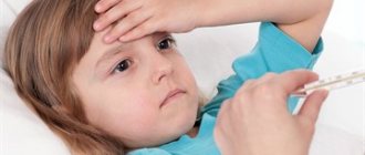
Signs of the disease
The first symptoms of the disease include a sharp increase in temperature to 39-40 degrees, chills, headaches, weakness, and red eyes.
The main symptoms of leptospirosis (except those mentioned) look like this:
- muscle pain - most often in the calves, thighs, and lumbar region. Sometimes the skin may hurt;
- enlargement of lymph nodes, as well as some organs, for example, the spleen or liver;
- rapid weight loss;
- problems with sleep and psychological state: insomnia, anxiety, irritability;
- redness of the throat is usually small;
- heart rhythm disturbances, decreased blood pressure;
- swelling in the facial area.
A rash with leptospirosis is an additional sign - it does not always appear. The same goes for symptoms such as jaundice, anemia, nausea or vomiting.
Are you experiencing symptoms of leptospirosis?
Only a doctor can accurately diagnose the disease. Don't delay your consultation - call
Methods of infection with leptospirosis
Humans can only become infected with leptospirosis from animals.
No sick person becomes a source of leptospirosis. Among animals, leptospirosis is transmitted through contaminated water and food or through fleas and ticks. These parasites can also transmit infection to humans.
There are two main foci of leptospirosis infection -
- natural
- agricultural.
The natural focus includes wild rodents and insectivores living in humid forest and forest-steppe zones.
The main animals of the natural focus of leptospirosis infection are:
harvest mouse; shrew; vole; gray rat; wood mouse; hedgehog; marmot; fox; less often - amphibians.
Infection with leptospirosis in an agricultural outbreak occurs due to domestic animals and small rodents living on livestock farms.
The main animals of the agricultural focus of leptospirosis infection are:
cattle (cow); small livestock (sheep, goat); horse; pig; dog; "domestic" black rat; house mouse; less often - poultry (ducks, geese).
All animals act as carriers and carriers of leptospirosis infection.
The parasite multiplies in the animal's body, remaining for a long time in the kidneys.
For many months (up to two years), leptospira are excreted in the animal’s urine into the environment, where they infect soil, water resources and surrounding objects.
The most dangerous are small bodies of water (ponds, swamps, small lakes and rivers), where a large concentration of Leptospira is created. In large bodies of water, the urine of infected animals is diluted with large amounts of water, reducing the concentration of Leptospira to a minimum.
Leptospirosis infection can enter the human body in several ways.
The main methods of transmission of leptospirosis infection are:
- contact;
- nutritional;
- aspiration;
- transmissible.
Contact route of infection with leptospirosis
The contact route of infection with leptospirosis is the most common. The parasite enters the human body through damaged skin and mucous membranes of the body that come into contact with contaminated surfaces and liquids. Infection occurs during agricultural work in wet meadows and pastures, on farms and meat processing plants. Infection with leptospirosis also occurs when caring for sick animals. There is a possibility of infection by contact through infected meat and blood of sick animals.
People who most often become infected with leptospirosis through contact are:
water meadow workers; rice farm workers; pet breeders; shepherds; milkmaids; meat processing plant workers; people working in slaughterhouses; veterinarians; livestock specialists; people working in dog kennels; people burying dead animals; taxidermists (people who make stuffed animals); deratizers (people who destroy rodents).
Superficial microtraumas (abrasions, scratches, small wounds) and undressed wounds become ideal entry points for leptospirosis infection. Even short-term contact of damaged skin with a source of Leptospira leads to infection of the body.
After infection, the infectious disease develops in several stages.
Alimentary route of infection with leptospirosis
The alimentary route of infection involves the penetration of infection into the human body through the oral cavity and gastrointestinal tract.
Infection with Leptospira is possible when using contaminated water for drinking, cooking, washing food and dishes. The infection persists for a long time on the surface of fruits and plants that are sprayed and watered with water from open reservoirs. Eating these foods may cause infection with leptospirosis.
Leptospira can also be found in food products themselves. They are found in meat and dairy products of infected animals. Or food contamination occurs through the urine of rodents and other sick animals.
Currently, infection with leptospirosis through contaminated food is quite rare, due to the low resistance (persistence) of bacteria. Leptospira survives in food products (milk, meat) for a short time - up to several days. And during the heat treatment of these products, bacteria die.
At the first stage of development of an infectious disease, leptospira enters the lymphatic and blood vessels through the mucous membranes of the digestive system (mouth, esophagus, stomach). What follows is a general picture of the infectious process.
Aspiration route of infection with leptospirosis
The aspiration method of infection with leptospirosis is observed less frequently than the alimentary method. It consists of the penetration of leptospira into the human body through the nasopharynx or oropharynx during aspiration of infected water.
Open water bodies are most susceptible to contamination by the urine of sick animals. When swimming in these bodies of water there is a high risk of aspiration and ingestion of water.
People who most often become infected with leptospirosis by aspiration are:
people involved in water sports (swimming, rowing); people who often swim in rivers and lakes; fishermen; flood victims; rescuers.
The development of an infectious disease begins with the penetration of leptospira into the blood and lymph through the mucous membranes of the respiratory tract (nose, trachea, bronchi) and oral cavity. Further stages of the disease are similar to the stages of contact infection.
Transmissible route of infection with leptospirosis
The vector-borne route of infection with leptospirosis is the rarest. The infection enters the human body through the bite of ticks and lice. Leptospires “use” blood-sucking insects and arthropods as vectors. From an infected animal, these parasites transmit the infection to other animals, and less often to humans.
Infection with bacteria is also possible through the bites of infected rodents, especially rats and mice.
Leptospira enters directly into the blood, from where it spreads throughout the body.
Symptoms of leptospirosis
The clinical picture of leptospirosis is very diverse. Symptoms and their severity depend on the form of leptospirosis (icteric or anicteric), as well as on the nature of the disease (acute or subacute).
Symptoms of leptospirosis include:
- fever and chills;
- rash;
- hemorrhages in the conjunctiva, nosebleeds;
- jaundice;
- damage to internal organs.
The following clinical periods of leptospirosis are distinguished:
- febrile period of the disease;
- peak period;
- recovery period.
The febrile period corresponds to general symptoms of intoxication - headache, aches, pain in the calf muscles, nausea, vomiting.
The dominant symptom is a temperature of 39 degrees Celsius and chills. Fever lasting more than 3 to 4 days is the first diagnostic sign of leptospirosis.
The peak period is also called the period of organ damage. This name reflects the essence of this period, since at this moment serious damage to the liver, kidneys and brain occurs. This period is characterized by increased mortality.
The recovery period corresponds to the restoration of vital functions, such as respiratory, cardiovascular and urinary.
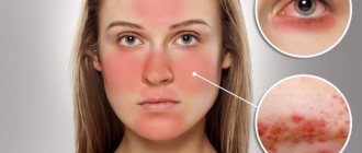
External diagnostic signs of leptospirosis include:
- puffy face;
- conjunctivitis;
- yellow coloration of the sclera;
- redness of the facial skin or icteric coloring of the face;
- enlarged lymph nodes located under the lower jaw and behind the neck;
- fear of light.
Prevention of leptospirosis
Leptospirosis infection can be prevented by limiting contact with carriers of the disease, which are infected rodent pests, farm and domestic animals.
Preventive measures to combat this disease can be divided into two groups.
- The first category includes rules for which the person himself is responsible.
- The second group includes activities, the organization and control of which is the responsibility of sanitary and epidemiological control authorities and health care institutions.
The rules for personal prevention of leptospirosis are:
- timely vaccination of animals;
- detection and treatment of disease in domestic animals;
- extermination of rodents and prevention of their appearance;
- implementation of preventive rules during agricultural work;
- compliance with safety rules when relaxing in nature;
- compliance with sanitary and hygienic requirements at the workplace in case of professional belonging to a risk group;
- immunization against leptospirosis.
Vaccination of animals against leptospirosis
Immunization of animals against leptospirosis is an effective measure that will protect humans and animals from this disease. Vaccination methods depend on the conditions in which the animal was acquired.
If the owner knows for sure that the animal is not infected with leptospira (when purchased from a nursery or from a breeder), a standard vaccination procedure is carried out.
If the animal was purchased by hand or picked up on the street, passive immunization is carried out, in which a special hyperimmune serum is placed before the vaccine.
Rules for spending time in open areas
During active recreation in nature, a person may encounter natural foci of leptospirosis, where pathogens circulate in the population of wild animals.
Zones of potential infection with Leptospira are located mainly on low relief of forests and river valleys.
Reservoirs with standing water and adjacent areas are also dangerous.
Places where there is a high probability of infection with leptospirosis are:
- wet grassy areas;
- thickets of sedge, cattail, reed;
- wet floodplain meadows;
- swamps;
- forest edges and wet deforestation.
Carriers of Leptospira in natural conditions are field mice, rats, moles, and shrews.
Infection of people in natural foci most often occurs in summer and autumn during fishing, hunting, hiking, and outdoor recreation.
Avoiding infection with Leptospira will help by following the rules of personal hygiene, refusing to use water from lakes and rivers for washing dishes or food, and wearing rubber shoes when fishing and hunting.
You should avoid swimming in bodies of water where prohibiting signs are posted on the banks.
You should also not swim in those lakes and rivers on the banks of which cows and other farm animals are grazing.
Taking precautions in the workplace
In addition to natural foci, there are economic zones in which the likelihood of infection with leptospirosis is increased.
The formation of such conditions is facilitated by the treatment, breeding and maintenance of animals that are carriers of Leptospira. Animals excrete pathogens along with their urine, contaminating the soil, water, pastures, feed and other environmental objects through which people become infected.
You can become infected with Leptospira through contact with animals, repairs and cleaning of the premises in which they are kept, or eating or drinking in the workplace, into which infected material may come into contact.
The incidence of leptospirosis in economic outbreaks has no seasonality.
The likelihood of infection is higher in those farms where rats and rodents are present. The danger also increases in facilities that are in unsatisfactory sanitary condition.
The high-risk group includes representatives of the following professions:
- employees of veterinary clinics;
- dog breeders;
- pig farmers, milkmaids, calf workers;
- employees of meat processing plants and livestock farms;
- people transporting animals;
- workers of circuses, zoos, equestrian sections.
Also, persons who, by the nature of their profession, often come into contact with synanthropic rodents (living nearby or together with people) are exposed to an increased likelihood of becoming infected with leptospirosis. The most common source of infection in this case is the gray rat.
This risk group includes such professions as miners, miners, employees of fish farms, workers in warehouses and wastewater treatment plants.
Measures to prevent leptospirosis infection in the workplace are:
- wearing rubber shoes and gloves, special overalls or gowns, rubberized aprons, hats;
- treating workwear at the end of the working day with special disinfectants;
- storage of work clothes and shoes in specially equipped rooms;
- refusal to take food and water in the workplace;
- storing drinking water and food in closed containers;
- disinfection and washing of hands with soap before eating and at the end of the working day;
- eating food in specially designated areas.
Persons constantly working on farms where the risk of contracting leptospirosis is increased should be vaccinated against this disease.
Immunization against leptospirosis
To form active immunity in a person who is at risk of infection with Leptospira, vaccination is carried out.
For vaccination, a drug is used that consists of several of the most common types of leptospirosis pathogens.
The vaccine is injected into the subscapular region twice. The volume of the first dose is 2 milliliters, the second (done 7–10 days later) is 2.5 milliliters.
The immunity of people who receive the vaccine lasts no more than a year. Therefore, people who are constantly in the territory of natural or economic outbreaks with an increased risk of infection must be vaccinated annually.
The interval between vaccination against leptospirosis and vaccines against other diseases should be at least 30 days for adults and at least 60 days for children. 24 hours before vaccination, you must give up alcohol and undergo an examination by a doctor, who will make a conclusion about admission to vaccination.
Head of the therapeutic department of outpatient department No. 1 - Sharonova I.N.
Danger of disease and complications
Diagnosis of leptospirosis is extremely important in the initial stages of the disease, since it can lead to a host of serious complications, including:
- internal bleeding;
- acute renal or liver failure;
- heart problems;
- pneumonia;
- meningitis;
- pancreatitis;
- paralysis and paresis of various types;
- toxic shock;
- and even death.
An infected person urgently needs medical help.
Symptoms of leptospirosis
The incubation period or the first phase (the period of time from the moment of introduction of the pathogen to the first clinical manifestations) lasts from 7-20 days, but on average - 10. At this time, leptospira, getting on the mucous membrane (of any area) or on the skin containing microdamages, begins multiply and spread through the blood (hematogenous spread) reaching various organs, but most often affecting the liver/kidneys/central nervous system/vascular endothelium. As soon as the concentration of the pathogen reaches a certain level, the next period occurs - clinical manifestations.
The period of clinical manifestations lasts up to 4 weeks, and this period includes generalization, peak and residual effects. The disease begins acutely, with general intoxication manifestations - chills and a rise in temperature to high numbers (lasting 4-5 days), myalgia (especially in the calf muscles due to focal necrotic and necrobiotic changes) and lasts about 7-10 days. After this, the second phase begins - the height, which lasted about 2 weeks, which occurs as a result of secondary bacteremia, and then secondary damage to internal organs and is characterized by toxinemia (third phase) due to the gradual death of leptospira and the release of endotoxin with their death with the subsequent development of ITS (infectious toxic shock) and multiple organ failure, systemic damage to the vascular endothelium also occurs in parallel, hemorrhages (bruising) occur on the skin, mucous membranes and internal organs.
Jaundice also develops, both due to hemolysis and destructive changes, resulting in liver failure. Due to vascular lesions in the kidneys, renal failure develops. After damage to the liver and kidneys, comatose states quickly develop, because they cease to perform one of their load-bearing functions - detoxification (excretion of toxic metabolic products). On objective examination, there is a “hood symptom”, which is represented by redness and puffiness of the face, redness of the skin of the neck and upper chest. Injection and icterus (yellowness) of the scleral vessels are noted, without signs of conjunctivitis.
As Leptospira spreads throughout the body, they spread through the blood-brain barrier, reaching the central nervous system, serous or purulent meningitis or meningoencephalitis occurs - meningeal symptoms become positive (stiff neck, Kering and Brudzinski symptoms)
The above listed symptoms (hemorrhages, renal and liver failure, ITS) are only collective images that hide the following:
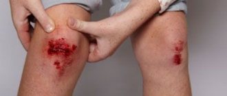
• Hemorrhages – hemorrhages on the skin, mucous membranes and, in severe cases, in internal organs. The rash has some features:
- on the skin these rashes resemble the appearance of measles/rubella/scarlatina-like rash; - localization on the chest, abdomen and arms; - during decompensation, nasal, abdominal (intraorgan) bleeding occurs, hemorrhages occur at the injection site - the rash may disappear after a few hours, leaving behind peeling and/or pigmentation
Hemorrhages with leptospirosis
• Renal failure - characterized first by a decrease (oliguria) and then by a lack of urination (anuria), as well as positive liver symptoms, uremia and disturbances in the concentration of electrolytes in the blood develop, which contributes to toxicity.
• Liver failure – will be manifested by jaundice and itching of the skin, icterus of the sclera, possible pain in the right iliac region. But jaundice also occurs due to the direct action of hemolysin (a pathogenicity enzyme) - it destroys red blood cells, releasing bilirubin.
• ITS develops as a result of the direct action of Leptospira exotoxin, and under the influence of endotoxin released during their death. Violation of the detoxification functions of the liver and kidneys leads to disruption of the elimination of toxic metabolic products. Also, the decay products of necrotic tissues formed as a result of destructive changes make their contribution to ITS.
Types of disease
There are several classifications of leptospirosis, let's name a few of them. The disease is classified according to its type, with and without jaundice, and according to the leading syndrome, there are 4 variants of the disease. There are cases when the kidneys are more affected, or the kidneys and liver at the same time, as well as the meninges. The variant with increased bleeding is also highlighted separately.
According to the severity of the disease, mild, moderate and severe degrees are distinguished. In the first case, the level of intoxication is mild, internal organs are not affected. In moderate cases, there are all the signs of leptospirosis and severe intoxication, but in severe cases, internal organs are already affected.
Also, the disease can occur without complications or with complications, and may or may not cause relapses.
Diagnostics
To make a diagnosis, not only visual and other signs of leptospirosis are taken into account. It is extremely important to conduct a number of studies:
- study a general and biochemical blood test, a general urine test;
- conduct PCR or RT-PCR tests, as well as diagnostics of RNGA and HCR;
- do a coagulogram, ultrasound, lumbar puncture (if necessary).
Clinical recommendations for leptospirosis involve differentiation (separation) of the disease from diseases that manifest themselves in a similar way. In this case, these are hepatitis of different types.
Causes of leptospirosis infection
The source is sick and recovered animals. Carriers – ticks, fleas. Carriers are rodents. Routes of infection:
• nutritional – through meat and other products from infected animals, through plant food infected with waste products of sick animals; • contact (in direct contact with a sick animal) and contact-household (in contact with contaminated household items); • aerogenic – when inhaling contaminated air; • transmissible – when bitten by infected ticks or fleas.
Treatment
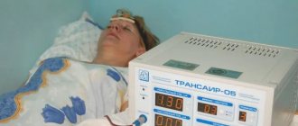
To effectively treat leptospirosis, the patient is first taken to the hospital - this is a prerequisite, since the disease can lead to fatal complications.
Next, patients are prescribed drug treatment, which includes at least four groups of drugs, each of which is responsible for its own tasks. It is necessary to administer a vaccine against the pathogen, relieve local symptoms, remove the consequences of intoxication, etc. Depending on the patient’s condition, complex procedures using special equipment can be used, for example, supporting the kidneys if they cannot cope with their functions.
Additionally, a diet is provided. If we are talking about kidney damage, then this is diet No. 7, for liver damage - No. 5.
Prevention
An important point is the prevention of leptospirosis, which allows you to avoid the disease or significantly reduce the risk of infection. Preventive measures include the following:
- vaccination against leptospirosis. Vaccination is carried out from the age of seven. In adults, it is indicated for people working in the agricultural sector and veterinary medicine;
- careful adherence to personal hygiene rules;
- refusal to swim in places where it is prohibited or simply not permitted. Avoid any untested bodies of water;
- working with animals only in compliance with personal safety measures. If possible, animals should be vaccinated with a special serum;
- timely treatment of any damage to the skin and mucous membranes with special means.
If symptoms of leptospirosis occur, you must immediately contact an infectious disease specialist, immunologist, or call an ambulance.
Animal leptospirosis
The disease affects various species of wild and domestic animals, of the latter pigs, cattle and dogs are most often affected; young animals suffer the most. Leptospira are excreted in urine, milk, semen, and discharge from the genital organs of sick and recovered animals; the duration of leptospir carriage ranges from several months to 2–3 years (in rodents, lifelong). Infection of animals with L. occurs through infected water during watering, bathing, and also through nutritional and sexual contact. Leptospira penetrate through damaged areas of the skin and mucous membranes. The disease manifests itself in the form of small epizootics or isolated cases. In sick animals, an increase in temperature, depression, abortions, the birth of non-viable offspring, a decrease in milk production, and atypical mastitis are observed. Mortality up to 20–70%, ch. perishes. arr. newborn young. L. often occurs in the form of asymptomatic leptospirosis carriage or immunizing subinfection (with the presence of specific antibodies in the blood in the absence of clinical signs and isolation of the pathogen). Treatment with antibiotics (streptomycin, tetracyclines), polyvalent hyperimmune serum, symptomatic. To prevent L. in domestic animals, vaccination is used.