Definition
It is a round formation of regular shape, located in the area of the foramina of Monroe, causing the development of hydrocephalus with signs of increased intracranial pressure.
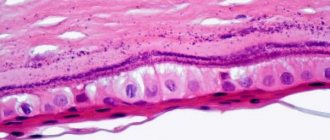
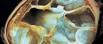
Macroscopically, the cyst looks like a thin-walled formation located in the area of the fornix or foramina of Monroe, attached to the choroid plexus or nearby brain structures. The size of the cysts ranges from 0.3 to 3-4 cm. The contents of the cysts are viscous, yellow-green in color. Microscopically, their inner wall is represented by one layer of cylindrical ciliated epithelium. Its cells produce mucin. Colloid cysts are characterized by a long course and a slow increase in their volume. With total removal, no relapses are observed, cerebrospinal fluid circulation is normalized and hydrocephalus is reduced.
Radiation diagnostics
On CT and MR tomograms, colloid cysts have a characteristic round shape, which distinguishes them from tumors of other histological structures.
CT semiotics
On CT, a colloid cyst is typically characterized by high density, although iso- or hypodense cysts that do not change after enhancement are rare. The reason for this increased density is iodine, which is contained in high concentrations in the colloidal contents of the cyst.
MRI semiotics
On T1-weighted MR images, the cyst usually appears iso- or hypointense (more often) compared to the medulla. In T2 mode, the intensity of the MR signal from the mass varies from hypointense, even lower than that of brain tissue, to hyperintense depending on the water and mucin content. Against the background of intravenous contrast enhancement, an increase in the intensity of the MR signal from the contents of the cyst and its walls is usually not observed. In this case, contrasting the septal veins bordering the colloid cyst is sometimes mistaken for contrasting its walls.
The ability to vary scanning parameters and obtain tomograms in any projection with MRI makes this method more informative than CT. At the same time, there are a number of observations where colloid cysts differed in varying degrees of density on CT and MRI signals, confirming the opinion that there are no absolutely specific diagnostic criteria in favor of one or another histological form of the tumor. In this case, an even more important differential diagnostic feature is the absence of contrast enhancement, which is not observed in cases of tumors of identical localization. Most often these are pilocytic astrocytomas.
Rice. 1. Colloid cyst of the third ventricle. CT. In the projection of the Monroe foramen, a round-shaped volumetric formation of increased density is determined (a). On MRI in T2 mode (b), the cyst has similar MR signal characteristics to the brain. On MRI in T1 mode (sagittal projection), the formation is hyperintense (c).
Rice. 2. Colloid cyst of the third ventricle. CT scan reveals an isodensity, round-shaped space-occupying formation in the projection of the foramen of Monro (a). On MRI in T2 (b) and T1 (c) modes, the cyst is detected only in T1 mode, as a focus of increased MR signal. In T2 mode, the formation is isointense with the brain.
Rice. 3. Colloid cyst of the third ventricle. On MR tomograms T2 (a) and T1 (b, c) in the projection of the foramen of Monroe, a small, round-shaped space-occupying formation is determined. The MR signal from the cyst is hypointense to the MR signal from brain tissue in the T2 mode and isointense in the T1 mode.
brief information
Colloid cyst of the third ventricle is a benign formation, the location of which is the third zone of the brain ventricle. It has a rounded spherical shape, surrounded by a thick protective layer consisting of connecting particles. Inside there is a jelly-like substance with a green-gray tint and is a product of the secretion of cavity cells. The volume of the tumor depends on the period of development of the abnormal process; in some situations it can engulf the entire perimeter of the organ. Deviation from the norm does not belong to the class of malignant neoplasms, as a result of which it does not provoke further spread of metastases. But since the cyst can increase in size, it naturally poses a huge danger to life. According to average statistical data, the presented form of pathology is diagnosed extremely rarely and accounts for no more than 1% of all cases of formations. This disease can strike at any age. The colloidal form occurs with equal regularity in both males and females.
Differential diagnosis
Differential diagnosis of a colloid cyst of the brain should be carried out with a wide range of tumors of the third ventricle . These tumors usually arise outside the third ventricle and are able to protect its lumen from external compression by the brain parenchyma. At the same time, these tumors themselves can cause a block of cerebrospinal fluid (CSF). Choroid plexus papillomas occur in the first 20 years of life in the lumen of the third ventricle. Also, 10%-30% of tumors found in the cavity of the 3rd ventricle can enter there from the lateral ventricles through the interventricular foramen of Monroe. Neurocytomas are intraventricular benign tumors of the nervous system, consisting of mature ganglion cells and occurring in children and young patients in the lateral and third ventricles of the brain.
Neurocytomas are often misdiagnosed as oligodendroglioma or ependymoma by light microscopy, so the true incidence of neurocytoma (a benign tumor of the nervous system composed of mature ganglion cells) may be higher than believed. Intraventricular meningiomas occur in 15%-17% of cases of meningiomas in children, and only in 1.6% of cases of a similar location in meningiomas in adults. By origin, meningiomas can be from the lumen of the lateral ventricles (rarely) or grow from the base of the skull into the bottom of the third ventricle (more often).
As mentioned above, the main damaging effect on the third ventricle comes from the brain parenchyma surrounding it. The majority of these lesions arise from glial tumors, including pilocytic astrocytomas, fibrillary astrocytomas, protoplasmic astrocytomas, subependymal giant cell astrocytomas, glioblastoma multiforme, and ependymomas. Tumor metastases (neoplasms) can involve the third cerebral ventricle through its roof, bottom, side wall or choroid plexus. Metastases from the lungs, colon, kidneys and breasts are most common. In such cases (tumor cell metastases), the prognosis is unfavorable and death occurs as a result of progression of the underlying disease.
Suprasellar germinomas and craniopharyngiomas can invade the bottom of the third ventricle from below from the base of the skull (middle cranial fossa). A suprasellar located pituitary macroadenoma can also invade the third ventricle. Reduced acuity and narrowing of visual fields, endocrine pathology and headache are the most common symptoms in such cases.
Other cysts in the anterior third ventricle include epidermoid cysts, dermoid cysts, and neurocysticercosis. Epidermoid and dermoid cysts are rare in the third ventricle, and neurocysticercosis is common in Eastern Europe, Asia, Central and South America, Mexico and Africa. The penetration of neurocysticercosis into the third ventricle is 15%-25% and leads to the subsequent development of hydrocephalus.
Inflammatory lesions, such as purulent abscess and granulomatous diseases such as tuberculosis and fungal infection, are much less likely to affect the third ventricle. Other lesions, such as sarcoidosis and histiocytosis, can affect the third cerebral ventricle through its floor and hypothalamus.
Also, a colloid cyst should be differentiated from vascular lesions of the brain, such as cavernous malformations and arteriovenous malformations.
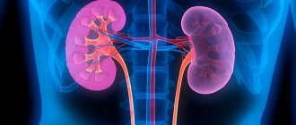
Colloid cyst of the brain. Colloid cyst of the thyroid gland
A colloid cyst is a benign tumor that grows slowly in the human brain. The cystic formation is filled with colloidal (gelatinous) fluid encapsulated by connective tissue cells. A colloid cyst can appear not only in the brain, but also in the patient’s thyroid gland.
Colloid cyst in the thyroid gland and brain - the difference in the development of the disease
- When a colloid cyst forms in the brain, the circulation of fluids is blocked. Because of this disorder, the process of development of hydrocephalus begins. Also, the constant pressure of the cystic formation on other parts of the brain can lead to sudden death;
- A colloid cyst in the thyroid gland has a more favorable course. Most often, these benign tumors contribute to the development of an abscess and inflammation of certain areas.
The main treatment method for colloid cysts is surgery.
What causes a colloid cyst?
Medical experts still could not give an exact answer about which cells in our body cause the development of a colloid cyst in the brain. However, they found that the cystic formation occurs during the embryogenesis stage during the formation of the child's central nervous system. Such a cystic formation may not show symptoms until the person becomes an adult. Usually by this age the cyst reaches such a large size that it begins to put pressure on the patient’s organs and tissues.
The formation of a colloid cyst is most often associated with hyperplasia and degeneration of its follicles.
The main reasons for the appearance of a colloid cyst:
- Lack of iodine in the human body;
- Sleep disturbance;
- Uneven daily routine;
- Smoking tobacco;
- Constant neuro-emotional stress;
- Frequent pregnancy;
- Constantly being in a cold space (living in the northern regions);
- X-ray irradiation.
Symptoms of a colloid cyst
Most often, a colloid cyst does not cause any discomfort in the patient. However, when the cystic formation begins to increase sharply, a person notices a sharp deterioration in his health:
- Constant headaches;
- Vomit;
- Loss of consciousness;
- A sharp decrease in visual acuity;
- Tinnitus;
- Mental personality disorders;
- Incontinence;
- Loss of coordination;
- Tremor.
Diagnosis of colloid cyst
When diagnosing a colloid cyst, the magnetic resonance imaging method is used. In rare cases, it happens that the cyst is an iso-dense MRI of the brain. In this case, the most effective diagnostic method is computed tomography. Thanks to MRI of the brain, you can easily determine the location of the colloid cyst and its location relative to the cerebral structures of the brain.
An MRI of the brain reveals a benign tumor. It is usually located in the anterior part of the third ventricle. In this place, a cystic formation blocks the opening of the Monroe, which causes the process of expansion of one of the lateral ventricles to begin.
How is a colloid cyst of the brain treated?
Unlike a colloid cyst of the thyroid gland, a colloid cyst of the brain cannot be left without attention and surgical intervention. If you use traditional methods and remedies as treatment, this will not affect the condition of the cystic formation in any way.
The presence of a colloid cyst of the brain is accompanied by constant pain and deterioration in the general condition of the human body. Unfortunately, to date, medical experts have not yet been able to discover an effective surgical method that would help get rid of the cyst without consequences.
Surgical options
Although there is no most effective surgical method for treating cystic tumors, doctors have discovered 4 safe ways to remove benign tumors:
- Transcallosal access is performed by craniotomy of the occipital region of the head. Most often, the right hemisphere of a patient whose left hemisphere of the brain is dominant is selected;
- Transcortical access – carried out through the middle temporal gyrus;
- Stereotactic approach is a neurosurgical method of brain surgery;
- Ventriculoscopic removal is an endoscopic method for examining the brain.
How does the operation proceed?
Typically, surgery to remove a cystic formation is performed using endoscopy. Surgeons gain access to the interhemispheric areas and hemispheres of the patient's brain.
If the cyst grows again, the surgeon prescribes a second operation, where a puncture is performed and the cavity of the cystic formation is emptied. Later, a sclerosant is injected into the cavity of the colloid cyst, causing its walls to stick together.
A colloid cyst can be a harmless benign tumor or a dangerous disease that can be fatal. This depends on the location of the cystic formation. But you should remember only one thing: if the cyst begins to grow sharply and causes you severe discomfort, then the only treatment option is surgery!