Pudendal nerve neuropathy: a patient’s history of recovery
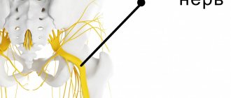
Complaints of pain in a sitting position, discomfort, numbness, burning in the pelvis can be explained by the anatomy of the pudendal nerve. This nerve leaves the spine at the level of 1-3 lumbar vertebrae and goes to the pelvis, passing between the muscles and ligaments (more precisely, between the piriformis muscle and the sacrospinous ligament). Note in Figure 1 where the pudendal nerve is labeled N.pudendus.
According to scientific research, most often the pudendal nerve is “pinched” either between two ligaments (sacrospinous and sacrotuberous), or between the piriformis muscle and the sacrospinous ligament. If this compression occurs, swelling will form around the nerve. This area begins to hurt, discomfort increases in a sitting position due to the location of the area of nerve compression. After the nerve has passed through these “danger zones”, it enters the dangerous canal - the Alcock canal (area of the ischium). Compression of the pudendal nerve in this canal is considered one of the reasons for the development of this pathology.
Why is this happening?
Risk factors for pudendal nerve compression include:
- Chronic microtraumas. They are provoked by unsystematic sports activities according to the principle: “rarely is better, but wear and tear” (cycling, training in the gym, team sports).
- Hormonal disorders (diabetes mellitus, hypothyroidism, etc.).
- Systemic connective tissue diseases (collagenosis, etc.).
- Chronic intoxication (alcohol).
- Genetic predisposition.
- Congenital anatomical features of the pelvis.
Pinching: reasons
Trouble may occur due to the piriformis muscle located in the pelvic cavity or due to compression between a pair of ligaments.
In addition, the pudendal nerve can be damaged after unforeseen situations, which include a car accident or a fall from a height. In such situations, a fracture of the pelvic bones may occur. Very often, the cause of chronic pain is damage to the nerve during childbirth or its involvement in the growth of a malignant tumor.
It is worth mentioning that certain types of human activities can also lead to pinching of the pudendal nerve over time. This includes riding a bicycle or horse.
Diagnosis of pudendal nerve neuropathy
Self-diagnosis
Answer yourself the question: Do you have these clinical symptoms:
- Pain in the area of innervation of the pudendal nerve: from the anus to the clitoris/head of the penis.
- The pain is felt worse when sitting.
- The pain does not cause awakenings at night.
- The pain is not accompanied by a decrease in tactile sensitivity.
Additional criteria:
- The pain is burning, shooting, stabbing, and may be accompanied by slight numbness.
- The pain worsens during the day and subsides after sleep.
- Mostly the pain appears on one side.
- Pain may be triggered by defecation.
If you notice that you have these symptoms, you should immediately consult a doctor.
Already at the initial appointment, the doctor will be able to conduct additional special manual tests.
If this is not enough, then electrophysiological studies are indicated. For example, the bulbocavernosus reflex will need to be assessed using electromyography.
All possible diagnostic methods are carried out at the Expert Urogynecology Clinic by highly qualified specialists in the field of pelvic floor pathology. We also carry out differential diagnosis with lesions of other pelvic nerves, including myofascial syndrome. The latter is characterized by painful sensations when pressing on various trigger points in the muscles.
| anonymously to the doctor, through the feedback form, we will try to help you. | Ask a Question |
Features of diagnosis and therapy
This type of neuropathy can cause serious problems with the genitourinary system. If it is not treated, the patient may experience problems in his sexual life (impotence, anorgasmia). Another unpleasant phenomenon that occurs is loss of control over urination.
Therefore, treatment of pudendal neuropathy is very important
To confirm the suspected diagnosis, methods such as:
- Ultrasound (during this, blood flow disturbances can be detected).
- Block of the pudendal nerve (for this disease, this procedure relieves discomfort, which indicates the correctness of the diagnosis).
The doctor must also examine the medical history and take into account complaints, especially the nature of the pain and cases of numbness. Neuropathy is also indicated by the fact that pain intensifies if the patient occupies a sitting position. To make such a diagnosis, the doctor must have sufficient experience to distinguish this pathology from other diseases.
Pinching
Treatment of this disease involves a complex effect
It is very important to identify the cause of its development and neutralize its impact. The main actions that the specialist takes are:
- Use of hormones and anesthetics (for blockade): Dexamethasone, Lidocaine.
- Relieving pain (most often drugs such as Lyrica and Tebantin are used).
- Taking muscle relaxants (they relax muscles, including the piriformis). The most famous remedy from this group is Mydocalm.
- Vitamin B. It promotes tissue repair.
Important Types of MRI examinations
In addition to drug therapy, you can use:
- Physiotherapy such as electrophoresis or phonophoresis. They stimulate blood flow and promote muscle relaxation.
- Kegel exercises, which involve training the muscles of the perineum. But their implementation is allowed only after the recommendation of a doctor.
- Help from a psychotherapist and taking antidepressants.
- Surgery may be required if traditional methods of therapy do not bring the desired results.
Pudendal neuropathy can cause severe pain and requires immediate treatment.
However, the difficulty is that it can easily be confused with other pathologies of the genitourinary system, so differential diagnosis through a comprehensive examination is necessary. With the right treatment tactics, the prognosis for the pathology is favorable.
Treatment of pudendal nerve neuropathies
Conservative treatment is aimed primarily at removing tension (decompression) of the nerve in “critical zones” and restoring normal trophism.
The first stage is recommended to carry out therapeutic and diagnostic blockades of these very problem areas, which we talked about at the very beginning (areas of ligaments and muscles).
The most common are blockades with local anesthetic (lidocaine, bupivacaine) and glucocorticoid (betamethasone).
Botulinum therapy.
Indicated in the presence of muscular-tonic syndrome, when the muscle needs to be relaxed. Botulinum toxin type A is successfully used specifically to target this pathological component.
When used correctly (according to indications and with good navigation via ultrasound), the risks of complications are minimal.
Manual therapy.
The main goal of treatment by an osteopathic neurologist is to eliminate biomechanical disorders and pathological changes in soft tissue structures.
When a person has a sore muscle, he tries to reflexively “save” it by changing his position, sitting or walking. This leads to the fact that these structures, which bear an additional “volume of work,” begin to experience additional stress and get sick. And this only strengthens the vicious circle of disease development. With the help of manual techniques, it is possible to return to its place what has been displaced and is involved in the pathological process.
Pudendal neuropathy
laesus_de_liro
The main symptom of pudendal nerve (PN) neuropathy is pain in one or more areas that are innervated by the n. pudendus (genital or pudendal nerve) or its branches. These are the areas of the rectum, anus, urethra, perineum and genitals.
One typical symptom is that the pain increases when sitting (usually the pain decreases when lying down) and progresses throughout the day. Also, pain during defecation and sexual intercourse is typical for PN neuropathy. Mild sphincter disorders may occur.
A few words about the anatomy of the PN.
The pudendal nerve contains both afferent (sensitive, sensory) and efferent (motor, motor) fibers, which causes sensory and motor disorders of the corresponding organs. Being the caudal part of the sacral plexus (S2(3) – S4) PN (n.
pudendus) exits through the foramen infrapiriforme (subpiriform opening), from the pelvic cavity along with a. pudenda interna (internal genital artery).
Then it bends around the back of the spina ischiadica (ischiadica) or sacrospinous ligament and through the foramen ischiadicum minus (lesser sciatic foramen) enters the fossa ischiorectalis (ischiorectal fossa), where it lies on the surface of the fascia obturatoria (obturator fascia) and then passes through Alcock's canal (which is formed by the split fascia of the obturator internus muscle). At the foramen ischiadicum majus (greater sciatic foramen) from n. pudendus branch – n. perforans ligamentum tuberososacrum, which pierces the corresponding ligament and goes down to the area of the ischial tuberosity, lying under m. glutaeus maximus (gluteus maximus muscle). Having reached the bottom edge of m. glutaeus maximus, the perforating branch goes around the muscle and is directed through the fascia to the skin of the gluteal region.
Important Hormones of the anterior, posterior and intermediate lobes of the pituitary gland and their functions: a table indicating the types of important regulators and their functions in the body
In the fossa ischiorectalis, at the ischial tuberosity, the following branches depart from the pudendal nerve: 1. Inferior rectal nerves, nn. haemorrhoidales inferiores (often depart from the pudendal nerve before it enters the foramen ischiadicum minus), short trunks, scattered into numerous branches directed forward and medially to m.
sphincter ani externus (external muscle of the anal sphincter) and to the skin around the anus (anus). 2. Perineal nerve, n. perinei, from its origin it goes forward and medially and is divided into nn. scrotales (labiales) posteriores (nerves of the scrotum or labia) - to the skin of the perineum and scrotum (large lips in women), and rami muscularcs (muscular branches) - to m.
transversus perinei superficialis, mm. bulbo – et ischiocavernosi. N. perinei anastomoses with n. haemorrhoidalis inferior and the perineal branch of n. cutaneus femoris posterior. 3. Dorsal nerve of the male penis (clitoris), n.
suspensorium penis, directed to the dorsum of the penis (clitoridis). Walking along with a. dorsalis penis (clitoridis) on the back of the penis, it gives off several branches to the skin of the penis and into the cavernous body and ends with several branches in the glans penis area. In women, the terminal branches of n. dorsalis clitoridis spread into the thickness of labia majora et minora.
Thus, the PN provides innervation to the levator ani and coccygeus muscles, the anal sphincter, the transverse perineal muscle, the bulbocavernosus muscle, the skin of the anterior part of the anus, the back of the scrotum or labia majora, the skin of the penis or clitoris, and the urethra. and the urethral sphincter.
The causes of PN neuropathy are still debated, but the most well known is compression of the pudendal nerve in the Alcock canal (which is formed by the split fascia of the obturator internus muscle). Also, compression of the PN can develop due to compression between the sacrospinous and sacrotuberous ligaments. As reported by A. Shafik (1991), R.
The role of the herpes virus is also actively discussed - indirect evidence is the effectiveness of acyclovir and valacyclovir in some cases of PN neuropathy.
There are the so-called Nantes criteria for PN, which were developed by JJ Labat, R. Robert, G. Amarenco (these criteria were discussed and ratified by a multidisciplinary working group in Nantes on September 23-24, 2006 and subsequently approved by SIFUP PP). Five main criteria have been identified:
1
.
pain in the area innervated by the pudendal nerve; 2
.
Predominant pain in the sitting position; 3
.
the pain does not cause sleep disturbance (i.e. does not cause the patient to wake up at night); 4
.
the pain does not cause serious sensory impairment; 5
. blockade of the pudendal nerve relieves pain.
Overactive bladder with bladder sensitivity
Enough has been written about this type of OAB; effective diagnostic and treatment methods have been developed.
The drugs of choice are M-anticholinergics, but quite often there are cases of insufficient effectiveness of these drugs. This may be due to the combination of OAB with DO and OAB with increased sensitivity of the bladder, which will be discussed below.
Clinically, this type of OAB (sometimes called “OAB without OAB”) manifests itself as a frequent or even constant feeling of the urge to urinate, but without urgency and episodes of urge incontinence in men, as in women. M-anticholinergics are usually ineffective.
Urodynamic manifestations:
- decrease in the volume of the first sensation of bladder filling;
- decreased volume of the first urge to urinate;
- decrease in maximum cystometric capacity;
- absence of detrusor overactivity and episodes of urinary incontinence;
- positive cold water test;
- positive test with lidocaine.
Important: How crazy people are treated. 1.2
Etiology:
- pudendal nerve neuropathy;
- myofascial syndromes;
- cross-sensitization of the urothelium in adenomyosis, IBS, adexitis;
- damage to the GAG layer of the urothelium.
Pathogenesis
Just as with painful bladder syndrome, there is an increase in the number of C-fibers and receptors affiliated with them. Moreover, sometimes the clinical and urodynamic manifestations of both conditions are identical. Perhaps these are manifestations of the same process that differ in severity.
Central sensitization also plays an important role. This is why the use of tibial neuromodulation is effective - the tibial nerve and the pudendal nerve have the same representation in the central nervous system.
Treatment:
- pregabalin;
- intravesical use of anesthetics, oxybutynin;
- intravesical electrophoresis of anesthetics, glucocorticoids, botulinum toxin;
- intravesical application of vanilloids (resiniferatoxin, capsaicin);
- transcutaneous electrical stimulation (TENS) with electrodes placed at the S2-S4 level (for example, Neurotrack Pelvitone, mode 10 Hz, 200 ms, 30 - 40 minutes per day);
- sacral neuromodulation;
- tibial neuromodulation;
- treatment of pudendal nerve neuropathy and myofascial syndromes;
- laser or electroablation of Lieto's triangle (A.I. Neimark, V. Gomberg);
- hydrobougation of the bladder.