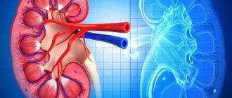
A kidney biopsy is an invasive diagnostic procedure that examines the cortex and medulla of the organ. The essence of the study is to obtain histological material, with the help of which it is possible to clarify the nature of the pathological process in the tissues of the organ. This study allows you to make a correct diagnosis, resolve the issue of locality or systemicity of the process, and prescribe the necessary treatment. Once a diagnosis has been established, a biopsy makes it possible to monitor the course of the disease and identify the need for organ transplantation or surgical treatment of the identified pathology.
- Types of biopsy
- Indications
- Contraindications
- Preparation
- How the research is carried out
- After the biopsy
- Complications
- The result of the procedure and its interpretation
- Which doctor should I contact?
Types of biopsy
There are several types of kidney biopsies, which differ from each other in the way they obtain material for research:
- Percutaneous needle biopsy, during which a long needle is inserted into the kidney tissue under ultrasound guidance and a biopsy is performed.
- An open biopsy involves taking histological material during surgery. Currently, it is used primarily for the removal of kidney tumors (rapid biopsy).
- Laparoscopic - performed using video control using an instrument that is inserted through small holes in the lumbar region.
The most common procedure is a percutaneous needle biopsy of the kidney. This procedure is minimally invasive, relatively safe and has a low risk of complications.
How to do a kidney biopsy
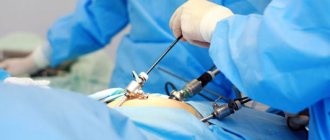
The main method is puncture. The procedure is performed in the operating room under local anesthesia and lasts 45 minutes. Doctors monitor the entire process visually using an ultrasound sensor or CT scanner. A small incision is made on the back in the lumbar region. A needle is inserted through it, which is advanced into the kidney 1–2 cm and a small column of kidney tissue is taken. Then the needle is removed and an aseptic bandage is placed on the puncture site. This wound then heals quickly. The collected tissues are sent to the laboratory for examination.
An open biopsy is only done during kidney surgery. Another method of kidney biopsy is endoscopic, when tissue is taken through a probe inserted into the urinary tract under general or spinal anesthesia. The method is used in pregnant women, the elderly, and patients after transplantation. Insertion of a probe through the jugular and renal veins and laparoscopic biopsy are possible, but these methods are rarely used.
Indications
A kidney biopsy is prescribed when it is necessary to obtain a sample of organ tissue, distinguish benign from malignant tumors and identify other changes at the cellular level. The list of indications for the study includes:
- Renal acute renal failure of unspecified etiology, especially if it is accompanied by systemic manifestations, symptoms of vasculitis and glomerulonephritis, anuria for 3 weeks or more.
- Nephropathy with symptoms of organic proteinuria, glomerular hematuria, nephrotic syndrome.
- Renal hypertension of unknown origin.
- Tubulopathy of unspecified etiology.
- Kidney transplant diseases: rapid decline in function or complete cessation of activity, the appearance of proteinuria and hypertension.
- Kidney tumors.
A kidney biopsy with the mandatory use of immunoluminescent and electromicroscopic analysis is carried out during the first 2 years after the diagnosis of chronic glomerulonephritis in order to select treatment tactics and monitor the effectiveness of prescribed therapy. Repeated biopsies, which monitor the correct choice of treatment tactics, are performed in patients with rapidly progressing glomerulonephritis and kidney recipients from 1 to 4-5 times a year, depending on the severity of the process.
Risk of bleeding after kidney biopsy in children: a systematic review and meta-analysis
03.04.2020
1550
0
Perinephric hematoma after kidney biopsy is found on average in 11% of children. But only a small proportion of patients require blood transfusions or additional interventions due to complications that develop after the procedure. This was shown by a systematic review of scientific studies over a period of 20 years, carried out by specialists from the Medical Center of Cincinnati Children's Hospital.
Kidney biopsy is an important screening tool for patients with kidney disease. Indications for the procedure include hematuria, proteinuria, nephrotic syndrome, acute nephritis, rapidly progressive glomerulonephritis (RPGN), chronic kidney disease of unknown etiology, systemic diseases that occur with kidney damage (for example, systemic lupus erythematosus), post-treatment observation and assessment of the condition of the transplanted kidney . Tissue sampling is carried out using biopsy needles with a spring mechanism under ultrasound control.
Like any other invasive procedure, a kidney biopsy can have unintended consequences. Complications of the procedure include hematuria (both microscopic and macroscopic), formation of perinephric hematoma, arteriovenous fistula, and inadvertent puncture of other internal organs or great vessels of the kidney. Hematuria, perinephric hematoma, and small arteriovenous fistulas may or may not be clinically significant.
When obtaining informed consent for a kidney biopsy, it is important to provide patients and their families with accurate information about the risk of complications and their clinical significance. However, studies on this topic are few and do not provide clear results.
To help clarify this issue, researchers from Cincinnati Children's Hospital Medical Center conducted a systematic review and meta-analysis of studies published between 1998 and 2017 that examined the incidence of bleeding after kidney biopsy in children.
A search for publications in English was carried out in the PubMed database using the terms: ultrasound, biopsy, child, kidney, ultrasound, imaging and pathology. Additionally, studies were manually selected from bibliographies of retrieved materials.
The review included retrospective and prospective observational studies and randomized controlled trials among pediatric patients undergoing native or transplant kidney biopsy in inpatient or outpatient settings. The primary objective was to determine the percentage of patients who developed a hematoma and who required a blood transfusion or additional intervention due to post-biopsy complications.
A total of 23 studies were included in the analysis, looking at a total of 5504 biopsies performed on 4188 patients. The proportion of patients who developed hematoma was 18% (95CI 9% to 35%) and 11% (95CI 7% to 17%) in different meta-analyses (3 studies were excluded from the second analysis (Riccabona et al ., Simckes et al. and Kersnik Levart), conducted between 1998 and 2001 and showing significantly higher results compared to the others).
The proportion of patients who received a blood transfusion was 0.9% (95CI 0.5% to 1.4%). Additional interventions due to complications after biopsy were required in 0.7% (95CI 0.4% to 1.1%).
Secondary analysis was not possible due to the lack of published data on laboratory parameters, needle sizes, patient age, and the qualifications of the specialists performing the procedure (staff physician or trainee).
Meta-regression analysis showed that the use of ultrasound guidance during biopsy did not affect the risk of hematoma, the rate of blood transfusion, or the rate of additional interventions due to complications arising after the procedure. The origin of the kidney (native or transplanted) was also not associated with the frequency of blood transfusions or additional interventions.
The review can be found in detail in the Clinical Journal of the American Society of Nephrology.
Source : Varnell CD Jr, Stone HK, Welge JA. Bleeding Complications after Pediatric Kidney Biopsy: A Systematic Review and Meta-Analysis. Clin J Am Soc Nephrol. 2019;14(1):57–65. doi:10.2215/CJN.05890518
Topics and tags
Urolithiasis disease
Comments
To post comments you must log in or register
Contraindications
There are a number of absolute and relative contraindications to percutaneous renal biopsy. It is strictly prohibited to prescribe the procedure in the following conditions:
- The only kidney.
- Polycystic disease, hydronephrosis.
- Renal artery aneurysm.
- Renal vein thrombosis.
- CHF (chronic heart failure), etc.
Nephrobiopsy is not performed in case of acute purulent inflammation of the organ and its tuberculosis. The presence of skin diseases with a microbial component, for example, bacterial eczema, with localization of rash elements on the skin of the intended puncture site also prevents the procedure.
Relative contraindications to kidney biopsy:
- Arterial hypertension that is difficult to control.
- Hypocoagulation and thrombocytopenia.
- Severe renal failure (serum creatinine more than 0.44 mmol/l).
- Nephrocalcinosis and periarteritis nodosa.
- Pathological mobility of the kidney.
- Severe atherosclerosis.
Additionally, doctors typically do not order kidney biopsies for women in the days leading up to their period. If possible, a date is chosen for the procedure during the first phase of the cycle.
Preparation
Before performing a kidney biopsy, the specialist collects a detailed medical history and examines the patient, identifying the presence of certain chronic diseases and allergic reactions. Also, in order to exclude possible contraindications, the patient is prescribed a laboratory and instrumental examination, which includes a general blood and urine test, assessment of the state of the coagulation system, intravenous urography, and MSCT of the kidneys. If urography is contraindicated, the patient is prescribed dynamic reoscintigraphy or ultrasound of the kidneys.
Before performing a biopsy, anemia (hematocrit above 35%) should be corrected and blood pressure should be normalized. Approximately 2 weeks before the procedure, in consultation with the cardiologist, you should stop taking anticoagulants. For dialysis patients, the procedure is carried out no less than 6 hours after the next hemodialysis, and the next session can begin no earlier than 24 hours after kidney puncture.
The evening before the test, the patient is recommended to have a light dinner no later than 8 hours before the biopsy. On the morning of the procedure, it is forbidden not only to eat, but also to drink.
How the research is carried out
A kidney biopsy is performed only in a hospital, in an operating room. On average, it takes 30-40 minutes to obtain histological material. Most often, local infiltration anesthesia is used; in rare cases, general anesthesia is used. During the procedure, the patient lies face down and a cushion is placed under his stomach.
Using an ultrasound sensor, the doctor determines the location of the kidneys. The skin of the intended puncture site is treated with an antiseptic, then a local anesthetic is injected. Next, the specialist pierces the skin and inserts a special needle along a predetermined trajectory. The procedure is carried out under ultrasound control.
The needle is inserted 10-20 mm into the organ to capture the cortex and medulla of the kidney. As soon as the volume of renal tissue required for analysis is obtained, the needle is removed. The puncture site is once again treated with an antiseptic solution and covered with a special plaster or sterile bandage.
Minimally invasive interventions in urology under ultrasound control.
There are three groups of minimally invasive interventions in urology with visualization using an ultrasound machine: percutaneous puncture biopsy, diagnostic and therapeutic puncture, puncture drainage.
Ultrasound-guided percutaneous needle biopsy provides a morphological sample and serves as an ideal reference method for non-invasive ultrasound examination. The relative safety of punctures significantly expands the scope of therapeutic effects, especially during drainage and sanitation of obstructive and purulent-destructive urological pathologies.
To perform a puncture under ultrasound control, a linear or convex sensor, a biopsy guide (biopsy adapter), and a biopsy needle are required. The design of the biopsy guide adapter varies by probe type and manufacturer. The main purpose of the adapter (guide) is a strictly defined direction of the puncture needle, coinciding with the dotted line indicated on the screen of the ultrasound machine.
It should be noted that today there are developments on the market of ultrasound devices and consumables that make it possible to clearly reflect the progress of the biopsy needle during manipulation.
For example, the VirtuTRAX and eTRAX bipsy kit from CIVCO. The biopsy navigation system provides clear and visualization of the biopsy needle using electromagnetic radiation.
Ultrasound-guided puncture has become one of the leading and widespread methods of differential diagnosis of space-occupying lesions of the kidneys and prostate.
Percutaneous puncture biopsy of the kidney under ultrasound guidance
A puncture biopsy of the kidney is used to diagnose acute and chronic rejection of a transplanted kidney, differential diagnosis of nephropathies, chronic glomerulonephritis, nephrogenic hypertension, diagnosis of difficult to recognize or controversial tumors of the kidney parenchyma.
Kidney biopsy is performed using aspiration and fine-needle methods. In urological practice, fine-needle biopsy is most often used, which allows one to obtain a sufficient amount of biomaterial for histology.
A kidney biopsy under ultrasound guidance is performed on the posterior outer surface of the lumbar region. Using an ultrasound sensor with a biopsy guide, the direction of the puncture is selected to the desired area, without affecting the pleural and abdominal cavities, neighboring organs, and unchanged kidney parenchyma.
After determining the entry point, direction and depth of the puncture, anesthesia is applied to the skin and along the puncture channel, which is continuously visually monitored with an ultrasound device. After anesthesia, they proceed directly to taking a biopsy sample. If the taken material is not enough, the procedure is repeated.
The most common complication of percutaneous renal biopsy is bleeding with the formation of intra- and pararenal hematoma; less commonly, macrohematouria occurs, which sometimes has a long intermittent nature and is a consequence of the formation of an arteriovenous shunt.
Contraindications to percutaneous puncture biopsy of the kidneys are: blood clotting disorders, high blood pressure.
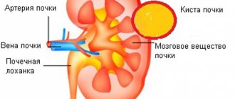
Ultrasound-guided diagnosis of cystic kidney disease
The technique of puncture of cystic formations of the kidneys under ultrasound control includes the following steps:
- treatment of the surgical field;
- selection of entry site, direction and depth of puncture;
- local anesthesia along the puncture channel;
- dissection of dense layers of skin with a pointed scalpel;
- puncture of the cyst with a puncture needle and mandrel;
- taking the contents of the cyst cavity for biochemical, cytological and bacteriological studies;
- performing x-ray cystography and its interpretation.
The diagnostic program for cyst puncture consists of a visual assessment of the irritated fluid, its cytological, biochemical and bacteriological examination, radiopaque cystography and its interpretation.
After completion of the diagnostic puncture program with visualization under the control of an ultrasound device, the therapeutic effect begins, the need and volume of which depend on the nature of the information obtained during the diagnostic puncture, the size and topical location of the cystic formation.
When puncturing cystic formations of the kidneys, the following groups of therapeutic effects are distinguished: aspiration of the contents of the cyst without sclerosis; aspiration of cyst contents and administration of sclerosing agents; drainage of the cyst cavity followed by step-by-step sclerosis; endoscopic dissection of the walls of the cystic formation.
Percutaneous puncture drainage in the diagnosis and treatment of obstructive and purulent-destructive diseases of the kidneys and perinephric tissue.
The main types of percutaneous puncture nephrostomy: improved Goodwin method, percutaneous puncture neurostomy using the Seldinger technique, combined technique.
Currently, the Seldinger method is most widely used.
Indications for puncture drainage are: diagnosis of disorders of the urodynamics of the UMP; obstructive purulent-inflammatory diseases of the upper respiratory tract; creating conditions for subsequent endorenal intervention, DLT or CLT; urinary diversion during OGTN. caused by supravesical retention; drainage of retroperitoneal purulent-destructive formations.
Complications of percutaneous puncture drainage under ultrasound guidance: traumatic injuries manifesting in the form of gross hematuria, hematoma or urinary leakage, inflammatory complications, exacerbation of chronic renal failure, discharge of nephrostomy drainage.
Invasive methods in the diagnosis and treatment of prostate and seminal vesicle diseases under ultrasound control
The location of the prostate is ideal for TRUS. However, there are difficulties in interpreting ultrasound data and in differential diagnosis of various prostate diseases. For example, a confident morphological diagnosis of prostate cancer can only be made on the basis of TRUS-guided prostate biopsy.
Ultrasound-guided therapeutic invasive interventions include puncture drainage of purulent-inflammatory formations of the prostate and seminal vesicles.
Prostate biopsy can be performed either perineally or transrectally. The latter method is currently the most widespread.
After obtaining an image using a transrectal sensor, the directions and areas for sampling are selected (at least 6-8 if prostate cancer is suspected). During perineal puncture, local anesthesia is applied to the skin and subcutaneous tissue in the direction of the puncture; for transrectal biopsy, local anesthesia is not required.
Then, under ultrasound control, a needle is brought to the border of the desired area to take a sample. All samples taken are carefully labeled in separate vials of fixative solution.
In case of purulent-destructive formations, invasive ultrasound intervention, from exclusively diagnostic, turns into therapeutic. This is done in various ways: aspiration of the contents using a puncture needle and the introduction of antibacterial agents; drainage followed by sanitation.
The technique of puncture drainage of seminal vesicles is used for obstructive vesiculitis. Despite the palliative nature of vesiculostomy, it can be used as a means of sanitizing the seminal vesicles before transurethral electroresection of the prostate for various prostate diseases with concomitant vesiculitis.
Sources:
1. Urology. National leadership. Edited by acad. RAMS Lopatkina N.A.
2. Presentation materials from Esaote and CIVCO
After the biopsy
After the material for histological examination has been obtained and the procedure has been completed, the patient is recommended to rest in bed for 8-10 hours. At this time, a urine test is prescribed to detect red blood cells, and blood pressure levels are monitored.
There may be minor pain at the puncture site, which in most cases does not require the use of analgesics. If there are no complications, the patient can leave the hospital the next day.
It is recommended to limit physical activity for several days after the biopsy. Heavy work and heavy lifting are prohibited for 2 weeks.
Complications after kidney biopsy
If the following complications occur after the procedure, you should immediately consult a doctor:
- Unable to urinate.
- Frequent or uncontrollable urination.
- Burning sensation when urinating.
- Urine is dark red or brown. Pink or slightly cloudy urine is normal for 24 hours after the procedure
- Blood or pus from the biopsy site (through the bandage).
- Increasing pain at the puncture site.
- Temperature increase.
- Feeling weak or dizzy.
- Pneumotrax. If the puncture is performed incorrectly, the needle may enter the pleural cavity and cause air to accumulate in it. This complication requires special treatment.
Kidney biopsy does not always give accurate results. Therefore, additional blood and urine tests, MRI, CT, ultrasound or x-ray of the kidney may be needed. A kidney biopsy may also give a false negative result.
Complications
A typical complication of kidney biopsy is microhematuria within 1-2 days after the procedure, which is observed in 20-30% of cases. Short-term gross hematuria is observed in 5-7% of cases.
Cases of the following severe complications were also recorded:
- Kidney infarction with prolonged hematuria and the development of bladder tamponade due to blood clots.
- Perirenal hematoma, which appears when bleeding develops in the kidney under its capsule.
- Post-biopsy paranephritis is infection of the resulting hematoma.
If the kidney biopsy technique is violated, injuries to nearby organs (spleen, liver) and damage to large vessels, for example, the inferior vena cava, may also occur. Such complications are extremely rare.
3. Symptoms, diagnosis
The most characteristic symptom of a kidney injury is intense specific pain, often radiating to adjacent areas (groin organs, intestines, chest) and caused by stretching of the renal capsule, acute local deficiency of blood supply (ischemia), mechanical pressure from the accumulated volume of blood, blocking the lumen of the ureter by coagulated blood clots . The purplish-bluish coloration of the skin in the lumbar region, edema, and swelling observed during external examination are not always found, and the absence of such visible signs does not exclude the presence of internal perinephric hemorrhage. A more characteristic symptom, which is also one of the main criteria for the severity of injury, is hematuria, i.e. the presence of blood in the urine (especially in the form of worm-shaped clots).
In clinical and diagnostic terms, the dynamics of the general condition is very important: in the first 1-2 days, symptoms can either gradually reduce or worsen into a life-threatening status with signs of sepsis and/or severe renal failure, unbearable pain, progressive hematuria, and shock.
In addition to collecting anamnestic information and complaints, examination and palpation (there are a number of diagnostically significant reactions), if a kidney hematoma is suspected, various methods of “examination from the inside” are used: cystoscopy, pyelography, angiography and other endoscopic, radiographic, ultrasound and tomographic studies, the choice of which is determined specific clinical situation.
About our clinic Chistye Prudy metro station Medintercom page!