What does blood in urine indicate during pregnancy? Having discovered bloody discharge after urination, you should not try to independently look for the cause of this phenomenon and take any therapeutic measures. Only specialized specialists can give an accurate answer to this question based on an examination of the expectant mother and the results of her tests. To prevent the appearance of blood when urinating, it is important to follow preventive measures, namely not to overcool, eat as little fatty, peppery foods as possible, and most importantly - maintain and strengthen the immune system, maintain genital hygiene.
Causes of blood in the urine of women during pregnancy
In medicine, it is customary to call the presence of blood in urine hematuria, which is a sign of various pathological changes in the body or indicates a malfunction of internal organs. The change in the color of urine in a pregnant woman depends on various factors, and the foods eaten can also affect it. Brown, pinkish or purplish-red discharge is observed.
Harmless
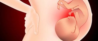
Hematuria in pregnant women does not always indicate serious illness; in most cases it is physiological in nature, which is not at all dangerous to the health of the expectant mother and her unborn baby. Blood appears after urination during pregnancy due to the pressure exerted by the fetus or the growing uterus on the bladder. In addition, pregnant women may experience red blood cells in urine when blood vessels rupture due to high intra-abdominal pressure and due to hormonal changes in the body that occur with the onset of pregnancy. However, the presence of blood in urine is not always safe and in some cases can hide dangerous pathologies. Only specialized doctors can determine the severity of hematuria during pregnancy, so the first thing you should do is go to a medical facility.
Hazardous to health
Bloody spots in the urine during pregnancy may indicate the following diseases:
- Improper functioning of the organs of the genitourinary system, caused by inflammation, infectious lesions in the genitourinary tract. Often during pregnancy, cystitis worsens, which is usually accompanied by frequent urination and severe pain. The appearance of blood after urination in pregnant women is also possible with glomerulonephritis, pyelonephritis, and sexually transmitted diseases.
- Urolithiasis, which is characterized by damage to the walls of the bladder with urinary fluid and the ureters due to the movement of sand and stones. As a result, blood enters the urine and its presence can be detected when urinating. The progression of stone formation is accompanied by sharp pain, and the urine takes on a pinkish or red tint.
- Pathologies in the kidneys and blood vessels requiring surgical intervention. In this case, we mean anatomical features that should be corrected surgically.
- Bladder cancer, in which blood clots are found in the urine, changing its color. This pathology is not accompanied by pain and the reason for its appearance is damage to the walls of blood vessels by malignant neoplasms.
Hematuria during pregnancy occurs with diabetes mellitus, anemia, trauma and tuberculosis of the skin with ulcers and nodules. The presence of blood in the urine of women in a position with premature birth, disturbances in the development of the intrauterine fetus, weak labor and complications after childbirth is dangerous. In this regard, at the first signs of discomfort, a pregnant woman should go for a consultation with a therapist or nephrologist.
M.M. Batyushin, D.G. Pasechnik State Educational Institution of Higher Professional Education Rostov State Medical University of the Russian Health Service, Rostov-on-Don
The concept of Hematuria is the leading symptom in the clinic of pathology of the kidneys and urinary tract. Hematuria refers to the appearance of blood cells (namely red blood cells) or their components (hemoglobin) in the urine. In this regard, the concept of hematuria includes erythrocyturia, hemoglobinuria and hemoglobin cylindruria (excretion of hemoglobin casts in the urine). Normally, during a general analysis, there are either no red blood cells in the urine, or their number when counted in Goryaev’s chamber does not exceed 1–2 elements in the field of view. Sometimes a higher number of red blood cells may be normal. This may be due, for example, to intense physical activity. Such episodes are short-lived. The appearance of 3 or more red blood cells in the field of view is considered erythrocyturia. There is no clear definition of persistent erythrocyturia, however, an increase in the number of red blood cells in the urine and persistence according to the results of three or more tests can be conditionally considered persistent erythrocyturia. In the case of erythrocyte adhesion and hemoglobin precipitation in the lumen of the renal tubules, the formation of erythrocyte and hemoglobin casts, which have a characteristic appearance and are stained red with hematoxylin, is possible. As part of the definition of hematuria, micro- and macrohematuria are distinguished. Microhematuria (more correctly, microerythrocyturia) refers to erythrocyturia that does not lead to a change in the color of urine, assessed by eye. With gross hematuria, the urine becomes red (from light pink to cherry). It may be diffusely colored or have blood clots. Thus, the concept of micro- or macrogematria is a qualitative symptom assessed visually. Previously, the phenomenon of “leached erythrocytes” was widely used in the differential diagnosis of erythrocyturia. It is now generally accepted that red blood cell leaching is related to the physicochemical properties of urine. Often, with undoubted renal origin of hematuria, fresh red blood cells are found; on the other hand, gradual leaching of red blood cells can occur when urine is stored for several hours before examination. In this regard, the phenomenon of “leached erythrocytes” should not be leading in differential diagnosis and retains a certain significance only when using phase-contrast microscopy. Hemoglobinuria develops due to the excretion of free extra-erythrocyte hemoglobin in the urine through glomerular filtration. Due to the oxidation of hemoglobin and its conversion into hemosiderin, which is a black pigment, urine with hemoglobinuria and hemosiderinuria may acquire a dark cherry color.
Prevalence The prevalence of erythrocyturia in the population ranges from 0.18 to 16.1%. According to our data obtained during a one-time study of 1446 patients in somatic hospitals in Rostov-on-Don (the study did not include patients from the departments of nephrology and urology), erythrocyturia was detected in 3.7% of cases. When examining young soldiers, P. Froom et al. found erythrocyturia in 39% of cases. However, it was transient in nature, since during subsequent examinations it occurred with a frequency not exceeding 16%. In postmenopausal women, erythrocyturia occurs with a frequency of 13%. In screening studies in pediatric populations, hematuria occurs with a frequency of 1–4% and increases with age, reaching 12–18% in adolescence.
Causes of hematuria and pseudohematuria Pseudogematuria Not every phenomenon of red urine is hematuria. Redness of urine not accompanied by erythrocyturia and/or hemoglobinuria is called pseudohematuria. Pseudohematuria can be caused by taking a number of medications, food nutrients, and chemicals that color it red (Table 1).
Table 1. Causes of pseudohematuria
| Urine color | Cause |
| Red | Antipyrine, amidopyrine, santonin |
| Pink | Acetylsalicylic acid in large doses, carrots, beets |
| Brown | Phenol, cresol, bears ears, activated carbon (carbolene), urates, porphyrins |
| Dark brown | Salol, naphthol |
In addition, it is possible to verify hematuria that does not actually exist due to a laboratory error or deliberate distortion of the result of a urine test, but the latter is extremely rare. This phenomenon should also be interpreted as pseudohematuria.
Hematuria of extraurinal origin If pseudohematuria is excluded, the next step in the differential diagnosis should be the exclusion of hematuria of extraurinal origin. In this case, we are talking about blood getting into the urine not from the kidney and urinary tract, but from other organs, as well as from the outside. The following types of hematuria of extraurinal origin include:
simulated hematuria:
- blood is added to the urine after urination from a wound inflicted by the simulator on a finger, lip, scrotum, etc., as well as the blood of another person;
- blood is introduced into the bladder through a catheter before urination;
- a foreign object injures the mucous membrane of the urethra before urination;
- the malingerer’s urine is mixed with the urine of a patient suffering from kidney pathology, manifested by hematuria.
hematuria of genital origin:
- taking a urine test during menstruation, as well as the day before or within 3-4 days after its end;
- bleeding from tumors of the uterus, vagina, atrophic colpitis;
- formation of vesico-uterine anastomosis (tumor, traumatic);
- preservation of menstruation with desquamation of the functional layer of the mucous membrane of the cervix during supravaginal amputation of the uterus;
- hematuria in pregnant women;
- postcoital hematuria.
hematuria of rectal origin:
- bleeding from a hemorrhoid;
- bleeding from anal fissure;
- rectal cancer;
- chronic proctosigmoiditis with the opening of a fistula in the perianal region, vesico-rectal anastomosis.
hematuria of perineal origin:
- boil, perineal carbuncle;
- perineal injuries.
During screening examinations of pregnant women, hematuria is observed in 20% of cases. Moreover, in 53% of cases it is detected before the 32nd week of pregnancy. Its appearance is a risk factor for the development of complications during childbirth and is usually due to obstetric reasons (threat of miscarriage, premature birth, premature abruption of the low-lying placenta, etc.). Postcoital hematuria is characterized by the appearance of erythrocyturia in a portion of urine given immediately after sexual intercourse. We observed 10 patients who had erythrocyturia after coitus. In 4 cases, erythrocyturia appeared after the introduction of foreign bodies into the urethra, and in one case gross hematuria was observed. It can be assumed that this cause of hematuria is not rare, but due to its delicacy it is rarely considered when carrying out differential diagnosis. In 6 cases, the cause of erythrocyturia was prostate disease and the patients were referred for examination and treatment to a urologist. As a rule, postcoital hematuria is combined with hemospermia. Isolated hemospermia and isolated postcoital hematuria are possible. The latter is an extremely rare option. Variants of isolated postcoital hematuria caused by a urethral polyp are described. Combined postcoital hematuria and hemospermia in young men in most cases is caused by the presence of benign papillary adenoma, in older men - prostate cancer. Isolated hemospermia in 40% of cases is caused by prostate infection; a malignant tumor also occurs; in some cases, the cause cannot be determined. Simulating pseudohematuria is not widespread due to the lack of difficulties in most cases in differentiating it from true hematuria. Their appearance is due to the motivation of malingerers and aggravators to enhance or direct the clinical manifestations of the disease in order to achieve some social benefits. Understanding the motive for malingering or aggravation is key to a successful differential diagnosis. Most often, elements of aggravation or simulation are observed in expert practice when conducting military medical, medical, social or forensic examinations.
True erythrocyturia There are many reasons for the development of erythrocyturia. There are no less attempts to classify this phenomenon. The appearance of red blood cells in the urine may indicate the development of pathology of the kidneys and urinary organs, as well as the genital organs. In 65% of patients, erythrocyturia is of extrarenal origin. The pathogenesis of renal anemia is poorly understood. A possible cause is the formation of damage in the basement membrane of the glomeruli, the so-called breaks or gaps. When the size of the defects exceeds 0.25 microns, red blood cells move through the membrane into the urinary space through them under the influence of intracapillary pressure and retraction forces. In this case, severe deformation and damage to the membranes of erythrocytes occurs, which explains the dysmorphism of these cells in the glomerular type of hematuria. Among the causes of hematuria, infectious, traumatic, autoimmune, toxic, tumor and mixed are distinguished. The classification of R.Cohen and R.Brown (2003) seems to us to be the most acceptable. When hematuria is detected, the diagnostic search is aimed at identifying these diseases. Noteworthy is the fact that hematuria is only one of the symptoms of the disease. The appearance of other symptoms often accompanies hematuria and forces the doctor to analyze their relationship and diagnostic significance. The classification of hematuria, distinguishing symptomatic and asymptomatic forms, is very conditional and is of an exclusively applied nature. Asymptomatic hematuria occurs in the adult population with a frequency of 0.19–21%. In approximately 10–12.1% of cases, it is caused by cancer of the kidneys and urinary tract. In this case, macrohematuria occurs in 3/4 of cases. As literature data show, in 39–90% of cases, the detection of asymptomatic hematuria is not subsequently accompanied by a diagnostic search. According to our data, only in 8% of cases the detection of asymptomatic hematuria led to further examination of patients (n=1446). In childhood, asymptomatic hematuria is most often caused by thin membrane disease (27.5% of all cases of asymptomatic hematuria) and IgA nephropathy (26.2% of all cases of asymptomatic hematuria). Of the glomerulopathies, the most common causes of gross hematuria in children are IgA nephropathy (54.2%) and Alport syndrome (25%). Among non-glomerular causes of hematuria, the most common are hypercalciuria (16%), urethrorrhagia (14.3%), and hemorrhagic cystitis (12.5%). An unidentified cause appears in 46% of cases.
Let's look at some of the causes of hematuria. 1. Urolithiasis Urolithiasis is the cause of 20% of all cases of hematuria. Hematuria with it is intermittent or persistent in nature and can be represented by both micro- and macrohematuria. In case of urolithiasis, Pasternatsky syndrome is described in the form of the appearance of erythrocyturia after walking or tapping the lumbar region. More often, hematuria is combined with pain in the lumbar region or flanks. The cause of hematuria is trauma to the mucous membrane of the urinary tract.
2. Tumors of the kidney and urinary tract Kidney tumors account for about 3% of all human tumors. 85–90% of all cases of oncological processes in the kidney are renal cell carcinoma, which develops from the epithelium of the proximal tubule. Of the benign neoplasms, angiomyolipoma, adenoma and oncocytoma predominate (6–8%). In 40–70% of cases, the disease is asymptomatic. In 5% of cases, the cause of microhematuria is cancer of the kidney and urinary tract. The risk of the latter increases with the patient's age, as well as in the presence of risk factors such as smoking, use of phenacetin, herbs containing aristolochic acid, and cyclophosphamide in high doses. Hematuria is a typical symptom of a tumor. Both micro- and macrohematuria, isolated or less often in combination with low proteinuria, can be observed. With gross hematuria, blood clots are often observed in the urine. Hematuria may be accompanied by pain (pain in the flank, hypochondrium, lumbar region, along the ureter), and may be asymptomatic. With kidney cancer in men, a varicocele may be observed, caused by compression of the testicular vein by the tumor. Paraneoplastic syndrome may develop, observed in 35% of patients. With prostate stromal tumors, hematuria occurs in approximately 14% of cases. The disease develops mainly in older men. The clinical picture of prostate cancer is caused by tumor growth and bladder outlet obstruction and is manifested by micro- and macrohematuria, chronic ischuria, stranguria, pain in the perineum and suprapubic region. Tumors of other locations are also possible: bladder, ureter, urethra.
3. Glomerulonephritis Hematuria is one of the clinical manifestations of acute, chronic and rapidly progressive glomerulonephritis. It is part of the nephritic syndrome. Acute post-streptococcal glomerulonephritis always manifests itself as isolated hematuria or hematuria in combination with proteinuria; urinary syndrome in chronic and rapidly progressive glomerulonephritis can occur without hematuria (proteinuric variant, nephrotic syndrome). Hematuria occurs in various morphological variants of nephritis, less often in minimal change disease and membranous glomerulonephritis. Hematuria with glomerulonephritis is usually preserved if other causes of hematuria are excluded, as well as in the presence of criteria for nephritic syndrome (hypertension, overhydration). Hematuria is observed both in idiopathic variants of glomerulonephritis and in its development as part of systemic connective tissue diseases (collagenosis, systemic vasculitis, arthropathy).
4. Connective tissue diseases Kidney damage in systemic vasculitis is varied and is represented by renal angiitis, interstitial nephritis and fibrosis with or without areas of necrosis, and various types of glomerulonephritis. The appearance of erythrocyturia in systemic vasculitis is evidence of the involvement of the kidneys or urinary tract in the pathological process. Differential diagnosis is usually aimed at excluding vasculitis, guided by criteria developed and widely used in rheumatological practice. In addition to systemic vasculitis, systemic lupus erythematosus, arthropathy such as gout, rheumatoid arthritis, and ankylosing spondylitis are often the causes of hematuria (Fig. 6).
5. Polycystic kidney lesions Polycystic kidney lesions may be accompanied by hematuria. It is usually asymptomatic. Clinical manifestations appear in the case of the development of infectious complications, as well as chronic renal failure.
6. Tubulointerstitial nephritis Interstitial nephritis of toxic origin Develops due to the effects of exo- and endotoxins on renal tissue. This group of kidney damage is diverse and includes exposure to industrial substances and household chemicals, medicinal and other substances.
Drug-induced interstitial nephritis The problem of drug-induced kidney damage is one of the pressing problems of modern nephrology. Approximately 6–60% of all cases of acute renal failure (ARF) are due to interstitial nephritis, as determined by nephrobiopsy. In half of the cases, the etiology of acute interstitial nephritis is drugs. Interstitial nephritis most often develops in response to antibiotics and nonsteroidal anti-inflammatory drugs (NSAIDs). NSAIDs are the cause of 44% of cases of acute interstitial nephritis, antibiotics – 33% of cases. Cases of interstitial nephritis have also been described during therapy with warfarin, thiazide diuretics, indapamide, mesalazine, ranitidine, and cimetidine. Erythrocyturia is present in 87–100% of cases when examining urinary sediment in acute drug damage and in 43–56% of cases in chronic cases.
Other drug-induced damage to the kidneys and urinary tract, accompanied by erythrocyturia
- Papillary necrosis - can be caused by NSAIDs, aspirin, analgin.
- Hemorrhagic cystitis - caused by cyclophosphamide, ifosfamide, mitotane.
- Urolithiasis - may occur when taking carbonic anhydrase inhibitors, dichlorphenamide, indinavir, mirtazapine, ritonavir, triamterene.
- Tumors of the urinary tract - can occur during therapy with cyclophosphamide, analgesics (phenacetin).
- Drug-induced vasculitis.
Interstitial nephritis due to herbal medicine Nephropathy due to the intake of Chinese herbs is known under the term “chinese herb nephropathy”. It is characterized by rapid progression of chronic renal failure (CRF) and is manifested morphologically by extensive interstitial fibrosis without glomerular damage. Occurs in women taking Chinese herbal supplements. Nephrotoxicity is determined by the presence of aristolochic acid in herbs. A cumulative dose of Aristolochia fangchi extract from the site of Stephania tetrandra has been shown to result in the development of CRF in 30.8% of cases. Erythrocyturia is one of the manifestations of the disease.
7. Congenital anomalies of the urinary tract Most often, hematuria is observed with nephroptosis, ureteral stricture, compression of the ureteropelvic segment or group of calyces by an aberrant renal artery, as well as with complete and incomplete duplication of the kidney, renal venous hypertension. The main cause of hematuria in patients with congenital anomalies is an increase in intrapelvic urine pressure due to compression and impaired urine outflow from the pelvis. As a result, microtraumatization of the mucous membrane of the pelvis develops with the development of microhematuria. With venous hypertension, venule ruptures with the development of microbleeding are observed. Typically, hematuria is recurrent and combined with renal colic or dull and weak flank and lumbar pain.
8. Infectious diseases Pyelonephritis, cystitis, prostatitis In some cases, erythrocyturia can be observed with pyelonephritis. It always occurs against the background of leukocyturia and is often caused by an unfavorable background in the form of a congenital anomaly of the urinary tract, urolithiasis, etc. With cystitis, erythrocyturia may be the only laboratory phenomenon. Prostatitis often leads to a combination of erythrocyturia and leukocyturia. In 10% of cases, isolated microhematuria is possible.
Septic nephropathy With sepsis, according to various sources, septic nephropathy develops in 10–45% of cases. It can be represented by septic acute glomerulonephritis, chronic glomerulonephritis, acute interstitial nephritis, tubular or cortical necrosis, renal carbuncle, apostematous nephritis and pyelonephritis, thrombosis of the renal arteries and veins. A wide range of possible injuries dictates the need for detailed differential diagnosis.
Tuberculosis With tuberculosis of the genitourinary system, epididymitis (58.1%) and kidney damage (16.3%) most often develop, less often than other organs: orchitis (9.3%), cystitis (7%), urethritis (4.7%) , damage to the prostate (2.3%), testicular membranes (2.3%). Tuberculosis of the kidney and urinary tract is manifested by erythrocyturia. Moreover, ultrasound or urographic imaging often reveals gross structural changes in these organs. Often a combination of tuberculosis of the kidneys and lungs.
Schistosomiasis An infection caused by a schistosome, most often Schistosoma haematobium. Distributed in the countries of Western (Benin, Burkina Faso, Gambia, Mali, Nigeria, Chad), Southern (Kenya, Madagascar, Mozambique, Tanzania, Zambia) and Northern (Egypt, Ethiopia, Sudan) Africa, also found among tourists who visited these countries . Less commonly, infection is caused by S. mansoni, S. japonicum, S. intercalatum and S. mekangi. Fever (44%), diarrhea (40%), itching (25%), chills (21%), and hematuria (20%) develop. Hypereosinophilia is observed in 82% of cases, increased liver enzymes – in 82%. Schistosomes are found in urine and stool in 60% of cases. With schistosomiasis, damage to the bladder is observed. Ultrasound examination reveals a change in the contours of the bladder wall, wall thickening of more than 5 mm, and visualization of a shadow in the form of a pseudopolyp. In this case, hematuria is a mandatory phenomenon.
Leptospirosis A zoonotic infection caused by Leptospira icterohaemorrhagia (50% of cases), L. canicola (41.4%), L. pomona (2.3%), L. grippotyphosa (5.5%), L. tarassovi (0.8% ). The most common sources of infection are rodents and dogs. Kidney damage in the anicteric form of leptospirosis is observed in 36.2% of cases and is represented by lower back pain (17.1%), a slight decrease in diuresis (21.3%), an increase in creatinine (12.8%), changes in urine tests (erythrocyturia , leukocyturia, low proteinuria). Morphological examination reveals interstitial nephritis. Jaundice forms are more severe and in 66.7% of cases are accompanied by the development of acute renal failure. Kidney damage in the icteric form is observed in 92.6% of cases. In this case, the clinical manifestations of kidney damage are most pronounced on the 5th–6th day of the disease.
Virus-associated nephropathies Kidney damage has been described in infections caused by hepatitis A, B, C viruses, HIV, parvovirus B 19, cytomegalovirus, Coxsackie virus B, Epstein-Barr virus, polyoma virus, hantavirus, adenovirus. Parvovirus infection is associated with focal segmental glomerulosclerosis, Coxsackie virus with IgA nephropathy, polyoma and hantaviruses with interstitial nephritis, hepatitis C virus with glomerulonephritis as part of cryoglobulinemic vasculitis.
9. Hematuria due to venous hypertension Nutcracker syndrome Nutcracker syndrome develops due to compression of the left renal vein between the aorta and the superior mesenteric artery [29]. The clinical manifestation of Nutcracker syndrome is hematuria, which develops as a result of venous intrarenal hypertension. According to WHO, varicocele occurs in 36% of men. Moreover, in 43% of cases of varicocele, hypertension is detected in the left renal vein [8]. The causes of varicocele development are aortomesenteric, less often retroaortic compression of the left renal vein, stenosis of the left renal vein. Changes in the kidney with venous hypertension are referred to as phleborenohypertensive nephropathy.
Montenbaker's hematuria ( Mountainbiker ), or physical effort hematuria Physical effort hematuria can be of renal and cystic origin. Most often develops after running. Cases of its development with minimal physical activity have been described.
10. Kidney infarction The main cause of kidney infarction is atherosclerosis of the aorta. The rupture of an unstable plaque is accompanied by the release of nuclear fragments rich in cholesterol into the blood, with the development of cholesterol embolism of the renal vessels. The absence of signs of cardiac pathology during renal infarction is confirmed by detailed examination in approximately 59% of cases. In this case, we are talking about idiopathic renal infarction. However, aortic atherosclerosis can occur without clinically manifested coronary atherosclerosis. Risk factors for cholesterol embolism are acute and chronic forms of coronary heart disease, including myocardial infarction, as well as atrial fibrillation of various origins, old age, diabetes mellitus, and a history of cerebral stroke.
11. Injuries to the kidney and urinary tract Traumatic injuries to the kidney and urinary tract are observed with impacts and compression of the lumbar region and pelvis. Micro- or macrohematuria is observed. Bleeding from the upper urinary tract can lead to the development of bladder tamponade. The appearance of blood clots is characteristic. Traumatic hematuria also develops during catheterization of the bladder, cystoscopy, catheterization of the ureter, urological operations, and kidney biopsy. In the latter case, microhematuria is observed the next day in 70% of patients, macrohematuria – in 3–5% of patients.
12. Coagulopathy Acquired coagulopathy is caused by the use of warfarin and direct anticoagulants. This occurs in approximately 3–15% of cases and usually resolves after reducing the drug dose. Renal bleeding is less common (0.5–3% of cases). The appearance of hematuria in DIC syndrome in the hypocoagulation phase is explained by the development of microinfarctions in the renal parenchyma and the capillary type of bleeding. Among congenital coagulopathies, hemophilia (A, B, C) and von Willebrandt's disease lead to micro- and macrohematuria. Administration of cryoprecipitate or concentrate of factor 8, 9 or 11 or fresh frozen plasma stops renal bleeding in hemophilia.
13. Rare causes of hematuria Tuberous sclerosis Tuberous sclerosis is a dominantly inherited disease with multifocal damage to the body. Hamartiasis (hamartomas) develops, affecting the skin, heart, kidneys, eyes, and brain. In approximately 80% of cases, a mutation of the TSC1 or TSC2 gene is observed. Kidney damage in tuberous sclerosis occurs in the form of angiomyolipoma, cysts or renal cell carcinoma). Renal manifestations are asymptomatic hematuria, less often - pain in the lumbar region, renal bleeding.
Sarcoidosis and kidney damage Most often occurs between the ages of 20 and 40 years. Frequency: 1 case per 2,500–10,000 people. Kidney damage is observed in 1% of cases, but during autopsy it increases to 20%. Three types of kidney damage have been described: nephrocalcinosis and nephrolithiasis, glomerulonephritis and granulomatous lesions (interstitial nephritis, destruction of renal parenchyma).
Psoriasis Kidney damage in psoriasis is rare. It is manifested by tubular dysfunction (47.5%), oxaluria (45%), uraturia (6%), leukocyturia (12%), according to some data, 50% of patients have a decrease in nitrogen excretory function of the kidneys (N.N. Panasyuk, 1988 ). Erythrocyturia is a common manifestation of psoriatic nephropathy. In the case of nephritis, IgA nephropathy is most often recorded.
14. Hemoglobinuria Hemoglobinuria is observed in hemolytic anemia in the case of the development of intravascular hemolysis. Acute intravascular hemolysis develops with the toxic effects of hemolytic poisons (viper venom, acetic acid, etc.), infusions of hypertonic or hypotonic solutions, sepsis, trauma (crash syndrome). In the first hours, the urine is red or pink, and later becomes brownish or black (hemosiderinuria). Under microscopy, erythrocyturia sediment is usually not observed. There may be cases of paroxysmal hemoglobinuria - paroxysmal cold hemoglobinuria and paroxysmal nocturnal hemoglobinuria (Marchiafava-Micheli disease). Thus, a wide range of pathological conditions that can cause hematuria requires a detailed diagnostic search with the involvement of urological and nephrological specialists. The most common mistakes when examining hematuria are the following.
- Lack of search for the causes of hematuria (in 92% of cases - own data);
- Incorrect interpretation of the cause of hematuria (chronic pyelonephritis, lack of interpretation);
- Refusal of endoscopic examination (cystoscopy, ureteroscopy);
- Refusal to perform renal nephrobiopsy with a probable diagnosis of chronic glomerulonephritis (12% of refusals – own data).
Conducting a narrow screening search If it is impossible to further examine a patient with hematuria in the medical institution where you work, the patient must be referred to a specialist with a wide range of diagnostic capabilities.
Conclusion In the process of managing a patient with the phenomenon of hematuria, it is necessary to use a wide range of studies, but their choice must be clearly related to the diagnostic hypothesis. Despite the great diagnostic capabilities of modern medicine, in approximately 5–9% of cases the cause of hematuria remains unclear. Often, long-term observation of such patients is not accompanied by verification of the diagnosis. The variety of diseases manifested by hematuria and the clinical features of the course and development of these pathological processes dictate the need for an integrated approach based on solid knowledge in the field of therapy, urology, oncology, gynecology, infectious pathology and toxicology. The wide occurrence of hematuria in outpatient therapeutic practice determines the relevance of the presented topic.
Literature 1. Alekseeva E.A., Antonova T.V. Kidney damage in anicteric and icteric forms of leptospirosis. Nephrology. 2002; 4:74–8. 2. Alyaev Yu., Krapivin A. Choice of diagnostic and therapeutic tactics for kidney tumors. Tver: Triad. 2005. 3. Berman R.E., Vaughan V.K. Pediatrics (director), book. 5. 1993; 314. 4. Eliseeva L.I., Varennikova E.I., Kurinnaya V.P., Shcherbinina I.G. Acute tubulointerstitial nephritis: diagnostic problems. Nephrol. and dialysis. 2002; 4 (2). 5. Mukhin N.A. (ed.) National Guidelines for Nephrology. M.: GEOTAR-Media, 2009. 6. Panasyuk N.N. Kidney damage in psoriasis. Ter. arch. 1988; 6:130–4. 7. Strakhov S.N., Spiridonov A.A., Prodeus P.P. and others. Changes in the renal and testicular veins in left-sided varicocele and the choice of surgical method in children and adolescents. Urol. and nephrol. 1998; 4:13–8. 8. Strakhov S.N., Burkov I.V., Spiridonov A.A. Nephropathy of phlebohypertensive origin and the choice of treatment method for varicocele in children and adolescents. Nephrol. and dialysis. 2001; 3 (4). 9. Tareeva I.E., Nikolaev A.Yu., Androsova S.O. Nephrology. 1995; 3. 10. Tareeva I.E., Androsova S.O. The effect of non-narcotic analgesics and NSAIDs on the kidneys. Ter. arch. 1999; 4: 17–22. 11. Abdel-Wahab MF, Ramzy I, Esmat G et al. Schistosoma haematobium infection in Egyptian schoolchildren: demonstration of both hepatic and urinary tract morbidity by ultrasonography. J Urol 1992; 148: 346–50. 12. Agbessi CA, Bourvis N, Fromentin M et al. La bilharziose d'importation chez les voyageurs: enquête en France métropolitaine. Rev Med Interne 2006; Jun. 13. 13. Albersen M, Mortelmans LJ, Baert JA. Mountainbiker's hematuria: a case report. Eur J Emerg Med 2006; 13: 236–7. 14. Aliabadi H, Cass AS, Gleich P. Utricular papilloma. Urology 1987; 29: 317–8. 15. Anagnostopoulos GK, Doriforou O, Sakorafas G, Missas S. Tuberous sclerosis associated with giant bilateral bleeding angiomyolipomas. Postgraduate Med J 2004; 80: 580. 16. Belfer MH, Stevens RW. Sarcoidosis: a primary care review. Am Fam Phys 1998. 17. Bolderman R, Oyen R, Verrijcken A et al. Idiopathic renal infarction. Am J Med 2006; 119 (4): 356. 18. Brown MA, Holt JL, Mangos GJ et al. 'Microscopic hematuria in pregnancy: Relevance to pregnancy outcome. Am J Kidney Dis 2005; 45 (4): 667–73. 19. Buysen JG, Houthoff HJ, Krediet RT, Arisz L. Acute Interstitial Nephritis: A Clinical and Morphological Study in 27 Patients. Nephr Dial Transpl 1990; 5:94–9. 20. Cameron JJ. Lupus nephritis. Am Soc Nephr 1999; 10: 413–34. 21. Cohen RA, Brown RS. Microscopic Hematuria. New Engl J Med 2003; 348:2330–8. 22. Collar JE, Ladva S, Cairns ThDH, Cattell V. Red cell traverse through thin glomerular basement membranes. Kidney Internat 2001; 59:2069–72. 23. Davison AM, Jones CH. Acute interstitial nephritis in the elderly: a report from the UK MRC glomerulonephritis register and a review of the literature. Nephr Dial Transpl 1998; 13(Suppl. 7): 12–6. 24. Edwards TJ, Dickinson AJ, Natale S et al. A prospective analysis of the diagnostic yield resulting from the attendance of 4020 patients at a protocol-driven haematuria clinic. BJU Int 2006; 97(2):301–5. 25. Enriquez R, Cabezuelo JB, Gonzalez C et al. Granulomatous interstitial nephritis associated with Hydroclorotyazide/Amiloride. Am J Nephrol 1995; 15:270–3. 26. Farrington K, Levison DA, Greenwood RN et al. Renal biopsy in patients with unexplained renal impairment and njrmal kidney size. QJ Med 1989; 70: 221–33. 27. Froom P, Ribak J, Benbassat J. Significance of microhaematuria in young adults. Br Med J (Clin. Res. Ed.) 1984; 288:20–2. 28. Gaughan WJ, Sheth VR, Francos GC et al. Ranitidine-induced acute interstitial nephritis with epithelial cell foot process fusion. Am J Kidney Dis 1993; 22: 337–40. 29. Gorospe EC, Aigbe MO. Macroscopic Haematuria in a 15-year old Male: A Case of Nutcracker Syndrome Managed by Endovascular Stenting. Scient World J 2006; 6:745–6. 30. Herawi M, Epstein JI. Specialized stromal tumors of the prostate: a clinicopathologic study of 50 cases. Am J Surg Pathol 2006; 30(6):694–704.
What to do if there is blood in your urine: treatment
Hematuria during pregnancy can be detected in both early and late stages. Regardless of this, when any disease is diagnosed, it will have to be treated. It should be understood that urine in the blood is only a symptom that accompanies the disease. Therefore, therapy will be aimed at eliminating the cause. It will depend on her how and how to treat hematuria. Only an experienced specialist can choose a medicine and prescribe dosages! Self-medication in this case is unacceptable!
However, there are some things you can do to help yourself at home. First of all, it is recommended, starting from the second trimester, to regularly take the knee-elbow position: it not only makes you feel better, improves blood flow in the pelvic organs and placenta, but also “unloads” the kidneys, preventing congestion. In addition, after consulting with your doctor, you can take diuretic fruit drinks and teas: cranberries, lingonberries. Always be attentive to your health and avoid hypothermia in the pelvic area in every possible way. We also must not forget that in any case, a healthy, balanced diet will be beneficial during pregnancy. And if there is a predisposition or risk of developing inflammation in the genitourinary organs, then even more so salty, hot, fried, spicy and synthetic foods should be excluded from the diet.
Otherwise, general recommendations remain relevant: daily walks, moderate physical activity, adequate sleep and rest, positive emotions.
Correction
Conservative therapy
There are no independent methods for correcting hematuria. To eliminate this laboratory phenomenon, treatment of the underlying disease is necessary. If hematuria develops while taking an anticoagulant, it is recommended to reduce the dosage or completely discontinue it. The following medications are used as conservative therapy:
- Antibiotics.
For the treatment of pyelonephritis, first-line drugs are penicillins and cephalosporins. For cystitis, fosfomycin trometamol is prescribed. Fluoroquinolones and macrolides are effective for prostatitis. - Glucocorticosteroids.
Preparations of adrenal hormones (prednisolone) are able to suppress the severity of inflammation in the glomerular apparatus and the autoimmune destruction of platelets during hemorrhagic diathesis. - Cytostatics.
Cytostatic agents (azathioprine, cyclosporine) are used for severe glomerulonephritis, especially for vasculitis and collagenosis. Also, combinations of chemotherapy drugs are prescribed for tumors of the kidney and bladder. - Alpha blockers (tamsulosin).
This group of drugs has a relaxing effect on the walls of the ureters and sphincters of the bladder, which promotes the spontaneous passage of small stones. - Alkaline solutions.
Potassium citrate or sodium bicarbonate are used to dissolve uric acid stones. - Cardiac drugs.
Patients with chronic heart failure and arterial hypertension are prescribed beta-blockers, ACE inhibitors, and potassium-sparing diuretics.
Surgery
Patients with urolithiasis undergo shock wave lithotripsy or laparoscopic stone removal; for polycystic disease, benign tumor, renal infarction, resection, nephrostomy or total nephrectomy are performed. In case of severe bilateral damage to the renal tissue with the development of end-stage renal failure, kidney transplantation can be performed for health reasons.
What should you be wary of?
During pregnancy, the expectant mother should immediately consult a doctor if red blood cells are visible in the urine with the naked eye, the urine becomes incomprehensibly pink, in some cases even brown. Women should be alerted to the appearance of blood clots in the urinary fluid. In addition, if there is blood in the urine of pregnant women, and in addition to it there is also pain in the lower abdomen, in the back, lumbar region or groin, then you should immediately go to the doctor. You should not hesitate to visit a nephrologist if there is a strong burning sensation when emptying through the urethra, body temperature has increased, nausea, vomiting has appeared, and the communication condition has significantly worsened.
Diagnosis of pathology
In particular, to accurately diagnose the problem, a glass urine sample is taken from the patient. This collection of biomaterial involves taking three urine samples - the initial, middle and final doses of the stream. They are collected in different containers. Next, when carrying out the analysis, the differentiation technique is used.
- So, if blood and its clots are found only in the initial stream of urine, then we are talking about the localization of the inflammatory process exclusively in the urethra. It can be caused by mechanical trauma, polyp growth, gonorrhea, syphilis or oncology.
- If blood is found in the middle portion of the urine stream, then the problem most likely lies in the neck of the bladder or in the back of the urethra (urethra).
- If red blood cells are detected in all three samples, it means that the pathology is hidden higher up - in the kidneys, parenchyma or bladder. In this case, the patient undergoes an ultrasound examination to determine the localization of the inflammatory process.
Causes of postrenal hematuria
Urolithiasis disease
The most common cause of hematuria in adults. The mechanism of hematuria in urolithiasis is associated with trauma to the renal pelvis or ureteral wall by the calculus. Blood appears during an attack of renal colic, accompanied by severe pain, nausea, and vomiting. The degree of hematuria depends on the extent of damage.
Microscopy often reveals a large number of different crystals - calcium oxalates, phosphates, ammonium urate crystals, uric acid. Due to the resulting obstruction to the outflow of urine, infection often occurs, as evidenced by the detection of leukocytes and bacteria in the urine. Outside of an attack, there is usually no blood. Hematuria completely stops after surgical removal of stones.
Lower urinary tract diseases
Hematuria with cystitis is quite rare; it is more often observed with polyposis or bladder stones. Hemorrhagic cystitis with severe hematuria is possible - with schistosomiasis (parasitic infestation) of the bladder or as an unfavorable side reaction to long-term use of the cytostatic drug cyclophosphamide.
With a bladder tumor, hematuria is considered a permanent symptom, and in people suffering from inflammation of the prostate gland or urethra (prostatitis, urethritis), it is extremely rare. In this case, hematuria is accompanied by leukocyturia, sometimes bacteriuria.
Features of treatment
The therapeutic course is selected individually and depends on the diagnosis. If blood in the urine during pregnancy is a consequence of cystitis, then antibiotics Ceftriaxone and Norfloxacin are prescribed. Antispasmodics and diuretics are also prescribed, which improve the outflow of urinary fluid. For vaginitis, it is recommended to use antibacterial solutions. To treat urolithiasis, they resort to crushing or removing stones, and in case of injuries to the genitourinary system, hemostatic medications are used.
Clinical picture of the pathology
The symptoms of a pathological condition in a pregnant woman completely depend on its underlying causes. And yet, most cases are characterized by the following symptoms:
- Painful sensations. Most often they are localized in the lumbar region and kidneys. Sometimes they can radiate to the stomach and labia. In this case, we may be talking about kidney stones. In order not to provoke a miscarriage from pain, it is better for the patient to go to the hospital for help or call an ambulance.
- Burning. It is observed mainly during urination and is characterized by the obvious presence of blood in the urine - gross hematuria.
- Temperature increase. It can be observed in 90% of cases in the presence of pain. This indicates an inflammatory process in the pregnant woman’s body.
- Vomiting and nausea. Appear as a consequence of previous conditions of pain and fever.
- Change in urine color. Its color can vary from pale pink to brown-red and even black with the inclusion of bloody clots.
Important: gross hematuria in most cases is asymptomatic. That is why expectant mothers have their urine taken for analysis once a month (and twice in the last month of pregnancy). Laboratory tests should not be ignored. Timely execution of tests will allow us to consider possible pathology at its very beginning and stop the condition until pregnancy is resolved.
Classification
By nature, the following types of hematuria are distinguished:
- Physiological (functional).
Hematuria can be detected in absolutely healthy people in many cases. In young children, hematuria is caused by increased permeability of the immature renal filter. In adults, hematuria is observed after overheating, hypothermia, checking Pasternatsky's symptom, intense physical activity or long walks (marching hematuria). - Pathological (organic).
Organic hematuria is associated with various pathological processes - kidney diseases of infectious or autoimmune origin, the presence of stones in the urinary tract, diseases with blood clotting disorders, etc.
According to the degree of severity, they are distinguished:
- Microhematuria.
Most common. Visually the urine is not changed. Red blood cells are detected only during microscopic examination. - Macrohematuria.
The appearance of a large number of red blood cells in the urine, which acquires a characteristic color. Thanks to this, hematuria can be suspected even when examining urine with the naked eye.
Based on the origin of hematuria, they are divided into:
- Renal.
The most common form. Blood in the urine appears due to various kidney diseases - pyelonephritis, glomerulonephritis, kidney tumors. - Prerenal.
The presence of blood in the urine is caused by congenital or acquired coagulopathies - immune thrombocytopenia, hemophilia, long-term use of antiplatelet agents or anticoagulants. - Postrenal.
Hematuria occurs with pathology of the lower parts of the urinary system - the presence of stones, polyposis of the bladder, hemorrhagic cystitis.
When studying the physicochemical properties of urine using test strips, it is possible to obtain false results for blood:
- False negatives.
A negative urine blood test can be obtained when the urine sample is insufficiently mixed or the concentration of ascorbic acid is high. - False positives.
False hematuria occurs when bacteria containing the enzyme peroxidase are present in the urine, when disinfectants with hypochlorite are deposited on the walls of a container for collecting urine, or when the antiseptic betadine gets into the urine. Also, a frequent cause of false hematuria is a violation of the preanalytical stage, for example, taking a urine test during menstrual bleeding.
Kidney stones are a common cause of hematuria
How to prevent hematuria?
It is quite clear that it is not possible to completely eliminate the risks of infectious and other diseases. But it is necessary to try to minimize such negative aspects. During pregnancy, it is recommended that women strictly monitor their own health status, because any pathology can negatively affect the formation of the fetus.
In order to prevent inflammation of the ureteral canals, it is recommended to wear warm clothes in cold weather and not to be in drafts. It will not be superfluous to follow a diet that benefits not only the expectant mother, but also her baby.
In order for possible infections to be identified in a timely manner, it is necessary to constantly visit a doctor, take tests, and undergo special examinations. In the intervals between submitting urine for testing, you can monitor its shade yourself. This is especially important in cases where there is a suspicion of some disease.
It is forbidden to take independent measures to eliminate the problem. Many medications and traditional herbs can cause miscarriage or have a negative impact on the general condition of the body.
When visiting a doctor, always remain attentive during consultations and tell them exactly what medications you are using. This will help prevent erroneous actions and quickly get rid of unpleasant diseases.
Remember that there is no cure for blood in the urine. This phenomenon is one of the signs of a disease that has begun to develop. When the organs return to normal and begin to work as before, blood cells from the biological fluid will immediately disappear.
When hematuria lasts long enough, anemia is likely to develop.
Cause: Bladder cancer
If there is blood in the urine, the causes may lie in malignant diseases. One of them is bladder cancer. This is a neoplasm that is formed from epithelial tissue. Men get sick more often than women. Cancer can be caused by the following reasons:
- exposure to carcinogenic substances;
- smoking;
- harmful production factors;
- schistosomiasis.
The disease occurs in 4 stages. In the first stage, only the mucous layer grows into the tumor. Stage two cancer is characterized by damage to all membranes, including the muscle membrane. In the third degree, the surrounding soft tissues are involved in the process. Stage IV cancer is characterized by the presence of distant metastases. This affects other organs.
Bladder cancer manifests itself with the following symptoms:
- intense aching pain in the lower abdomen;
- difficulty in the outflow of urine;
- skin itching;
- weight loss;
- false urge to go to the toilet;
- the presence of fresh blood in the urine.
Dangerous symptoms
The appearance of blood in the urine during pregnancy rarely goes away on its own, except perhaps after childbirth, if abnormalities in the tests were caused by physiological reasons. There may not be any characteristic manifestations. If blood impurities during urination arose as a result of pathological changes in the body, the pregnant woman may additionally experience the following symptoms:
On the topic: Crocheting a cord: patterns and description. Cord "Caterpillar". Crochet threads
- pain in the lumbar region or bladder;
- pain when urinating;
- weight loss;
- weakness, dizziness, increased fatigue;
- sudden jumps in blood pressure and temperature;
- lack of appetite.
If pain in the lower abdomen or lower back is accompanied by fever, nausea, vomiting and a general deterioration in the body’s condition, the pregnant woman should urgently consult a doctor.
Causes of renal hematuria
Glomerulonephritis
The main reason for the appearance of blood in the urine in children. With glomerulonephritis, red blood cells enter the urine through the wall of the capillaries of the glomeruli of the kidneys, damaged by the inflammatory process. As they pass through the glomerular capillaries, blood cells become deformed, which is considered a characteristic feature of urine microscopy. In chronic glomerulonephritis, moderate or minor hematuria is noted.
With exacerbation of inflammation, the number of red blood cells in the urine is very high, up to gross hematuria with a change in the color of the urine. A combination with leukocyturia and proteinuria is often observed. After specific therapy, hematuria disappears quite quickly. Glomerular pathologies include:
- Poststreptococcal glomerulonephritis.
- IgA nephropathy (Berger's disease).
- Glomerulonephritis with collagenosis:
lupus nephritis with systemic lupus erythematosus, scleroderma kidney with systemic scleroderma. - Glomerulonephritis with systemic vasculitis:
Henoch-Schönlein hemorrhagic vasculitis, polyarteritis nodosa, Wegener's granulomatosis. - Kidney damage due to hepatitis B and C.
Pyelonephritis
Hematuria is observed in approximately 30% of patients with acute or exacerbation of chronic pyelonephritis. The inflammatory process in the renal pelvis leads to blood entering the renal tubules. However, the number of red blood cells is insignificant or moderate (up to 15-20). Hematuria is always accompanied by leukocyturia, bacteriuria, and a positive test for nitrites. After antibiotic therapy, blood, as a rule, is not detected in the urine.
Necrotic kidney lesions
Necrotic destruction of renal tissue is accompanied by severe hematuria. This condition can be caused by acute thrombosis of the renal veins, embolism of the renal artery by thrombotic masses formed in the cavities of the heart during atrial fibrillation or infective endocarditis. Papillary necrosis also occurs, a specific complication of severe pyelonephritis in patients with diabetes mellitus.
Hematuria usually occurs acutely, against the background of dull or aching pain in the lower back, and is combined with leukocyturia. After thrombolytic, anticoagulant therapy, surgical removal of a blood clot or an entire kidney, a small amount of blood may be present in the urine for some time.
Other reasons
- Polycystic kidney disease;
- Kidney tuberculosis;
- Kidney tumor;
- Tubulointerstitial nephropathies;
- Congenital anatomical defects of the urinary system;
- Hemolytic-uremic syndrome;
- Essential cryoglobulinemia;
- Alport syndrome.
Prevention of hematuria
During pregnancy, every patient must understand that now the life of her baby depends on her well-being and health in general, so she must especially monitor her life and avoid all sorts of pathological factors that can provoke any deviations in pregnancy, including hematuria. To exclude the possibility of hematuria, it is necessary to avoid hypothermia, drafts, sitting in the cold, etc. A gentle diet, which has a positive effect on the general course of pregnancy, will not hurt. To do this, you need to give up fatty foods, smoked and salted foods, hot seasonings, and excessively sweet foods.
The main condition for prevention is regular visits to an obstetrician-gynecologist, passing all prescribed tests and hardware diagnostic procedures, monitoring the condition, etc.