Laser vision correction is a procedure to completely restore the functionality of the eyeballs.
Eliminates both poor vision in the distance and near. The procedure requires permission from an ophthalmologist.
If the patient has absolute contraindications, the operation is unacceptable. In case of relative contraindications, the deviation in health status is first treated, then the procedure is performed. After surgery, it is possible to restore the patient's vision up to 100%.
Indications for laser vision correction
The procedure is carried out to correct the following deviations:
- myopia (myopia) – a decrease in the quality of patients’ vision for objects located far away, since the light beam is projected not on the retina, but in front of it;
- farsightedness (hypermetropia) – a decrease in the patient’s visual acuity for objects located nearby, which is associated with the projection of the beam behind the retina;
- astigmatism is a violation of the shape of the eyeballs, which leads to a decrease in visual acuity and impaired perception of the shape of objects;
- aberration disorders - distortions of the cornea, leading to refractive errors, the patient’s inability to correctly distinguish the shape of an object.
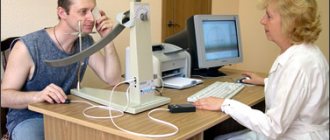
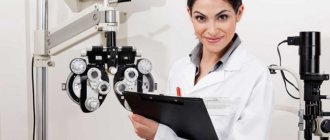
Before the procedure to restore visual acuity, the doctor conducts diagnostic tests, determining the condition of the eyeballs, and makes an accurate diagnosis.
Contraindications
There are many contraindications that make the procedure impossible:
- Infectious and viral diseases in the acute stage. Vision correction is possible after complete recovery, elimination of all symptoms, and repeated laboratory tests.
- Diseases affecting the immune system. These include rheumatism, rheumatoid arthritis, HIV, AIDS. If a patient with this diagnosis undergoes laser correction, the tissue will take a very long time to heal.
- Progressive decrease in visual acuity. Carrying out the procedure is irrational, since after it the vision will reappear.
- Minor age. Doctors recommend performing the procedure after 25 years, since by this time the eyeball will have fully grown. If vision correction is performed earlier, visual acuity will decrease again due to age-related changes. The correction will be irrational and a repeat operation will be required.
- Pregnancy and lactation. It is recommended to perform the operation 6 months before pregnancy. This is due to the fact that after exposure to the laser, the retina may detach during childbirth, as the woman actively pushes. If a caesarean section is planned, there are no contraindications to the procedure. Laser correction during breastfeeding is not recommended, as it is very stressful for the woman; local anesthetics that pass into breast milk are used.
- Thinning of the cornea. High risk of rupture. This will lead to a sharp deterioration in the quality of vision, up to complete blindness. The procedure is completely contraindicated for keratoconus, when the cornea protrudes forward, which leads to its thinning.
Since there are many contraindications to the procedure, it is recommended to undergo timely consultation with all specialists in order to identify deviations in health status.
Types of laser correction
Today, laser vision correction is represented by several types of operations: PRK (photorefractive keratectomy), LASIK (laser keratomileusis), as well as its various modifications. Vision correction performed using the SuperLASIK method deserves special mention. The SuperLasik technique, or customized ablation, involves drawing up an individual program based on abberometric data obtained using a WaveScan wavefront analyzer. This program allows you to create a personalized scheme of laser action on each point of the cornea to simulate its ideal shape. As a result, visual acuity can be raised to 1.5 or higher. This method makes it possible to provide patients with the highest possible visual acuity.
Pros and cons of the operation
The procedure has many positive aspects:
- improvement in vision quality up to 100%;
- no need to continue wearing contact lenses for glasses;
- the operation takes no more than 20 minutes;
- a minimally invasive method for surgical intervention, requiring only an incision on the cornea;
- low percentage of postoperative complications;
- relatively low cost, which is in different ranges;
- quick result, which depends on the type of operation.
The procedure has some negative sides:
- the possibility of hypocorrection, when the patient’s vision is not restored sufficiently, as a result of which it is necessary to undergo repeated surgery or wear glasses;
- temporary overcorrection, when a person’s eyes see objects too well, this property is eliminated within 1 month;
- discomfort during rehabilitation;
- increased risk of retinal tears for women during vaginal childbirth;
- inability to undergo the procedure for free under the policy.
The number of disadvantages decreases if the patient and doctor choose a more modern method of vision restoration using expensive devices.
Laser correction methods
Laser correction methods are characterized by the following features:
PRK is one of the first methods of laser correction, which served as the beginning of the development of modern methods of laser vision restoration. Photorefractive keratectomy (PRK) corrects minor astigmatism, nearsightedness and farsightedness. The operation is carried out in stages - first, the upper layer of corneal tissue (epithelium) is removed, the next step is to form the necessary curvature of the cornea. After the operation, the injured surface of the cornea heals for several days (or weeks), during this period the patient experiences pain and discomfort, lacrimation is noted, and there is a risk of infections. Since the method has certain disadvantages, it is currently rarely used.
LASIK - the technique showed effective results in correcting eye refraction in the early 90s. For that time, these were acceptable results in terms of patient comfort and possible risks. In LASIK technology, the top layer is first cut off with a blade (microkeratome), then the excimer laser works.
FemtoLASIK is an advanced LASIK technique. When carrying out correction, two types of laser are used - femtosecond and excimer. During the operation, a corneal flap is formed with a femtosecond laser, then the cornea is given a new shape - the refractive power of the cornea changes.
EpiLASIK – before the correction, a thin corneal flap is formed, which actually consists of the corneal epithelium. During the operation, an excimer laser and microkeratome are used. Used to treat vision in patients with thin corneas.
SuperLASIK or “personalized” (Custom View) – when carrying out treatment, the individual characteristics of the optical system of the patient’s eye are taken into account. Astigmatism, myopia, and farsightedness are effectively corrected using this method. In fact, it differs from the above methods only by an additional preoperative examination using an abberometer.
Smile (ReLExSMILE) is the most advanced, safe and patient-friendly correction method. During the operation, a small incision is made through which the lenticule pre-formed with a femtosecond laser is removed. The technology was developed by Professor Walter Secundo (Germany), who, by the way, operates in Moscow.
The technology has received recognition in all countries of the world, is widely used for the treatment of myopia and myopic astigmatism, and is one of the innovative advanced technologies of ophthalmology. The method is characterized by microinvasiveness, good preservation of corneal tissue, high-precision correction, painlessness, fast rehabilitation period, does not require the formation of a corneal flap, the treatment is suitable for patients with dry eye syndrome.
The operation requires a VisuMax laser (femtosecond laser); with the help of a laser, an optical lens (lenticule) is very quickly formed, which is removed through a small incision in the cornea. The method is suitable for the correction of high and moderate myopia, preserves the mechanical strength of the eye frame, and reduces the development of postoperative complications. The operation is painless, after the operation there is slight pain for a short time, healing occurs quickly, which creates a certain comfort for the patient.
Preparing for surgery
Before undergoing surgery, the patient must undergo laboratory and instrumental examination methods:
- general blood and urine analysis, blood biochemistry;
- bacterial cultures from the eyes and to eliminate the risk of postoperative infection;
- analysis for HIV and hepatitis;
- fluorography of the lungs;
- electrocardiography;
- identifying the quality of vision using a diagnostic table, refractometer, ultrasound of the eye and other necessary techniques.
Immediately before the operation, the patient must follow the following rules:
- 1 week before the procedure, it is recommended not to use new medications that the attending physician has not been warned about;
- 2 days before surgery you should not drink alcoholic beverages;
- 2 weeks before the procedure, avoid wearing contact lenses and replace them with glasses;
- the procedure is carried out on an empty stomach, so the last meal should be 8 hours before;
- on the day of surgery, the patient takes a shower, his skin and face should be clean;
- Women should not wear makeup before correction.
In the surgeon's office, the patient is given disposable sterile underwear and a cap under which the hair is hidden. This reduces the risk of infection in the eyes. All rules must be followed exactly.
Features of the event
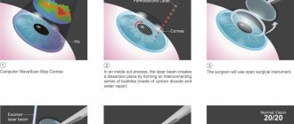
LASIK or LASIK is an abbreviation of the name of the operation in English (Laser-Assisted in Situ Keratomileusis). An approximate translation is “laser keratomileusis”. This is a vision correction technique using a “cold” excimer laser. It generates powerful ultraviolet light to correct the shape of the cornea so that the image is focused on the retina.
Operations using this technique have been carried out since the late 80s. XX century It has been well developed and has long proven its effectiveness. Now LASIK is the most popular method of laser vision correction. The operation itself takes place in 3 stages:
- Separation of the corneal flap.
- Laser correction of the cornea.
- Returning the corneal flap to its original position.
All necessary steps are repeated for each eye.
It is preceded by consultations with an ophthalmologist and diagnostics. Also during the consultation, the ophthalmologist talks about the essence of the method, the expected results, how the eyes will “feel” after LASIK surgery. After a detailed clarification of the medical history and clarification of the question of the absence of contraindications, the person signs an agreement with the clinic for the provision of medical services and voluntary informed consent to the operation.
Diagnostics to determine the parameters of the visual organs are carried out using high-tech equipment
The date is set so that people who wear contact lenses can go 2-3 weeks before surgery without them. You also need to have time to pass a set of tests.
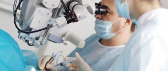
The success of LASIK surgery depends on the clinic and the doctor. The medical institution must have a license for this type of activity and certificates for the equipment used. The surgeon is required to have a diploma and experience in this field.
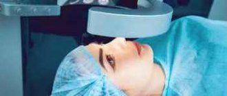
On the appointed day, the person comes to the clinic. The ophthalmologist conducts a follow-up examination and may suggest taking a mild sedative. The client is then escorted to the surgical department. There he changes into special clothes. An anesthetic is instilled into his eyes several times at short intervals.
After about 15 minutes, the person sits in a chair, and a special “eyelid dilator” is installed on the operated eye, which prevents involuntary blinking. The surgeon aligns the laser unit relative to the eye.
A corneal flap is formed only when using a vacuum ring, which ensures that the correct position of the eyeball is maintained relative to the laser and prevents its movement. During operation of the microkeratome (mechanical automated microsurgical instrument), a slight noise, buzzing, and buzzer are heard.
A man looks at a bright luminous point while the laser is working. During the operation of the microkeratome, complete blindness occurs for about half a minute, then light appears in the field of vision again.
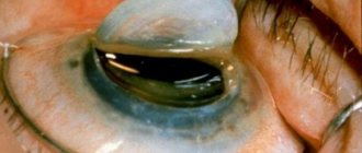
As a result of manipulation on the surface of the cornea, it is as if a lid is being opened. The formed flap, about 0.1 mm thick and 7-8 mm in diameter, is raised and fixed in this position. The excimer laser “evaporates” the tissue underneath to a certain depth to restore the desired corneal configuration.
The LASIK operation ends with the return of the corneal flap to its place. It fixes itself, “sticking” back to the “main surface” under the influence of adhesion forces due to the high content of collagen in the tissues. No stitches are required.
The eye is washed with an antiseptic solution and drops are instilled to prevent the development of inflammation. Sometimes special “bandage” contact lenses are worn.
After LASIK laser vision correction, a person remains in the clinic for about half an hour. The ophthalmologist once again reminds him of all the restrictions during the rehabilitation period, gives him 2-3 types of eye drops, gels and describes the scheme for their use.
A follow-up inspection is required the next day. Then you can start working or studying. But ideally, it is recommended to stay at home for at least 2-3 days.
Carrying out the operation
Surgery to restore vision is carried out in several stages:
- the patient lies down on the couch, his hair should be tucked under a hat;
- the doctor drops an anesthetic into the patient’s eyes, thanks to which he does not feel pain;
- dilators are inserted into the eyes, eliminating the risk of eyelid closure;
- an incision is made on the cornea using a surgical scalpel or laser;
- part of the cut cornea is removed to the side;
- laser eliminates eye defects that reduce visual acuity;
- The corneal flap is installed back, no stitching is required, the tissue will heal on its own.
The whole procedure takes 15-20 minutes. After it, the patient can go home immediately if no complications arise. It is recommended to leave by taxi or with the help of relatives and close friends, as there may be a temporary decrease in visual acuity immediately after the operation.
Video about laser correction in TV shows
Konovalov Mikhail Egorovich is often invited to participate in television programs dedicated to eye health. These programs discuss various vision problems. And in particular, several issues were devoted to topics such as myopia, farsightedness and astigmatism, and how vision can be restored using laser correction.
“Live Healthy!” program. Prof. Konovalov talks about Laser vision correction
How to improve your vision. While visiting the “Live Healthy” program, Professor, Doctor of Medical Sciences Mikhail Egorovich Konovalov will discuss the issues of vision restoration. We will find out how the operation goes, watch a video of laser vision correction and get answers to your most pressing questions.
Program “Health with Elena Malysheva”
Treatment of myopia in 30 seconds.
Program “Live Healthy” from September 07, 2010
Myopia in children (watch from 02.30 minutes of the video).
Rehabilitation and recovery period
Immediately after the procedure, small hemorrhages will appear on the cornea. The eyes will be inflamed, pain, burning, and discomfort will occur as the anesthetic wears off. It is recommended to follow the following rehabilitation rules:
- wear sunglasses outside for the first 1-3 days after the procedure;
- the first day after the procedure, stay in a dark room to eliminate the effect of bright sunlight on the eyes;
- It is forbidden to touch, rub, press or perform other actions in relation to the eyeballs;
- you cannot wear makeup for the first 2 weeks after surgery;
- lack of physical activity in the first 2 weeks after surgery, then you can engage in some sports with the doctor’s permission;
- It is forbidden to visit the bathhouse, sauna, or sunbathe under the open sun for 1-2 months after the procedure, so as not to cause bleeding;
- periodic visits to an ophthalmologist to perform a general examination and identify abnormalities, if any.
If you follow all the rules of the rehabilitation period, the risk of complications will decrease. If the hemorrhage does not go away over time, periodically reappears, the cornea has detached, the pain worsens for more than two days, the patient feels unwell, you must immediately consult your doctor.
Types and prices of laser correction
There are many types of laser vision correction. Each of them has its own advantages and disadvantages. When choosing the type of operation, the doctor tells the patient how much it will cost and how long the rehabilitation period will last.
Lasik
This is the very first type of visual acuity correction. To carry out this procedure, a surgical scalpel is used, with which the doctor independently cuts the cornea. The flap has a large thickness and uneven edges, which significantly increases the risk of complications.
The rehabilitation period lasts 1-1.5 months. The approximate cost of Lasik is 25,000 rubles.
Femto Lasik
Method for restoring visual acuity using a femtosecond laser. First, the doctor calculates the thickness and curvature of the corneal flap on a computer. This data is entered into a laser, which splits the corneal tissue rather than cutting it. A thin flap with precisely specified parameters is formed. This significantly reduces the risk of postoperative complications.
The rehabilitation period lasts 14 days. Using the technique, hypercorrection of vision is possible. The cost of Femto Lasik reaches 50,000 rubles.
Super Lasik
The technique is designed for patients with aberration disorders and high-degree astigmatism. Corneal parameters are calculated individually for each patient. After completion of the technique, operational violations are eliminated.
The rehabilitation period lasts 2-3 weeks, depending on the state of vision and the degree of development of the disease. The approximate cost of Super Lasik is 35,000 rubles. If the patient has a severe degree of the disease, it can increase to 45,000 rubles.
PRK
This is a basic technique intended for people who have a contraindication to Lasik technology. The price varies depending on the degree of violation, ranging from 25,000 to 36,000 rubles.
There is a variation of the procedure called Super PRK. It is necessary for people who have contraindications to the Lasik technique and who have severe visual impairment in their eyes. Therefore, they need an individual calculation for the incision on the cornea. The price of the technique can reach up to 50,000 rubles.
Lasik - video for those interested in the process
A video that you can watch on the website of Professor Konovalov’s clinic will help you learn more about the Lasik operation. Many patients at a laser vision correction clinic in Moscow are interested in what the difference is between PRK and Lasik, not to mention such procedures as Epi-Lasik, Super-Lasik and Intra-Lasik. Videos dedicated to PRK and Lasik operations make it clear how these procedures occur and what is the meaning of each of their stages. If you are uncomfortable watching videos about Lasik surgery, schematic drawings will help you get acquainted with the general progress of the operation. Whatever the differences between the above methods, all types of excimer laser vision correction are united by the same process: under the influence of an excimer laser, layers of the cornea are evaporated. The fundamental difference between Lasik and PRK is the effect of the laser on the internal, and not on the surface layers of the cornea. In addition to the video dedicated to Lasik, on our website you will find a lot of useful information about the structure of the eye and ophthalmological diseases.
Studying videos about Lasik surgery, you can see how at the initial stage a microkeratome is used to form a thin flap from the surface layer of the cornea, which is folded back and exposes the inner layers to the laser. At the second stage, the laser performs the same manipulations as with PRK, after which the flap is put in place and healing occurs. to explain the process of this operation to treat farsightedness and myopia, but the best way to understand Lasik is a video that gives a clear visual picture of what is happening. The advantages of the Lasik method are the painless course of the operation, the stability of its results, and the complete and rapid restoration of vision after laser correction. If the video on Lasik does not convince you enough, a professional ophthalmologist will do so during a consultation.
Generally speaking, there are plenty of videos about Lasik surgery on the Internet: there are some rather unpleasant naturalistic ones, some sketchy ones like a cartoon, and just interviews with doctors and reviews from patients. However, no matter what video you watch about Lasik, you can decide to undergo this operation only after a complete examination and approval of the attending physician. Many videos dedicated to Lasik surgery talk about such important contraindications as an insufficiently thick cornea, impaired immunity, unstable refraction and corneal diseases.
“Live Healthy!” program. Prof. Konovalov talks about Laser vision correction
How to learn to look into the distance and how to get rid of myopia. While visiting the “Live Healthy” program, Professor, Doctor of Medical Sciences Mikhail Egorovich Konovalov will discuss the issues of vision restoration. We will find out how the operation goes, watch a video of laser vision correction and get answers to your most pressing questions.
Detailed video of laser vision correction
In this video you can watch the full progress of the laser correction operation.
Laser vision correction LASIK
Reminder for those preparing for Lasik surgery
Scheme of laser vision correction surgery.
Tags: laser eye surgery, video of laser correction
Possible complications
With modern devices, vision correction reduces the risk of complications. The following deviations occur extremely rarely:
- hypocorrection of vision – the laser exposure was insufficient, so the patient retained farsightedness or myopia to a slight extent;
- overcorrection is often short-term, but the formation of a permanent violation is possible, this causes discomfort in the patient, constant headaches due to eye strain;
- decreased visual acuity up to complete blindness;
- violation of the shape of the eye;
- retinal detachment;
- intraocular bleeding;
- decreased color perception function;
- the development of night blindness, that is, the elimination of vision function during dusk and complete darkness.
Some complications cannot be prevented, as they develop due to the anatomical features of the eyeballs. But most of them can be avoided by choosing the right doctor and surgical technique.
Laser telemetry for vision correction: complete surgery with commentary (not for the faint of heart)
Now I will show something that doctors usually never show patients. More precisely, all this is shown in the form of a beautiful rendering, from which it in no way follows that there will be a piece of iron sticking out right in your cornea for a couple of minutes. Fortunately, you won’t feel it because of the painkiller premedication, you won’t recognize it or remember it, because the gland will be out of focus.
Inserting a spatula to cut the remaining tissue bridges into the corneal incision after laser treatment, but before removing the lenticule
Go. So, watch the video, and I will show still frames with comments. This is a real operation on a patient in a German clinic, the recording was made on a device like the “black box” of the VisuMAX device. In this case, the patient consented to the use of the recording for educational purposes; usually access to such recordings is strictly limited.
First, let’s briefly repeat the previous post about why it is necessary to cut out such a lens inside the eye and then take it out:
In short, operations of previous generations formed such an unpleasant “lid”, which during healing was held on by one flap. The flap “lid” is removed, the lens is evaporated, and the “lid” is put back.
The invasiveness is high, the “lid” never completely takes root (during injuries and physical stress, it sometimes comes off with extremely unpleasant consequences), a lot of heat is used for incisions, passing into the tissues of the eye, the Bowman’s membrane is severely damaged both by the cut and by thermal fronts.
The next generation's idea is to cut the lens right inside the cornea, and then remove it through a thin incision without this “cover.” The effect is possible because lasers allow focusing within the tissue rather than on its surface. That is, you can make a cut according to this scheme:
Now let's look at the rendering of the operation:
First, the lower surface of the lens inside the cornea, then the upper, then a small incision on the side - and remove the lens with tweezers.
This is what any clinic will show you so you don't get scared. The real operation looks like this (now telemetry):
First, the patient is asked to look at a flashing green light. The lamp does not just light up, but blinks so that the person catches it in focus often enough. The eye makes an incredible number of micro-movements-saccades per second, but it is precisely this pace that allows the surgeon to make the correct capture of the eye a little later.
Here you can see the swab out of focus (in the video it moves on the left side of the eye) - this is the surgeon wiping the surface again to insure against microclumps of fat floating on it. The second purpose of this touch is to check how the premedication went. The patient must answer whether he felt something. He shouldn’t feel anything, the nerves have already been turned off by tetracaine.
Here the capture is approaching the eye, this is clearly noticeable by the appearance of the iris closer to the sharpness zone:
The pneumatic gripper is centered and fixed. The very moment of capture can be seen by a slight rotation of the eye, and this effect is very important to control when correcting astigmatism, because a rotation of 30 degrees negates the entire operation in the case of astigmatism. Therefore, the surgeon corrects the alignment.
After successful capture, we switch the observation range from visible to infrared in order to control the process and not blind the patient:
A slowly expanding ring from the edges of the eye to the center is millions of femtolaser micropulses that form a cut along the upper surface of the lenticule lens inside the cornea. The surface of the cornea is not cut through. Let me remind you that the main effect is the evaporation of several cells inside the corneal layer, and then the release of a relatively large amount of gas, which gently pushes the tissue apart.
The bottom surface of the lens is finished:
The second circle, which expands from the center of the eye to the edges, is the upper surface of the lens, which is also cut inside the corneal layer, without directly “piercing” the surface:
At approximately 01:02, a cut is formed through the surface of the cornea to the cut out lenticule lens, but it is extremely difficult to see in the video. The lens will be removed through this incision.
Now switching views (and the end of the video from the laser) - the patient is placed under the CNC control under the surgeon’s hands (in fact, he moves under the device from under the laser closer to the doctor’s hands). The first step is to wet your eye:
Switching the microscope lens, fixing the eye so that it does not twitch, using tweezers. This is a tuck, but the patient does not feel it (due to pain relief) and does not even really see the tweezers (more precisely, he sees the surgeon’s hand above his head):
The incision for extracting the lenticule is carefully separated with a tool - a spatula:
This is a very interesting place; the first small spatula is used to separate first the upper edge of the lens and surrounding tissues, and then the lower edge of the lens (in the frame below):
The spatula moves in subtle jerks - this is the resistance of micro-adhesions that arise due to the discrete structure of the cut; in some places individual fibers are not “unstuck” and need to be moved apart. You can’t just pull the lenticule by the edge, it can get caught, so a full pass is always made with a spatula over the surface so that air enters the cavity between the upper edge of the lens and the cornea and the lower edge of the lens and the cornea. The worst part looks like this:
The surgeon's hand rests with the elbow on a special support, and the wrist rests on the patient's forehead. The spatula is designed in such a way that even if the surgeon is pushed (slightly) or the patient is distracted by a loud bang, the eye will not be damaged. However, all such factors must be strictly excluded.
So, the lens is divided and ready to be removed from inside the cornea. The surgeon takes tweezers and hooks the lenticule:
All. You can finish here, but you still need to rinse your eyes. Here you can see how the fluid supply works:
The next stage is the supply of saline solution directly into the formed cavity inside the cornea:
As can be seen further, it will later flow out through the same incision through which the lenticule was removed. Previously, a second incision was made opposite the first (at the other pole of the eye) to remove this fluid, but it turned out that this was not necessary. Moreover, the smaller the incision, the better.
Smoothing to avoid micro wrinkles:
Then - removing the fixing films and finishing. You can blink a couple of times. The patient rests for a few seconds and then slowly gets up and walks into the hallway. In every clinic, there is a clock at the end of the corridor for monitoring. Next is a dialogue to reassure a worried patient:
- What time is it over there? — Six minutes past eleven. “Would you have been able to distinguish a clock from such a distance before?” - Oh. Exactly. - Well, everything is fine.
The patient calms down and goes to rest. After 2-3 hours the eye no longer feels anything. You can’t read, write, or use a phone or tablet/laptop for a day or two to promote rapid healing (after all, reading is “jogging” for the eyes); then you can’t go to a hard sauna or pool for two weeks (until the incision to remove the lenticule has completely healed, so There was no infection.) And that’s it.
I hope you haven't run away in horror yet when you see the spatula. Despite the frightening, I note that this is one of the simplest operations. Collecting the eye piece by piece after an accident with removing glass shards and other side delights for several hours, or precise manual work with a microscalpel is much more difficult and stressful.
To questions about how exactly a pneumatic gripper works, how it is possible to accurately cut a lens of the desired shape and diameter, what happens to the corneal tissue at the cut boundary, and why it is generally so difficult to cut a lens inside the eye (and why it is the safest). Plus how the operating room is prepared, how the surgeon calms the patient, how premedication is performed, what kind of protection there is from unforeseen situations (power outages, a passing truck) and so on, I will answer in the next big post. And about the promised comparison of lenses and operations later too. All of these are separate topics for discussion, and alas, it’s not possible to write much about them a little later. In the first post in the discussion, many topics are superficially touched upon.
How to minimize side effects after surgery
To reduce the risk of complications, it is recommended to adhere to the following rules:
- undergo all health examinations before surgery;
- choosing a qualified doctor and modern techniques;
- use all recommendations for rehabilitation;
- use anti-inflammatory, antibacterial, moisturizing eye drops;
- Visit your ophthalmologist again to have your vision checked.
Application of the above rules will not completely eliminate the risk of eyeball pathology, but will significantly reduce it.
Reviews from doctors
Nikolay, 42 years old, ophthalmologist: For my patients, I often recommend Femto Lasik surgery to restore visual acuity. It is based on a femtosecond laser, which cuts the cornea according to precise calculations. This reduces the risk of complications.
Elena, 38 years old, ophthalmologist: My patients often consult me about laser surgery to restore vision. I recommend not using Lasik technology, but resorting to the Femto Lasik procedure.
Patient reviews
Vyacheslav: A year ago I underwent a laser vision restoration procedure. I used Super Lasik technology. My vision has been restored to 100%, I don’t need to wear glasses. The rehabilitation period was completed within 2 weeks.
Oleg: A month ago I had laser vision correction. I did it using Femto Lasik technology, since regular Lasik was not recommended to me. The eyes were very inflamed, red, the pain persisted for 2 days. But after that, all the unpleasant symptoms quickly disappeared, and now I see well.