Causes and classification Symptoms Diagnostics Treatment Our advantages Treatment prices
Entropion of the eyelid (synonym “Entropion”) is a pathology in which the edge of the eyelid is turned towards the eyeball throughout its entire length or in a certain area. In this position, the eyelashes mechanically irritate the mucous membrane of the eye (cornea and conjunctiva), which causes irritation and can subsequently cause the development of inflammatory and dystrophic changes in the cornea.
Causes and classification
Depending on the cause of entropion, there are:
- Involutional (senile, senile) entropion of the eyelids - occurs against the background of age-related weakening of the orbicularis oculi muscle in elderly patients
— Cicatricial (post-traumatic) entropion of the eyelid – develops due to deformation of the cartilage of the eyelid after injuries or burns of the eyelid or eye
— Congenital entropion of the eyelid – diagnosed in children with congenital pathologies of the orbicularis oculi muscle or eyelid cartilage, including developmental defects (Larsen syndrome)
— Spastic inversion of the eyelid is a consequence of spasm of the fibers of the orbicularis oculi muscle
— Atonic or paralytic entropion of the eyelid develops with paresis or paralysis of the orbicularis oculi muscle
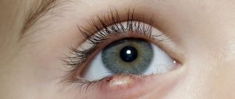
Inversion of the lower eyelid in a child
Entropion is common in children. This unpleasant disease causes discomfort, as the edge of the eyelid or eyelashes constantly rub against the conjunctiva. As a result, redness, irritation, and pain occur. This threatens ulceration and clouding of the cornea. To avoid dangerous complications, it is necessary to treat the disease in a timely manner.
In patients of the younger age category, congenital entropion of the eyelid often occurs , which occurs due to genetic disorders: improper development of the muscle group that is responsible for the correct position of the upper eyelid. Underdeveloped nerve endings can also cause the development of the disease.
As for the acquired type of volvulus, it develops due to injury or burn to the inflamed conjunctiva. As a result of the damage, a scar is formed on the inner membrane, which fuses the conjunctiva with the mucous membrane of the eye, and the eyelid folds.
A child with entropion is easy to recognize: to look at an object, he raises his head, since the boundaries of his field of vision are narrowed. Over time, vision deteriorates.
It is preferable to treat entropion in children with a special plaster ; it tightens the eyelid and prevents eyelashes from rubbing against the mucous membrane of the eye. You can protect the cornea with contact lenses made of soft materials. With the help of keratoprotectors, epithelializing drugs, and moisturizing drops, the damaged cornea is restored.
Surgery is the most radical method used for severe entropion.
Four types of entropion: an extremely uncomfortable disease! Pathology treatment methods
When the eyelid is inverted, it turns inward ; when it is everted, it hangs outward . Eversion is accompanied by lacrimation, inflammation and irritation of the skin, and there is a feeling that there is a foreign body in the eye.
Code in ICD-10
Acquired volvulus refers to diseases of the eyelids, lacrimal ducts and orbits and, according to the international classification of diseases, has code H 02.0 . Congenital entropion refers to congenital developmental anomalies, coded according to ICD 10 as Q 10.2.
Causes of entropion of the lower eyelid in humans
The following reasons can lead to the disease:
- congenital pathology;
- inflammatory conjunctivitis;
- burn and injury to the orbit;
- neoplasms and the development of their complications;
- weakening of the muscles that give shape to the eyelid.
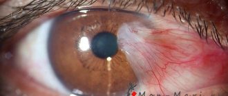
Photo 1. A burn to the cornea of the eye often causes the development of entropion of the lower eyelid.
Types and nature of the disease
Volvulus is divided into several types.
Senile
With age, the skin loses its elasticity, sags, and the muscles designed to maintain the shape of the eyelid weaken. Senile entropion almost always manifests itself in the lower eyelid, and is often expressed simultaneously in both eyes and progresses more and more with age.
Scarred
This type of disease occurs after injuries, burns or inflammation of the visual organs, leading to tissue growth in the eye.
It progresses slowly, and the severity is influenced by the location of concentration and the size of the scar.
Mechanical
Appears when various neoplasms occur. If tumor tissue begins to grow, this leads to eye pathology.
Congenital
During intrauterine development, abnormal formation of the visual organs occurs. This pathology is called congenital entropion. May appear on both eyelids.
In most cases, congenital entropion of the upper eyelid is accompanied by a small size of the eyeball.
Inversion of the lower one provokes improper formation of the muscle that fixes the shape of the visual organ .
Features of the disease in a child
To treat children, it is necessary to use a special patch. It needs to be glued under the sore eye so that the skin is pulled back, preventing the eyelashes from rubbing against the mucous membrane.
It is recommended to wear special lenses made of soft materials to protect the eyes from friction.
To heal the damaged surface of the cornea, it is necessary to instill drops that moisturize and nourish the epithelium.
Reference! When treating bloat in a child, such a radical method as surgery is resorted to only if the most severe form of the disease is diagnosed.
Symptoms of entropion
With entropion, a person suffers from the following symptoms:
- Itching and burning.
- Constant sensation of foreign body and sand.
- Redness of the mucous membrane.
- The blood vessels become inflamed and visible.
- Pain.
- Copious discharge from the eyes.
- Fear of bright light.
- Dryness and discomfort.
Diagnostics
To diagnose and determine the nature of the disease, use:
- Visual and slit lamp inspection.
- Visometry - testing visual acuity. The ophthalmologist checks to see if rubbing the cornea with eyelashes has led to decreased vision.
- Biomicroscopy is a procedure that allows you to see the presence of changes, adhesions or erosions in the eye tissues.
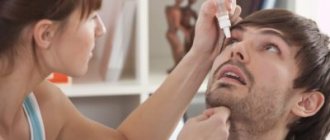
Photo 2. The visometry procedure is necessary to check visual acuity, since eyelashes can damage the cornea.
Symptoms
Mostly, patients with entropion complain of eye discomfort, which is caused by irritation of the mucous membrane of the eyeball by the edge of the eyelid turned towards it and the eyelashes located on it (pseudotrichiasis). Irritation is accompanied by a sensation of a foreign body in the eye, pain when blinking, swelling and redness of the conjunctiva, possible lacrimation, burning, itching.
If entropion has been formed for a long time, pathology may affect the cornea (corneal erosion or dystrophy, ulcerative keratitis). Such complications threaten vision loss.
How to recognize
An inversion of the eyelid in a person is easy to recognize visually. The eye looks as if eyelashes are growing from the inside, the eyelid is thickened and pushed forward. This phenomenon is accompanied by other symptoms. Eyelashes are in constant contact with the cornea of the eyeball, irritating it, which causes the following complaints from patients:
- sensation of a foreign body in the eye;
- intense lacrimation;
- photophobia;
- pain and itching when blinking.
Entropion is easy to recognize by the patient’s appearance; in addition, due to the constant contact of the cornea with the eyelashes, the patient suffers from severe discomfort - itching, burning, stinging, lacrimation
Further manifestations of the disease vary depending on the type of disease.
Cicatricial entropion
Cicatricial volvulus always occurs as a consequence of another ophthalmological pathology - chronic conjunctivitis, complicated by damage to the cornea and subsequent tissue scarring, trauma, thermal or chemical burn, after which healthy tissues are also replaced by dense connective tissue. The primary disease can be completely cured, but the scar between the eyelid and conjunctiva will remain. This can lead to gradual deformation of the eyelid. It will thicken, take on the shape of an arc, and eventually curl inward.
How vividly the symptoms will be expressed depends on the location of the scar. Sometimes, already in the early stages, the patient feels significant discomfort, which intensifies when blinking and closing the eyes. But it also happens that the eyelid is completely turned inward, but unpleasant sensations, irritation, and lacrimation only occur periodically. A symptom such as photophobia indicates that lesions in chronic conjunctivitis have reached the cornea.
Involutional inversion of the eyelids
With this type of volvulus, there are no scar adhesions, which makes it different from other forms of the disease. The eyelid may roll inward uncontrollably when the muscles spasm. But the patient will effortlessly return it to its normal position with his finger. As the disease progresses, the orbicularis oculi muscle will thicken and form a ridge. With atonic entropion, the lower eyelid protrudes forward, while the upper eyelid, on the contrary, fits tightly to the eyeball.
Surgery to correct entropion (on one eye)
Inversion of the eyelids, entropium palpebrarum (gr. en - inward + trope inward inversion, lat. palpebra eyelid) - inversion of the edge of the eyelid or part of it inward, towards the eyeball. Entropion is an anatomically incorrect arrangement of the eyelids, manifested in the fact that the edges of the eyelids (upper and/or lower) are “turned” inward, as a result of which eyelashes and hair, in direct contact with the eyeball, cause various pathological processes (Fig. 1).
Figure 1 – Inversion of the lower eyelid (eyelashes injure the cornea, thereby causing inflammation and clouding)
.
How does entropion develop?
Inversion of the eyelids in animals is due to the fact that the edges of the eyelids, which normally should fit well to the eyeball, turn inward, and the delicate cornea of the eye comes into contact with eyelashes and fur. Because of this, constant mechanical irritation of the eye. As a result, you may notice that the animal's eye is watery, the cornea is severely irritated, and conjunctivitis often appears. Over time, untreated entropion leads to perforation (perforation) of the cornea and the formation of ulcers.
Which animals are prone to drooping eyelids?
1. Congenital entropion in cats can be noticed when kittens open their eyes, which occurs on the 10-15th day of their life. In dogs, signs of entropion appear during the first three months of life. 2. Acquired volvulus can occur at any age.
Entropion in dogs is most common in breeds such as the Chow Chow and Shar Pei. This eye disease in dogs of these breeds is caused by their natural “folding” of the skin. However, entropion can occur among St. Bernards, Great Danes, Cocker Spaniels, Bulldogs, Boxers, Pugs and Pekingese. Among cats, this eye disease is most often observed in Sphynxes, Rexes and Persians. But sometimes outbred cats can suffer from entropion. Entropion of the eyelids can be congenital or acquired.
ATTENTION!!! Entropion can occur on both the upper and lower eyelids, and sometimes on both at the same time.
Why does entropion of the eyelids develop in adult animals?
1.
Weight problems often lead to entropion of the eyelid. Fat that is deposited under the skin stretches it, thickens it and makes it heavier. As a result, the skin of the eyelids cannot cope with such a load and turns towards the cornea.
2.
Any injuries or other eye diseases can also cause entropion. Volvulus is often preceded by conjunctivitis or a foreign body entering the animal's eye. All this is due to the fact that the muscles of the eyeball can become overstrained and move the eye inside the orbit. As a result, it turns out that the size of the eye does not correspond to the anatomical shape of the orbit, and the eyelid, in turn, begins to turn inward.
ATTENTION!!! At the initial stage of the disease, you can try to treat entropion with ointments, drops and other drugs. Carrying out such therapy is advisable only in cases where entropion of the eyelids occurs periodically, and its cause can be completely eliminated. But in the end, unfortunately, you have to resort to surgery. Of course, while using local medications, you can notice an improvement in the condition of the animal’s eyes, but after the end of treatment, all the symptoms of the disease return.
Operations are more often planned. At the SQ-lap clinic and the SQ-lap veterinary hospital, they are carried out after a preoperative examination of the animal, which includes ultrasound, ECG, General and Biochemical Blood Tests, and an 8-hour diet is required. Before the operation, the doctor and the owner draw up a document on informed consent for the operation, after all the risks of these activities are explained to the owner.
It is very important that the owner brings a warm blanket to the operation; it will be needed to warm the animal after the operation, disposable absorbent diapers, and napkins.
REMEMBER! POSTOPERATIVE CARE CAN BE PERFORMED IN A HOSPITAL CONDITION.
How is the operation performed?
The operation is performed using combined anesthesia (anesthesia + local anesthesia). The width of the removed strip of skin is determined by the skin fold. To do this, grab the skin with tweezers at a distance of 3-5 mm from the edge of the eyelid and parallel to it along the entire length of the area of the eyelid turned inward. The strip of skin to be removed is isolated by two incisions: parallel to the edge of the eyelid at a distance of 3-5 mm from it and an arcuate incision. Then the strip of skin is slightly lifted and cut off from the subcutaneous layer with scissors. This operation is first performed on the upper, then on the lower eyelid of one eye, then the dog is fixed in the opposite (right or left, respectively) lateral position and similar actions are performed on the other eye. The edges of the skin at the incision sites are connected with knotted sutures (atraumatic non-absorbable suture material is used).
How to care for an animal after surgery?
After surgery, antibacterial therapy is prescribed (locally in the form of drops and/or systemically). It is very important to prevent animals from scratching their eyelids; for this purpose, wear a protective collar. The seams must be treated with antiseptic agents daily.
How to prevent entropion of the eyelids?
Prevention of entropion involves timely diagnosis and treatment of inflammatory diseases of the eyelids and eyes, as well as avoidance of eye injuries. To prevent further consolidation of signs predisposing to the development of entropion in those breeds whose representatives have this pathology, sick animals should not be allowed for breeding. This recommendation, unfortunately, is not followed by the majority of breeders and owners, which leads to an even greater spread of this disease.
Entropion in humans: symptoms, surgery, photos of the upper and lower entropion in adults and children
Entropion is an abnormal position of the eyelid; the edge of the eyelid, together with the eyelashes growing on it, is completely or partially turned towards the eyeball. As a result, the friction of eyelashes on the cornea causes irritation, tearing, redness, and in especially advanced cases, it becomes covered with ulcers.
Entropion has four forms:
- Cicatricial (occurs after illnesses).
- Spastic or senile (occurs most often in older people due to sagging skin).
- Mechanical (consequence of mechanical trauma, chemical or thermal burn of the cornea).
- Congenital (caused by a genetic disorder in the structure of the eyeball).
ICD-10 code: H02.0 Entropion and trichiasis of the eyelid
How to recognize an entropion of the eyelid?
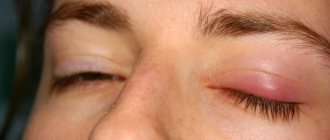
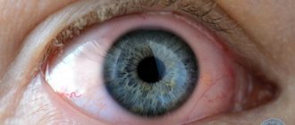
Entropion of the lower eyelid in an elderly patient
The clinical picture for all forms of entropion is almost the same.
It is worth highlighting the following symptoms:
- irritation resulting in swelling of the eyelid;
- pronounced hyperemia (redness of the skin) and micro damage;
- sensation of a foreign body in the eye;
- tearfulness;
- photophobia;
- burns;
- itching;
- decreased vision as a result of corneal clouding.
It should be noted that the manifestation of symptoms increases with blinking and closing the eyes.
Causes
Appearance of the eye when the lower eyelid is turned in
The most common reason is the stretching of the skin that occurs with age and the weakening of the muscles that hold the physiologically correct position of the eyelid.
The following reasons:
- suffered severe inflammation;
- burns and injuries to the eyeball;
- genetic disorder of the structure of the eyeball;
- laxity of the muscles that fix the position of the eyelid;
- impaired development of the eyeball (its size is significantly smaller than normal);
- allergic eye diseases.
How to diagnose entropion?
Most often, patients themselves discover an entropion of the eyelid due to the constant sensation of a foreign body and the presence of other symptoms characteristic of this disease. But only an ophthalmologist can confirm this diagnosis after an external examination of the patient, determination of visual acuity, and biomicroscopy (evaluation of the tear film).
Treatment
Depending on the degree and severity of the disease, appropriate treatment methods are used:
Operative (surgical) intervention
Its essence is to excise a strip of skin or skin and orbicularis muscle; operations to shorten the lower eyelid retractor by creating its fold; partial removal of cartilage. Such operations are performed under local or general anesthesia.
Folk remedies
The simplest and most accessible method of treatment, but it is not always effective. When turning up the eyelid, it is recommended to comply with the following hygiene requirements:
- regularly rinse the conjunctival sac with an antiseptic solution;
- use vitamin and antiseptic ointments;
- be sure to take care of all facial skin;
- make special eye baths with tea;
- Rinse your eyes regularly with cold water;
- add slices of fresh cucumbers;
- make lotions from linden infusion (pour 2 teaspoons of linden with 1 glass of boiling water, leave for 10–15 minutes).
Drug treatment
Aimed at alleviating the symptoms of the disease. It is recommended to constantly use special preparations to moisturize the eyelid (erythromycin, tetracycline ointments).
Sometimes injections are prescribed for temporary akinesia (immobility) of the eyelid. Contact lenses may be prescribed to avoid irritation.
It is important to remember that only a doctor can determine the correct and most appropriate treatment method.
Forecast
With proper and timely treatment, doctors give a positive prognosis, preserving the patient’s life and ability to work. In rare advanced cases, irreversible damage to the cornea may develop (formation of a cataract, ingrowth of newly formed vessels), resulting in decreased visual acuity and disability, but this happens extremely rarely. Full recovery is possible within a month.
Complications
Untimely and incorrect treatment can lead to a number of complications, including:
- dry eyes and irritation;
- damage to the cornea of the eye, resulting in complete or partial loss of vision;
- the appearance of painful ulcers on the cornea;
- eye infections, which can lead not only to loss of vision, but also to damage and death of the eye.
Prevention
There is no specific prophylaxis to prevent entropion and the risk of the disease cannot be completely prevented. However, thanks to a number of simple manipulations, you can reduce the risk of this disease.
Necessarily:
- Maintaining hygienic conditions.
- Regular (every two weeks) eye baths with tea.
- When returning home from the street, wash your eyes to remove dust and dirt with boiled water.
- Wash your eyes with cool water in the morning.
- Take a walk in the fresh air.
- Do eye exercises regularly.
- Monitor your eye pressure.
- Stop smoking.
- Protect your eyes from the harmful effects of the sun and computer technology with special glasses.
- Avoid stress.
- Do a special eye massage.
- Use anti-allergic cosmetics.
By following simple rules and undergoing regular examinations at the clinic, you can prevent and avoid the development of eye diseases. And timely professional treatment will help maintain visual acuity and avoid negative consequences.
Source: https://glazam.info/zavorot-veka/
Pathogenesis of the disease
Degenerative processes in the tissues of the eyelid cause the following processes to occur:
- Weakness of the eyelids in the horizontal direction, which occurs due to stretching of the tendon plates located in the corner of the eye.
- Instability of the eyelid in the vertical direction. Develops due to weakening, splitting and rupture of the tendons of the retractor muscles. Such phenomena can be recognized by a decrease in the excursion of the eyelid when looking down.
- Shifting the lower border of the tendon plate forward from the eye, moving its upper border towards the eye. This ultimately leads to the folding of the eyelid margin inward.
Symptoms
Signs and symptoms of entropy occur as a result of the eyelashes and outer eyelid rubbing against the surface of the eye. The patient may experience the following symptoms:
- foreign body sensation;
- redness of the eyes;
- irritation;
- pain;
- dry eye syndrome (present in 72.1% of patients with the involutional form of the disease);
- punctate epithelial erosions were noted in 61.9%;
- sensitivity to light, wind;
- excessive tearing;
- deterioration of visual perception.
There may be mucus discharge from the eyes. Symptoms of entropion are aggravated by blinking. The patient cannot read, work or play sports normally. You cannot put off visiting an ophthalmologist.
Features of the disease in a child
To treat children, it is necessary to use a special patch. It needs to be glued under the sore eye so that the skin is pulled back, preventing the eyelashes from rubbing against the mucous membrane.
It is recommended to wear special lenses made of soft materials to protect the eyes from friction.
To heal the damaged surface of the cornea, it is necessary to instill drops that moisturize and nourish the epithelium.
Reference! When treating bloat in a child, such a radical method as surgery is resorted to only if the most severe form of the disease is diagnosed.
Diagnostics
Entropion is usually diagnosed through a routine physical examination of the eyes. During this, the doctor may pull on the eyelids, ask you to blink or close your eyes. This helps him assess eyelid position, muscle tone and density. If the abnormal disease is not caused by scar tissue, previous surgery, or other conditions, the doctor will also examine the surrounding tissue.
A patient with suspected entropion should undergo a thorough ophthalmologic examination. The physician should pay close attention to the structures of the eyelid margin to evaluate for trichiasis and distichiasis. These conditions may simulate entropion but do not cause edge misalignment.
It is necessary to examine the cornea with fluorescein for the presence of abrasions, scars, thinning and neovascularization of the cornea. Careful examination of the conjunctiva is important because partial entropion of the lateral or medial eyelid may not affect the cornea.
The ophthalmologist should pay attention to symblepharon, shortening of eyelashes and edge abnormalities, if any, as these findings may indicate an inflammatory process.
In addition, histological studies have shown that chronic mechanical damage can cause chronic inflammation, which can lead to squamous metaplasia of the conjunctiva.
What else do you need to know
Eversion of the third eyelid is sometimes observed in pets, for example, Maine Coons, Shar Peis, and Chow Chows. British and hairless breed cats also have a predisposition to this pathology.
The cause of development is an abnormally long eyelid or an eyeball that is set too deep; in this case, the irritant is the animal’s fur. In advanced cases, ulcers and corneal clouding develop as a result of scarring of abrasions.
The main symptoms of the disease in cats and dogs:
- constantly closed eyes;
- damp hair in the corners and around the eyes;
- purulent discharge;
- redness and inflammation of the eyeball.
Entropion of the eyelids in pets can only be treated surgically, even in 2-3 month old kittens and puppies. The operation is performed under general anesthesia, the doctor makes an incision in the long eyelid and performs plastic surgery on the affected areas. In the postoperative period, the animal will require a course of antibiotic therapy.
A rolled eyelid in a child, as a rule, is of the congenital type; its development is caused by genetic disorders that affect not only the external elements of the visual apparatus, but also the structures of the eyeball. Underdevelopment of the nerve endings of the eyes in premature babies can also provoke this pathology.
They try to correct such a defect for children without a scalpel. Since the child’s skin and muscle tissue are very soft and elastic, it is possible to return the eyelid to its normal position by pulling it back and fixing it with a plaster or by applying sutures.
Schoolchildren are additionally fitted with soft contact lenses to protect the cornea from contact with eyelashes.
But in severe cases of entropion, surgical intervention is performed regardless of the age of the small patient, even in newborns.
Entropion of the eyelids is a pathology from which it is almost impossible to protect yourself, since its development is influenced either by etiotropic factors or genetic abnormalities. It is only possible to partially stop the progression of the disease if you do not neglect the rules of personal hygiene, treat periodically occurring ophthalmological diseases in a timely manner, and monitor the condition of the circulatory system.
The quality of oxygen supply and nutrition of the structures of the organs of vision depend on the condition of the blood vessels, so this point is also very important. The general prognosis of the pathology is good; very rarely, entropion of the eyelid becomes the cause of decreased visual acuity and other serious ophthalmological diseases. If you need to resort to surgery, in most cases it is tolerated easily and without complications.
After operation
Artificial tears are prescribed for minor eyelid inversion and after surgery. The drugs help restore the condition of the tear film. The following medications are considered effective:
- Visine;
- Artificial tear;
- Lacrisin.
These medications relieve irritation and redness.
Keratoprotectors are prescribed. They speed up recovery and prevent the development of dry keratoconjunctivitis. After surgery, medications with dexpanthenol are prescribed.
Spastic inversion of the eyelids
The disease is characterized by excessive stretching of parts of the lower eyelid, which, in the case of age-related enophthalmos, causes instability of the eyelid. In this case, the circular muscle moves to the edge of the eyelid with simultaneous thickening. Often this type of entropion occurs along with blepharospasm.
The treatment uses a combination of shortening the external ligament of the eyelid in the horizontal plane with retractor plastic surgery and excision of a piece of skin from the lower eyelid.
This pathology often relapses even after successful treatment.
Types of pathology
Entropion of the lower eyelid is more common. This is easily explained if you remember the structure of the eye. Both eyelids are supported by special cartilage, which ensures their elasticity and firmness. And the cartilage of the lower eyelid is half the size of the upper eyelid, therefore, it is more susceptible to the disease.
You can also see an inversion of the upper eyelid. Usually this pathology is combined with the so-called microphthalmia, which means a small eyeball. These pathologies are congenital and can only be treated surgically.
The following types of inversion are distinguished:
- spastic;
- cicatricial;
- age;
- congenital.
Ectropion in the postoperative period
Some patients may experience eyelid inversion after blepharoplasty. This happens because the doctor removed too much skin.
As a result of unsuccessful manipulations, the following is observed:
- deformation of the lower eyelid;
- drying of the mucous membrane of the operated eye.
Sometimes eversion after blepharoplasty is double. Ectropion also occurs as a result of a person ignoring medical recommendations regarding the care of the affected areas.
Correcting a defect surgically can result in:
- hemorrhages;
- divergence of applied sutures;
- penetration of infection;
- rough scarring of the skin;
- development of cysts;
- malfunction of the lacrimal glands;
- turn of the century;
- deterioration of visual acuity.
Source: https://alternativa-mc.ru/glaza-bolezni/zavorot-veka.html
Treatment of entropion
Treatment of the disease should be aimed at correcting the existing eyelid defect and preventing the development of corneal pathology. Entropion of the eyelid is difficult to treat conservatively; surgical treatment is the most effective.
Drug therapy for entropion relieves symptoms of eye irritation and prevents damage to the cornea. For this purpose, constant use of moisturizing drugs (artificial tears), keratoprotectors (dexpanthenol) is indicated. It is possible to prescribe antibacterial eye ointments (erythromycin, tetracycline). For spastic entropion, injections are used for temporary akinesia of the eyelid.
Surgical treatment of entropion of the eyelid may include various techniques depending on the cause of the entropion: application of everting sutures, excision and plastic surgery of spasmodic muscle fibers with spastic entropion, strengthening of the eyelid with atonic and senile entropion, transplantation of a skin flap with cicatricial entropion.
What is entropion? How dangerous is this?
Entropion of a dog's eyelids
(
entropion
) - a change in the position of the eyelid, in which the free edge of the eyelid and eyelashes come into contact with the eyeball.
Entropion of the eyelids in dogs
(with a small degree) most often causes lacrimation and conjunctivitis. But if left untreated, the degree of the defect may increase.
A complication is often a corneal ulcer, which appears due to friction of eyelashes and hair on its surface. At the same time, the owner sees that the eye becomes cloudy, and lacrimation and spasm of the eyelids intensify, and purulent discharge appears.
I would like to draw your attention to the fact that entropion of the eyelids of dogs (especially when severe) is not a cosmetic defect, but a serious pathology that can lead to partial or complete loss of vision.
Thus, correction of entropion is not just “plastic surgery”, but a surgical intervention for medical reasons aimed at preserving vision and the eye. Sign up for a consultation with veterinary ophthalmologist, candidate of sciences Elena Sergeevna Bodryagina online.
What clinical signs suggest that a dog has entropion?
When the eyelids turn up, the dog experiences unpleasant or even painful sensations – “sand” in the eyes.
We all know how unpleasant it becomes when just one speck or eyelash gets into the eye. The eye immediately closes, it hurts, tears flow. What if you imagine that your eye constantly catches not one, but dozens of eyelashes?
Yes….! This is how terrible a dog with twisted eyelids feels.
From the outside, you can notice that the dog always either periodically covers his eye or squints it slightly. There are tear tracks and sometimes mucous discharge visible on the dog's face.
Differences in entropion of the eyelids in cats.
How is entropion of the eyelids treated?
The main method of treatment is surgery – surgical correction of entropion.
For different breeds of dogs, veterinary ophthalmologists have developed various methods of surgical correction of entropion. For each dog, the veterinary ophthalmologist, after a thorough examination, draws up an individual surgical plan.
The task of a veterinarian ophthalmologist is to correct the position of the eyelids and make the shape of the dog’s eyes beautiful, natural, taking into account the breed standard.
Preparation for eyelid correction surgery in dogs. General anesthesia
The operation is performed under general anesthesia. If the dog is “aged” or suffers from other diseases, additional studies are carried out before surgery, based on the results of which drug treatment is selected for the dog before surgery.
Preparation for surgery is a twelve-hour fasting diet.
Contraindications to surgery are general somatic diseases of the animal, for which it cannot be given anesthesia.
If surgery is contraindicated, temporary non-surgical correction of the eyelid position is performed using autohemotherapy. Using a syringe, the blood of the same dog mixed with medications is injected into the thickness of the eyelids using a special technique.
This procedure unfolds the eyelid and brings it into the correct or almost correct position. The effect is achieved within 10-14 days. The procedure can be repeated.
What are the reasons for entropion of the eyelids?
It can be assumed that entropion of the eyelids in dogs is caused by hereditary factors. Since this disease occurs mainly in purebred animals, especially with inbreeding. But it is impossible to say directly that this is a genetic defect, since it is not possible to identify one specific gene that causes entropion of the eyelids. After all, the appearance of this defect is influenced by many factors at the same time - this is the shape of the dog’s skull; shape and location of the eyeball; the presence and severity of skin folds on the face; length, elasticity of the eyelids themselves.
Sometimes entropion of the eyelids in dogs can be caused by an injury to the eyelids or a chronic eye disease, when the dog constantly squints the eye and the eyelids turn in over time. But these cases are quite rare.
Classification
The classification of forms of entropion of the upper and lower eyelids is determined by the causes of the disease. There are two types of entropion - congenital and acquired.
- Congenital is diagnosed from childhood, it manifests itself if there is a genetic tendency;
- Acquired volvulus can occur for a number of reasons. These are injuries, tumors, weakening of the eyelid muscles, and a number of diseases of vital systems.
There are 4 types of acquired entropion:
- Mechanical - appears as a tumor complication. New growths cause tissue to grow and distort the eyelids. When treating mechanical entropion, the main goal is tumor removal. Then you can correct the shape of the eyelid, and it will take an anatomically natural position.
- Cicatricial - with this form of the disease there is a scar that appears between the eyelids and the mucous membrane of the eye (conjunctiva). The cause of cicatricial volvulus is trauma, burns (including chemical ones), and severe inflammation. The condition gets worse quickly, and this form of eyelid disease progresses markedly if left untreated.
- Spastic - occurs only on the lower eyelids. It occurs from constant inflammation of the mucous membrane in combination with contraction (spasm) of the retractor of the eyeball. The cause of spastic entropion may be a decrease in the palpebral fissure, the cause of which was surgery or the application of a tight bandage on the eyes. Spastic entropion may be almost invisible at a quick glance at a person. The deformation is visible only when the eyeballs move and with a certain position of the eyes (if they are deep-set).
- Senile - can appear in older people who have severely weakened muscles and cartilage that hold the eyelids in a normal position. With age, the skin stretches, the muscles become weaker - as a result, the eyelash edge turns inward. Entropion is more common in the lower eyelids of both eyes.
Entropion of the lower eyelid is diagnosed more often than the upper eyelid (the causes of the disease do not affect the statistics). This happens because the cartilage on the lower eyelids is almost twice as weak and smaller as compared to the upper ones. That is, they are easier to injure and more vulnerable to deformation.
general description
Entropion of the century (ICD 10, H02.0) is an inversion of the eyelid.
The edge of the eyelid is turned inward towards the eyeball. Volvulus can occur due to:
- spastic contraction m. orbicularis (spastic volvulus),
- cicatricial contraction of the conjunctiva and cartilage (cicatrical volvulus).
Spastic volvulus occurs almost always on the lower eyelid. For its formation, a minimal convulsive contraction of m is required. orbicularis and some prerequisites from both the eyelid itself and its relationship to the eyeball, for example, eyes deeply sunk into the orbit (in older people). In young people, spastic volvulus occurs with severe inflammation. Cicatricial volvulus develops as a result of cicatricial wrinkling of the conjunctiva and cartilage, usually a consequence of trachoma.
Advantages of treatment at MGK
The earlier an entropion is diagnosed and treatment is prescribed, the more favorable the prognosis will be. By contacting the Moscow Eye Clinic, you will receive a high-quality examination using modern imported equipment. Based on the examination results, the doctor will recommend the most effective treatment methods. Reception is by appointment, the examination takes place in comfortable conditions and takes a minimum of time.
An important factor in recovery is the professionalism of the attending physician. Specialists at the Moscow Eye Clinic have extensive experience in treating various diseases of the visual system.
The clinic is open seven days a week, from 9 a.m. to 9 p.m., seven days a week. There is a day hospital for patients, where every patient can receive timely help.
Treatment methods
Entropion must be treated without fail . In addition to the fact that the affected human eye does not look aesthetically pleasing, entropion leads to decreased vision and negatively affects the visual organ.
Attention! As a result an ulcer , leading to complete destruction of the cornea.
The form of treatment for the disease depends on what type of volvulus the patient has.
Temporary relief of symptoms
Skin retraction patches can be used as temporary methods to combat entropion .
The patch can help get rid of bloat only if the degree of entropion development is low.
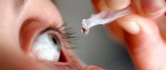
Contact lenses can be used as a barrier between the eyelashes and the cornea itself, and moisturizing drops can help improve the condition of the cornea.
Correction by surgery
If we talk about the most effective method that guarantees the correction of pathology, this is surgical intervention. The type of operation depends on the form of the disease, it can be: excision of the skin, dissection of the muscle, or sutures that retract the eyelid.
There are a number of contraindications for surgical intervention:
- if there are inflammatory processes on the skin;
- malignant neoplasms;
- pregnancy and lactation;
- elevated temperature;
- systemic blood diseases.
Important! It is strictly forbidden to perform surgery on patients with a pacemaker and metal implants in the facial area. The prognosis after surgery is very favorable; relapses usually occur only with senile entropion.
Entropion of the eyelids in Chinese Shar Pei and Chow Chow.
These breeds have extensive excess skin folds on the face that hang over the eyes and contribute to the formation of entropion. In addition, in these breeds, the skin of the eyelids is elastic, but does not have sufficient strength. Veterinary ophthalmologists take these features into account when performing operations. In parallel with the correction of the eyelids themselves, in some cases, skin folds on the forehead are fixed or partially removed (Fig. 2, 3, 4).
Inversion of the upper lower eyelid in a Shar Pei puppy. (Figure 2.)
The same Shar Pei puppy after eyelid surgery. (Fig. 3.)
Additional fixation of skin folds after correction of the position of the eyelids in a Shar Pei puppy. (Fig. 4.)
Almost all Shar Peis exhibit entropion of the eyelids, usually bilaterally. According to localization, the lower eyelid, upper eyelid and external commissure of the eyelids can be rolled up. The degree of the defect is different for all dogs. Sometimes we have to perform operations on very young puppies (1-2 months old), in which, due to the severe degree of the defect, the eyes are no longer visible (Fig. 5).
A 5-week-old puppy immediately after eyelid surgery (Fig. 5.)
Some Shar Peis with severe entropion require multiple surgeries to completely correct the defect.
Entropion of the eyelids in a Central Asian and Caucasian shepherd dog.
Until the 90s, we practically never encountered this defect in Central Asian and Caucasian shepherd dogs. In recent years, entropion of the eyelids in Central Asian Shepherds and entropion of the eyelids in Caucasian Shepherds have become a widespread pathology. This is due to the use of inbreeding (related breeding), and the appearance of larger, “raw” dogs.
These breeds usually have entropion of the lower eyelid, less commonly external commissure of the eyelids, and even more rarely of the upper eyelid. In this case, the lower eyelid is usually deformed. During operations, the entropion is eliminated and the shape of the lower eyelid is restored.
Inversion and eversion of the eyelids in the Cane Corso.
In the Cane Corso, as in other dogs (American bulldogs, Ca de Bou, etc.), inversion of the lower eyelids is usually combined with inversion of the lower eyelids
. Sometimes the external commissure of the eyelids and the upper eyelid are folded (Fig. 6).
Inversion and eversion of the lower eyelid in a Cane Corso. (Fig.6)
When performing corrective operations, both entropion and eversion of the eyelids are eliminated.
Entropion of the eyelids in pugs and Pekingese.
These dog breeds have physiological exophthalmos (protrusion of the eyeball is normal), in addition, they have an extensive skin fold in the nose area. These two factors lead to inversion of the inner corner of the lower eyelid (Fig. 7).
Inversion of the medial angle of the lower eyelid in a pug. (Fig.7)
Inversion and inversion of the lower eyelid in English bulldogs.
The English Bulldog has a pronounced nasal skin fold, which causes the inside of the lower eyelid to roll up. Also, in English Bulldogs, inversion of the external commissure of the eyelids and upper eyelid is often observed. Skin folds on the forehead contribute to an increase in the degree of inversion of the upper eyelid. In these dogs, along with volvulus, ectropion of the lower eyelid
(Fig.8).
Entropion of the eyelids in an English bulldog. Deep parenchymal ulcer of the cornea. (Fig. 8.)
Is it difficult to care for a dog after entropion surgery?
Postoperative care takes quite a long time. We ask owners to pay attention to this! There is no point in having surgery if the owners cannot provide adequate post-operative care.
When correcting entropion in dogs, we use fairly thin suture material. This is good because after the operation there is not the slightest trace left, but! the dog can remove one or more stitches with one movement of the paw. Therefore, a protective collar is put on the dog immediately after surgery so that it cannot damage the stitches. The dog wears a protective collar constantly until the stitches are removed. The collar should be 4-5 cm longer than the tip of the dog's nose. Otherwise, the dog may rub its eye on the corners of the furniture and pull out the seams. Also, during the postoperative period, the dog is isolated from other animals.
On the first and second days after surgery, 2-3 times a day, the suture is treated with a 0.05% solution of chlorhexidine bigluconate using a sterile gauze pad. Also, once a day, before removing the stitches, you must carefully lubricate the nodules with a cotton swab moistened with 70% alcohol. When processing, we “comb” each seam from the knot to the ends in the direction from the eye so that the ends of the threads do not touch the cornea. The postoperative suture should be in perfect condition, without crusts of dried blood and other discharge.
Eye drops after surgery
In addition, the veterinarian ophthalmologist prescribes certain eye drops and general antibiotics to prevent inflammation individually for each case.
The sutures are removed after 2 or 3 weeks; this period depends on the surgical technique, which is selected individually for each dog.
Thus, the success of the operation depends on the precise work of the ophthalmologist and the responsible care of the dog owners.
We would like to note with gratitude that most of the owners with whom we collaborated followed all recommendations with the utmost precision, which led to an ideal result.
Treatment
: normalization of the relationship between the anatomical and topographic structures of the eyelids and the eyeball.
Treatment tactics Non-drug treatment: · table No. 15; · mode 3.
Drug treatment: Antibacterial for the prevention of inflammation of the anterior segment of the eye: sodium sulfacetamyl - eye drops 30%, 2 drops 2-3 times a day for 7-10 days; · chloramphenicol – eye drops 0.25%, 2 drops 4 times a day for 5-7 days. Anti-inflammatory drug for the prevention of postoperative inflammation of the anterior segment of the eye · dexamethasone - eye drops 0.1%, 2 drops 2-3 times a day for 5 days. Anesthetics: · oxybucaine - eye drops 0.4%/5ml; 2 drops three times before surgery; · lidocaine – injection solution 2%, 0.5 ml once during surgery.
Entropion and trichiasis of the century, ectropion of the century
- print version
- Download or send a file
Recommended by the Expert Council of the RSE at the PVC "Republican Center for Healthcare Development" of the Ministry of Health and Social Development dated September 30, 2020 Protocol No. 10
Entropion is the turn of the century. The edge of the eyelid is turned inward towards the eyeball partially or throughout its entire length [7,8].
Trichiasis is an abnormal position of the eyelashes, they are turned towards the eye [7,8].
Ectropion is an inversion of the eyelid. The edge of the lower eyelid lags behind the eyeball or is turned down, the mucous membrane of the eyelid is turned outward [7,8].
Protocol name – Entropion and trichiasis of the century, ectropion of the century
Protocol code:
code(s) :
H02 Other diseases of the centuryH02.0 Entropion and trichiasis of the centuryH02.1 Ectropion of the century.Q10.2 Entropion of the century
Abbreviations used in the protocol: none.
Date of protocol development/revision: 2015.
Patient category: adults, children.
Users of the protocol: therapists , pediatricians , general practitioners, ophthalmologists, ophthalmic surgeons.
Mobile application “MedElement”
Download the application for ANDROID / for iOS
Mobile application “MedElement”
Download the application for ANDROID / for iOS
Clinical classification (Fedorov, Yartseva, Ismankulov/“Eye diseases”/Moscow/2005) [8].
· by etiological factor: · involutional (senile) entropion, ectopion; · cicatricial (due to cicatricial changes in the conjunctiva, burns, injuries); · spastic (due to surgical trauma, eye irritation, blepharospasm).
· congenital. List of basic and additional diagnostic measures.
Basic (mandatory) diagnostic examinations carried out on an outpatient basis [3,4,5]: · visometry (without correction and with correction)*; · biomicroscopy; · measurement of the palpebral fissure width*; · fluorescein test*;
The minimum list of examinations carried out to prepare for surgical treatment:
· general blood test; · general urine test; · biochemical blood test (total protein, its fractions, urea, creatinine, bilirubin, ALT, AST, electrolytes, blood glucose); · microreaction to syphilis; · coagulogram (PTI, fibrinogen, FA , clotting time); · blood test for HIV using ELISA. Additional diagnostic examinations performed on an outpatient basis: · ophthalmoscopy; · Schirmer test; * · West test.*
Diagnostic criteria for diagnosis.
Complaints and anamnesis: · lacrimation – with trichiasis due to mechanical irritation of the cornea; with entropion due to non-immersion of lacrimal openings in the lacrimal lake; · photophobia - with trichiasis and entropion due to contact of eyelashes with the cornea, irritation of the cornea occurs; · decreased vision - due to damage to the cornea and tear formation; · abnormal growth of eyelashes, unevenness width of the palpebral fissure of both eyes; Physical examination: · external examination of the eyelid (turned towards the eye or turned away from the eye, eyelashes do not grow correctly); · measurement of the width of the palpebral fissure (the palpebral fissure is narrowed or widened); Laboratory tests: no Instrumental studies: · visometry - decreased visual acuity; biomicroscopy - the position of the eyelids and the location of the eyelashes in detail. Indications for consultation with specialists: therapist - to assess the general condition of the body before surgery; pediatrician - to assess the general condition of the body before surgery; neurologist - to exclude systemic and neurological diseases; · oncologist – if the presence of malignant tumors is suspected. Differential diagnosis Table - 1. Differential diagnosis
| Diagnostic sign | entropion | trichiasis | ectropion |
| eyelids | the edge of the eyelid is turned (inverted) towards the eye | eyelids are positioned correctly | the edge of the eyelid is turned outward |
| eyelashes | come into contact with the conjunctiva of the eyeball | come into contact with the conjunctiva of the eyeball | distant from the eye |
| Symptoms | foreign body sensation, lacrimation, hyperemia | foreign body sensation, lacrimation, hyperemia | foreign body sensation, lacrimation, lacrimation, hyperemia |
| One-way/two-way process | Single sided or double sided | Most often unilateral | Single sided or double sided |
| Conjunctival hyperemia | Expressed | Expressed | Weakly expressed |
| Nosology | Symptoms not typical for entropion and trichiasis |
| Keratoconjunctivitis | Reduced tear production, pericorneal conjunctival injection |
| Blepharitis | Swelling of the eyelids, ulcers, films |
| Iridocyclitis | Ciliary pain on palpation, congestive conjunctival injection, pupil constriction, corneal precipitation, enlarged lymph nodes. |
| Acute attack of glaucoma | Severe congestive injection of the conjunctiva, dull pain radiating to the occipital region, increased intraocular pressure, pupil dilation, lack of reaction to light. |
Treatment Goals
: normalization of the relationship between the anatomical and topographic structures of the eyelids and the eyeball.
Treatment tactics
Non-drug treatment: · table No. 15; · mode 3. Drug treatment: Antibacterial for the prevention of inflammation of the anterior segment of the eye: · sodium sulfacetamyl - eye drops 30%, 2 drops 2-3 times a day for 7-10 days; · chloramphenicol - eye drops 0.25% 2 drops 4 times a day for 5-7 days. Anti-inflammatory agent for the prevention of postoperative inflammation of the anterior segment of the eye · dexamethasone - eye drops 0.1% 2 drops 2-3 times a day for 5 days. Anesthetics: · oxybucaine - drops ophthalmic 0.4%/5ml; 2 drops three times before surgery; lidocaine – solution for injection 2%, 0.5 ml once during surgery. Other types of treatment. Other types of treatment provided on an outpatient basis: · fixation of the eyelids with adhesive tape.
Surgical intervention
The purpose of the surgical intervention: normalization of the relationship between the anatomical and topographic structures of the eyelids and the eyeball. Type of surgical intervention:
· eyelash hair removal (ICD9-08.93.)
Indication for surgical intervention: trichiasis. Contraindications: no
Diathermocoagulation of eyelashes ( ICD9-08.91.)
Indication for surgical intervention: trichiasis. Contraindications: no
· Eyelid surgery (ICD-08.36)
Indications for surgical intervention: · entropion, ectropion. Contraindications: · somatic condition of the patient; · uncompensated diabetes mellitus; · oncological diseases.
Surgical intervention provided on an outpatient basis:
· eyelash epilation; · diathermocoagulation of eyelashes; · eyelid plastic surgery.
Surgical intervention provided in an inpatient setting: not performed
Further management:
· observation by an ophthalmologist; · control examination 2 times a year (control of eyelash growth, eyelid position)
Indicators of treatment effectiveness:
· Correct ratio of the anatomical and physiological structures of the eyelids and the eyeball.
| Dexamethasone |
| Lidocaine |
| Oxybuprocaine |
| Sulfacetamide |
| Chloramphenicol |
Indications for hospitalization. · planned hospitalization – not carried out; · emergency hospitalization – not carried out. Preventive measures : · exclusion of injuries; · timely treatment of inflammatory diseases of the eyelids and conjunctiva.
- Minutes of meetings of the Expert Council of the RCHR of the Ministry of Health of the Republic of Kazakhstan, 2015
- References: 1) Mahvan TD, Hornecker JR, Buckley WA, Clark S. The Role of Besifloxacin in the Treatment of Bacterial Conjunctivitis/Ann Pharmacother. 2014 Feb2) American Academy of Ophthalmology. Guideline.Conjunctivitis – 20133) Clinical Knowledge Summaries.
Prodigy Guidance. Conjunctivitis – infectious. 2004. National Institute for Health and Care Excellence. 4) Prodigy Knowledge. Prodigy Guidance. Conjunctivitis – allergic.20045) International Council of Ophthalmology. Guideline Conjunctivitis. 2010.6) Sheikh A., Hurvitz B., Antibiotics Versus placebo for acute bacterial conjunctivitis. Cochrane Database Syst Rev.2012;9:CD0012117) Evidence-based medicine. Annual directory. 2003 – Part 7. 2314-2321. Fedorov S.N., Yartseva N.S., Ismankulov A.O. Eye diseases: A textbook for medical students. – 2nd ed. – M.: 2005.– 440 p.
List of protocol developers : 1) Uldanov Oleg Galimovich – Candidate of Medical Sciences, Associate Professor of the Department of Ophthalmology of the Kazakh National Medical University. S.D. Asfendiyarov, (Almaty).
2) Dzhumataev Erik Asylkhanovich – Candidate of Medical Sciences, doctor of the highest category, ophthalmologist at the Consultative and Rehabilitation Department of the Kazakh Research Institute of Eye Diseases (Almaty).
3) Niyazov Ilzat Alimzhanovich – doctor of the highest category, ophthalmologist at the prosthetics office of the Kazakh Research Institute of Eye Diseases. (Almaty).4) Mazhitov Talgat Mansurovich – Doctor of Medical Sciences, Professor of Astana Medical University JSC, clinical pharmacologist of the highest category, general practitioner of the highest category.
Conflict of interest – none.
Reviewer: Irina Anatolyevna Dolmatova – Doctor of Medical Sciences, head of the ophthalmology course at the Kazakh-Russian Medical University
Conditions for reviewing the protocol:
Review of the protocol 3 years after its publication and from the date of its entry into force or if new methods with a level of evidence are available.
If you are not a medical professional:
- By self-medicating, you can cause irreparable harm to your health.
- The information posted on the MedElement website and in the mobile applications “MedElement”, “Lekar Pro”, “Dariger Pro”, “Diseases: a therapist’s guide” cannot and should not replace a face-to-face consultation with a doctor. Be sure to contact a medical facility if you have any illnesses or symptoms that concern you.
- The choice of medications and their dosage must be discussed with a specialist. Only a doctor can prescribe the right medicine and its dosage, taking into account the disease and condition of the patient’s body.
- The MedElement website and mobile applications “MedElement (MedElement)”, “Lekar Pro”, “Dariger Pro”, “Diseases: a therapist’s reference book” are exclusively information and reference resources. The information posted on this site should not be used to unauthorizedly change doctor's orders.
- The editors of MedElement are not responsible for any personal injury or property damage resulting from the use of this site.
Source: https://diseases.medelement.com/disease/%D1%8D%D0%BD%D1%82%D1%80%D0%BE%D0%BF%D0%B8%D0%BE%D0%BD -%D0%B8-%D1%82%D1%80%D0%B8%D1%85%D0%B8%D0%B0%D0%B7-%D0%B2%D0%B5%D0%BA%D0% B0-%D1%8D%D0%BA%D1%82%D1%80%D0%BE%D0%BF%D0%B8%D0%BE%D0%BD-%D0%B2%D0%B5%D0% BA%D0%B0/14394